Introduction
Embryo production by in vitro fertilization (IVF) is a well established tool to evaluate oocyte competence. IVF, however, depends on the viability of spermatozoa (Zheng, Reference Zheng2007). For capuchin monkeys (Cebus apella), in vitro capacitation of spermatozoa is challenging because of their unique seminal coagulum (Dixson & Anderson, Reference Dixson and Anderson2002). Although motile spermatozoa from capuchin monkeys can be obtained after liquefaction of the semen coagulum in coconut water-based solution (Oliveira et al., Reference Oliveira, Miranda, Leão, Brito, Santos and Domingues2011), it does not guarantee that harvested spermatozoa will be able to fertilize oocytes. To evaluate the quality of the spermatozoa, embryo production via IVF (Morgan et al., Reference Morgan, Boatman and Bavister1991) or by intracytoplasmic sperm injection (ICSI; Yamasaki et al., Reference Yamasaki, Iwatani, Tsuchiya, Okahara, Sankai and Torii2011) can be applied. However, embryo development is influenced by the oocyte quality. Therefore, parthenogenesis appears to be an excellent option to investigate oocyte quality (Paffoni et al., Reference Paffoni, Brevini, Gandolfi and Ragni2008). Parthenogenetic activation (PA) is usually performed using activating agents such as ionomycin associated with protein kinase inhibitors, i.e. 6-dimethylaminopurine (6-DMAP) or roscovitine (Mitalipov et al., Reference Mitalipov, Nusser and Wolf2001; Grupen et al., Reference Grupen, Gilchrist, Nayudu, Barry, Schulz, Ritter and Armstrong2007). Usually in primates, females are stimulated with hormones to harvest in vivo matured oocytes (Marchall et al., Reference Marchall, Wilton and Moore1998; Mitalipov et al., Reference Mitalipov, Nusser and Wolf2001; Grupen et al., Reference Grupen, Gilchrist, Nayudu, Barry, Schulz, Ritter and Armstrong2007) because of the higher rates of cytoplasmic and nuclear maturation when compared with in vitro matured oocytes (Zheng et al., Reference Zheng, Wang, Bavister and Ji2001; Domingues et al., Reference Domingues, Caldas-Bussiere, Petretski, Ohashi, Lima, Santos, Cordeiro and Castro2010). Recently, we have shown the feasibility of the approach of in vitro matured oocytes collected from hormonally unstimulated ovaries of capuchin monkeys (Domingues et al., Reference Domingues, Caldas-Bussiere, Petretski, Ohashi, Lima, Santos, Cordeiro and Castro2010). In this former study, however, we have performed in vitro maturation (IVM) for 36 h only, using pregnant mare serum gonadotrophin (PMSG) and human chorionic gonadotrophin (hCG) in association, and act as follicle stimulating hormone (FSH) and luteinising hormone (LH) stimulators, respectively. It has been suggested that FSH and LH might be more efficient for oocyte IVM than PMSG and hCG (Zheng, Reference Zheng2007). Furthermore, the ability of in vitro matured oocytes to be either fertilized or to form parthenotes has not been evaluated, as yet, in capuchin monkeys. We aimed to establish an optimal IVM protocol for capuchin monkeys by testing two different culture lengths (36 and 40 h) and the effect of FSH and LH on the IVF and PA of oocytes collected from unstimulated females. PA was tested in the presence of ionomycin in combination with either 6-DMAP or roscovitine. Finally, we assessed spermatozoon quality after recovery from seminal coagulum using an acyl carrier protein extender ACP-118®.
Materials and methods
All chemicals were purchased from Sigma-Aldrich (Sigma Chemical Co., St. Louis, MO, USA), unless otherwise stated.
Animals
All experimental protocols were approved by the Ethical Committee in Animal Research (no.045/2008/CEPAN/IEC/SVS/MS) and by the Brazilian Institute for Wildlife and Environment (IBAMA). Nine healthy, sexually mature capuchin monkey specimens (six females and three males), aged 10–20 years were used in this study.
Oocyte collection and preparation
At days 7–11 of the menstrual cycle, antral follicles that contained immature oocytes were punctured from unstimulated capuchin monkeys by laparotomy (Domingues et al., Reference Domingues, Caldas-Bussiere, Petretski, Ohashi, Lima, Santos, Cordeiro and Castro2010). Aspirated oocytes were divided into three groups: (i) denuded oocytes (DO); (ii) oocytes surrounded by 1–2 layers of cumulus cells (partially intact oocytes; PIO); and (iii) oocytes surrounded by two or more layers of cumulus cells (intact oocytes; IO). Oocytes were submitted separately to IVM for 40 h. To this end, oocytes were washed twice in TCM199 HEPES medium and subsequently cultured in 100 μl droplets (10 oocytes/100 μl) of TCM199 supplemented with 50 μg/ml LH (Bioniche Animal Health, Bellevile, Ontario, Canada), 0.5 μg/ml hFSH (Bioniche Animal Health), 0.5 mg/ml epidermal growth factor, 0.22 mM sodium pyruvate, 50 mM β-mercaptoethanol, and 10% fetal bovine serum (FBS).
In vitro maturation procedure
IVM was performed under mineral oil at 38.5°C in a humidified atmosphere of 5% CO2 in air. Cumulus cell expansion was assessed before (0 h) during (36 h) and after (40 h) IVM. The degree of expansion was assessed according to a subjective scale from 0–4 (Morgan et al., Reference Morgan, Boatman and Bavister1991). According to cumulus cell expansion, IVM was performed for 40 h and oocytes were submitted to IVF (n = 10) or PA (n = 32). For IVF, used semen was collected and freshly manipulated as described by Oliveira et al. (Reference Oliveira, Miranda, Leão, Brito, Santos and Domingues2011), applying ACP-118® (Biotecnologia, CE, Brazil) as extender. After liquefaction of the seminal coagulum, samples were drawn into 0.25 ml plastic straws (Minitüb, Tiefenbach, Germany), sealed with a metal bead and cooled gradually from 36°C to 4 °C (4°C/min) and stored at 4°C. Spermatozoa were selected for IVF by swim up. Insemination was carried out by adding a 5-μl aliquot of sperm (5 × 10⁶ sperm/ml) to an 80-μl droplet of IVF medium that contained 10 oocytes. The fertilization medium consisted of TALP medium (Parrish et al., Reference Parrish, Susko-Parrish, Winer and First1988), supplemented with hypotaurine, epinephrine and penicillamine to induce hyperactivation (Wolf et al., Reference Wolf, Thormahlen, Ramsey, Yeoman, Fanton and Mitalipov2004). Incubation was performed under mineral oil, for 30 h at 38.5°C in a humidified atmosphere of 5% CO2 in air. For PA, oocytes were denuded with 0.5 μg/ml hyaluronidase and the extrusion of the first polar body was recorded. Subsequently, oocytes were exposed for 5 min to 5 μM ionomycin in TALP/HEPES medium supplemented with 1 mg/ml bovine serum albumin (BSA), followed by washing in TALP/HEPES medium 30 mg/ml BSA added for 3 min. After activation with ionomycin, oocytes were divided into two groups, i.e. treated with 2 mM 6-DMAP (n = 20) or 50 μM roscovitine (n = 12), transferred to SOF medium and incubated for 3 h at 38.5°C in 5% CO2 in air. Subsequently to IVF and PA, zygotes that displayed pronuclei were transferred to SOF medium, supplemented with 6 mg/ml BSA and 10% FBS and cultured on a cumulus cells monolayer. Extrusion of the second polar body and pronuclear formation were evaluated after 24 h and embryo in vitro development was assessed. For this process, cleavage was assessed and the nuclei of embryos were labelled with Hoechst 33342 and counted under fluorescence microscopy (Eclipse TE300, Nikon, Japan).
Statistical analysis
All data were reported as mean ± standard error of the mean (SEM). The Kolmogorov–Smirnov test (BioEstat 5.0, Belém, Pará, Brazil) was performed to verify normality. The analyses described as follows were performed using StatView software (SAS Institute Inc., Cary, NC, USA). The meiotic competence among DO, PIO and IO was tested by Kruskal–Wallis test. For the cumulus cells expansion, data were statistically evaluated by using the Mann–Whitney test to compare oocytes within the same classification (PIO and IO) and time of culture. The Kruskal–Wallis test was used to verify differences between cumulus expansion (I, II, III, IV) in the same oocyte classification and within the same time of culture. Analysis of variance (ANOVA) was used to evaluate the effect of treatment (IVF, roscovitine, 6-DMAP) in the percentage of second polar body extrusion, pronuclear formation, and cleavage. Differences between means were determined with Fisher's protected least signficant difference post-hoc test. Differences were considered to be statistically significant when p-values were <0.05.
Results
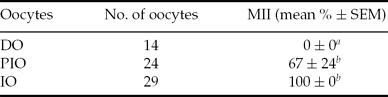
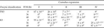
A total of 67 oocytes were retrieved from 14 ovarian puncturings (4–5 oocytes/female), from which 14 were denude, 24 were partially intact (PIO) and 29 were intact (IO) (Table 1). Although 87% of the oocytes (100% of the IO and 67 ± 24% of the PIO) reached MII, only IO presented complete expansion of cumulus cells (87 ± 8% grade 4), while PIO remained at grades II (64 ± 15%) and III (23 ± 10 %) of expansion of the cumulus cells (Table 2).
Percentages of denuded oocyte (DO), partially intact oocytes (PIO) and intact oocytes (IO) capable to reach MII after 40-h IVM
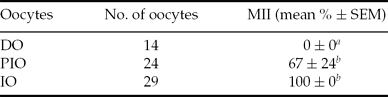
a ,b Different lower-case letters within columns indicate values that are significantly different.
IVM, in vitro maturation; MII, metaphase II; SEM, standard error of the mean.
Degrees of cumulus cells expansion in intact oocytes (IO) and partially intact oocytes (PIO) at two different IVM time periods: 36 and 40 h

a,bDifferent lower-case letters indicate differences between IO and PIO within the same cumulus expansion degree and time of in vitro maturation (IVM).
A–C Different upper-case letters indicate differences between cumulus expansion degree (I, II, III, IV) within the same oocyte classification and time of IVM.
E,FDifferent upper-case letters indicate differences between IVM time in the same cumulus expansion degree and oocyte classification.
Collected semen presented 80% of motility and vigour 4 after dilution in ACP-118® and cooling for 6 h at 4°C. Spermatic capacitation in TALP took 30 min. After 30 h of IVF, extrusion of the second polar body was observed in 40 ± 16% of the oocytes, and 20 ± 13% of the formed zygotes were able to cleave to the 4-cell stage. Parthenotes were able to cleave only when oocytes were activated by 6-DMAP, while roscovitine-treated oocytes formed haploid zygotes and no cleavage was observed (Table 3).
Percentages of second polar body (2PB) extrusion, pronuclear formation (one or more) and cleavage after in vitro fertilization and parthenogenetic activation of in vitro matured oocytes

a,bDifferent lower-case letters within columns represent significantly different values.
6-DMAP, 6-dimethylaminopurine; IVF, in vitro fertilization.
Discussion
This study is the first one to describe in vitro embryo production in capuchin monkeys. It is important to note that we used unstimulated females, whereas most studies that report in vitro embryo production from other NHPs have been performed using in vivo matured oocytes collected from hormonally stimulated primate females (Marchall et al., Reference Marchall, Wilton and Moore1998; Mitalipov et al., Reference Mitalipov, Nusser and Wolf2001; Grupen et al., Reference Grupen, Gilchrist, Nayudu, Barry, Schulz, Ritter and Armstrong2007). Complete cumulus expansion was obtained after 40 h IVM, and most of the cultured IO reached MII. In a previous study, we obtained a mean of 55% MII oocytes after 36 h IVM, using PMSG and hCG (Domingues et al., Reference Domingues, Caldas-Bussiere, Petretski, Ohashi, Lima, Santos, Cordeiro and Castro2010). In the present study, we showed that FSH and LH are also efficient in the in vitro maturation of oocytes from capuchin monkeys. Moreover, in contrast with oocytes from rhesus monkeys that reach MII in vitro independently of the cumulus expansion grade (Morgan et al., Reference Morgan, Boatman and Bavister1991), cumulus expansion interferes in the meiotic competence of oocytes from capuchin monkeys. This discrepancy may reflect species-specific differences in the cumulus expansion mechanism.
The semen from capuchin monkeys is characterized by its high spermatic concentration on its coagulum (Oliveira et al., Reference Oliveira, Miranda, Leão, Brito, Santos and Domingues2011). We have obtained viable spermatozoa from diluted and cooled semen in ACP-118®. This extender was able to maintain spermatozoon integrity and fertilization capacity even after 6 h. As far as we know, this report is the first time that oocytes from capuchin monkeys have been fertilized in vitro with spermatozoa harvested from seminal coagulum using ACP-118®. Spermatozoon quality was preserved when chilled semen was liquefied in ACP-118®. Similarly, Silva et al. (Reference Silva, Peixoto, Santos, Castelo, Oliveira and Silva2011) have shown the importance of ACP in processing and cryopreservation of spermatozoa from agouti. These authors suggested that ACP compounds such as proteins and indole-3-acetic acid enhance sperm motility and preservation at low temperatures.
Successful IVF and parthenogenesis means that embryo competence is not compromised by the chemical exposure. The applied protocol to produce in vitro embryo from capuchin monkeys allowed the cleavage to 4-cells when matured oocytes were submitted to IVF or activated by ionomycin/6-DMAP. Use of 6-DMAP resulted in a better rate of embryo production than the use of specific inhibitors of MPF, e.g. roscovitine. Similar results have been reported previously in a study using Macaca mulatta (Mitalipov et al., Reference Mitalipov, Nusser and Wolf2001). It has been shown that when PA protocols result in haploids parthenotes, e.g. ionomycin/roscovitine, initial cleavage will be prejudiced (Schramm and Bavister, Reference Schramm and Bavister1999). Embryo development was stopped at the 4-cell stage, similar to that observed previously in Callithrix jacchus treated with ionomycin/6-DMAP (Grupen et al., Reference Grupen, Gilchrist, Nayudu, Barry, Schulz, Ritter and Armstrong2007). However, M. mulatta was submitted to the same treatment and blastocyst formation was obtained at a similar rate in embryos produced by ICSI (Mitalipov et al., Reference Mitalipov, Nusser and Wolf2001). As observed by other groups (Mitalipov et al., Reference Mitalipov, Nusser and Wolf2001, Grupen et al., Reference Grupen, Gilchrist, Nayudu, Barry, Schulz, Ritter and Armstrong2007), the most advanced embryo stages were observed after IVF. We suggest that our PA protocols may lead to failure in the development of the parthenotes (Grupen et al., Reference Grupen, Gilchrist, Nayudu, Barry, Schulz, Ritter and Armstrong2007).
We have established the optimal IVM length (40 h) and the positive effect of FSH and LH on IVF and PA in oocytes collected from unstimulated female capuchin monkeys. Spermatozoa from capuchin monkeys obtained from semen dissolution in coconut water (ACP-118®) were competent to fertilize in vitro. Comparison between those treatment allows us investigate the effects of oocyte development on meiotic and development oocyte competence.
Acknowledgements
The authors thank CENP for logistic support.