Introduction
Extramedullary plasmacytoma is a rare, malignant proliferation of plasma cells which occurs without affecting the bone or bone marrow.
Tumours may be multicentric, develop in any tissue, and occur as part of the multiple myeloma complex. Tumours most commonly occur in the upper respiratory tract (in the paranasal sinuses, nasopharyngeal region, nasal cavity and tonsils), but can appear in the lymphatic ganglions, parotid glands, lungs, pleura, muscle, thyroid, gastrointestinal tract, liver, spleen, pancreas, testicles, breast and skin.Reference Alexiou, Kau and Dietzfelbinger1–Reference Gormley, Primrose and Bharucha3 The vague symptoms associated with this disease can delay diagnosis, with potentially disastrous consequences for the patient.
We believe that the patient presented below represents the first reported case of multicentric extramedullary plasmacytoma presenting on different occasions.
Case report
A 53-year-old man initially presented to the service 30 years previously. At that time, during a routine anaesthetic for an unrelated procedure, he was noted to have a laryngeal lesion on the right aryepiglottic fold and false vocal fold, which measured approximately 1.75 cm in diameter.
Microlaryngoscopy was performed and the lesion excised endoscopically. Histological examination revealed a plasmacytoma, with clear surgical margins.
The patient remained disease-free for the following sixteen years.
Sixteen years after the initial procedure, he re-presented with a firm, lobulated mass at the base of the tongue involving the left vallecula. Endoscopic excision was performed and, again, a histological diagnosis of plasmacytoma was made. The surgical margins of the excision were tumour-free. Serum electrophoresis excluded multiple myeloma, and a skull base computed tomography (CT) scan showed no signs of destructive change.
The patient re-presented once more, nine years later, with a further mass in the left post-nasal space. Magnetic resonance imaging of the skull base showed fullness of the left pharyngeal wall (Figure 1).
Axial magnetic resonance imaging scan of the skull base, showing fullness of the left pharyngeal wall (arrow). L = left

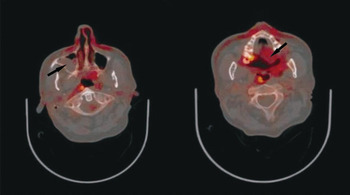
Endoscopic excisional biopsy of the lobulated lesion again showed recurrent plasmacytoma, with clear surgical margins. Investigation included analysis of bone marrow aspirates and trephine biopsy, in addition to positron emission tomography CT. There was no evidence of systemic plasma cell disease. The positron emission tomography CT showed focal increased fluorodeoxyglucose uptake at the inferior right maxillary antrum, at the alveolar plate (maximum Standardized Uptake Value (SUV) 8.4), and also at the soft palate on the left side (maximum SUV 5.7), without any evidence of active malignant disease elsewhere (Figure 2).
Axial positron emission tomography computed tomography scans, showing focal increased fluorodeoxyglucose uptake (arrows) at the inferior right maxillary antrum, at the alveolar plate, and also at the soft palate on the left side.

Multiple biopsies from the soft palate and the right maxillary antrum were performed. Histological examination showed no evidence of field change.
At the time of writing, the patient remained well, under both otolaryngological and oncological follow up.
Discussion
Plasmacytoma is a rare neoplasm which originates in B lymphocytes.Reference Loh4–Reference Kapadia, Desai and Cheng7 The worldwide annual incidence is 3 per 100 000. Eighty per cent of cases occur in the upper respiratory tract, and 10 per cent of cases are multiple.Reference Waldron and Mitchell6
Plasma cell tumours may develop in any tissue, be multicentric, and occur as part of a multiple myeloma complex.Reference Batsakis8, Reference Malpas, Bergsagel and Kyle9 Between 80 and 90 per cent of extramedullary plasmacytomas are located in the head and neck.Reference Gormley, Primrose and Bharucha3, Reference Waldron and Mitchell6 Of these, 40 per cent are located in the nasal cavities and paranasal sinuses, approximately 20 per cent are nasopharyngeal, and approximately 18 per cent are oropharyngeal. Rarely, plasma cell tumours can occur in the tongue, maxillary sinus and larynx.Reference Gormley, Primrose and Bharucha3, Reference Waldron and Mitchell6, Reference Malpas, Bergsagel and Kyle9, Reference Mendenhall, Thar and Million10
Schridde described the first case of plasmacytoma of the upper respiratory tract in 1905, and only approximately 300 cases have since been published.
Diagnosis of a solitary extramedullary plasmacytoma is complex and requires radiological, haematological, biochemical and histological investigation.Reference Gormley, Primrose and Bharucha3, Reference Mendenhall, Thar and Million10–Reference Kyle14 It can only be confirmed when the presence of systemic disease is excluded via serum and urinary protein electrophoresis, skeletal survey, bone scan and bone marrow trephine.Reference Waldron and Mitchell6–Reference Malpas, Bergsagel and Kyle9
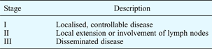
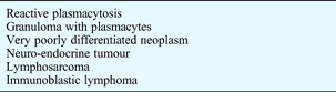
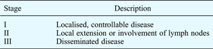
Once the diagnosis is confirmed, extramedullary plasmacytoma can be staged as shown in Table I.Reference Waldron and Mitchell6 The differential diagnosis is summarised in Table II.Reference Gormley, Primrose and Bharucha3, Reference Galieni, Cavo and Pulsoni5, Reference Abemayor, Canalis, Greenberg, Wortham, Rowland and Sun11
Stages of extramedullary plasmacytoma
Differential diagnosis of extramedullary plasmacytoma
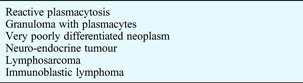
Treatment is determined by the degree of local extension and the presence or absence of systemic involvement. Combined therapy has been proven effective; the alternatives are surgery plus radiotherapy or chemotherapy.Reference Hardwood, Cummings and Fitzpatrick15–Reference Kost18 Some authors prefer radiotherapy alone when the tumour is located in an anatomical region with restricted surgical access; however, others report that the radio-sensitivity of plasmocytoma is variable.Reference Malpas, Bergsagel and Kyle9, Reference Mendenhall, Thar and Million10, Reference Hardwood, Cummings and Fitzpatrick15
Primary extramedullary plasmacytoma can progress either to multiple myeloma (in 17–33 per cent of cases)Reference Gormley, Primrose and Bharucha3, Reference Wax, Yun and Omar19 or multiple plasmocytoma (in 10 per cent).Reference Waldron and Mitchell6
The typical presentation of multiple myeloma is anaemia and bone pain. Biochemical investigation may reveal hypercalcaemia, renal dysfunction, and monoclonal spikes on electrophoresis. Plasmacytoma is often essentially a diagnosis of exclusion, and bone scintigraphy, X-ray imaging and exclusion of Bence–Jones protein is required to confirm the diagnosis.Reference Abemayor, Canalis, Greenberg, Wortham, Rowland and Sun11, Reference Knowling, Harwood and Bergsagel20, Reference Soutar, Lucraft and Jackson21
The real incidence of extramedullary plasmacytoma is difficult to quantify, due not only to the rarity of the disease but also to its long latency period and the degree of overlap between it and other plasmacyte dyscrasias.Reference Gormley, Primrose and Bharucha3, Reference Waldron and Mitchell6, Reference Kost18
The prognosis for patients with extramedullary plasmocytoma is generally favourable compared with that for multiple myeloma.Reference Galieni, Cavo and Pulsoni5, Reference Batsakis8 The five-year survival rate is excellent, at 90 per cent, and recent data suggest that local regression does not necessarily indicate a poorer prognosis.Reference Waldron and Mitchell6
• Extramedullary plasmacytoma is a rare tumour of the upper respiratory tract; the diagnosis is often one of exclusion as there is no specific macroscopic appearance or symptom profile
• The condition can present with either solitary or multifocal disease
• The head and neck are the most common primary sites, although tumours may occur anywhere in the body
• Diagnosis requires thorough biochemical, haematological, immunohistochemical and radiological investigation
• A multidisciplinary approach to treatment is required
• The tumour has a tendency to disseminate widely; careful, long-term follow up is thus required
Our case is notable as being within the 10 per cent of cases which present with tumour at multiple sites: our patient presented on different occasions with tumour in three separate sites within the head and neck, over a protracted course of 30 years. Our patient was managed surgically on all occasions, and at the time of writing remained disease-free. After reviewing the literature, we believe that our patient represents the only reported case with this extremely unusual presentation.