Introduction
Studies on mammalian oocyte cryopreservation date back to the end of 1970, when a rapid freezing/thawing protocol was applied (Whittingham, Reference Whittingham1977). It is not yet clear when the application of oocyte cryopreservation started in the human reproductive medicine contest, but today it is known that the news of the first child birth from a cryopreserved oocyte was published in 1986 (Chen, Reference Chen1986). Twenty-two years later, oocyte cryopreservation still represents a theoretical option for the management and preservation of female fertility and for the improvement of ART results (Imai et al., Reference Imai, Furui and Yamamoto2008).
It is known that the human oocyte is a very complex cell, highly sensible to intrinsic and extrinsic perturbations, including modest temperature modifications that are able to cause lethal damage during the preimplantation and postimplantation phases. To prevent the damage associated with cryopreservation, it is possible to use substances that are capable of protecting the oocyte. On the other hand, however, these same substances have cytotoxic effects and are able to determine total or partial oocyte degeneration, which affects meiotic spindle, pericortical mitochondria, calcium pathway regulation, cortical granule release and zona pellucida structure (Zeron et al., Reference Zeron, Pearl, Borochov and Arav1990; Fabbri et al., Reference Fabbri, Porcu, Marsella, Rocchetta, Venturoli and Flamigni2001; Jones et al., Reference Jones, Van Blerkom, Davis and Toledo2004).
Recent advances in cryopreservation techniques partially overcome the biological vulnerability of the metaphase II oocytes and increase the survival rates of frozen oocytes, with satisfactory fertilization and cleavage rates and good embryonic morphology following intracytoplasmic sperm injection (ICSI) (Gook, Reference Gook and Edgar2007). Despite these encouraging biological results, in most current oocyte cryopreservation programmes clinical outcome still remains unsatisfactory (La Sala et al., Reference La Sala, Nicoli, Villani, Pescarini, Gallinelli and Blickstein2006; Oktay et al., Reference Oktay, Cil and Bang2006).
Previous studies (Capmany et al., Reference Capmany, Tailor, Braude and Bolton1996; Payne et al., Reference Payne, Flaherty, Barry and Matthews1997; Scott, Reference Scott2003; James et al., Reference James, Hennessy, Reggio, Weimer, Larsen and Cohen2006; Nicoli et al., Reference Nicoli, Valli, Di Girolamo, Di Tommaso, Gallinelli and La Sala2007; Gianaroli et al., Reference Gianaroli, Magli, Ferraretti, Fortini and Greco2003, Reference Gianaroli, Magli, Ferraretti, Lappi, Borghi and Ermini2007) on zygote morphological evaluation have tried to create a new non-invasive tool to substitute and/or improve the embryo selection for embryo transfer (ET). This evaluation includes: (i) the observation of the position of the pronuclei in the zygote cytoplasm; (ii) the number, dimension and distribution of nucleoli within the pronuclei; and (iii) the orientation of polar bodies in the axis of pronuclei (Gianaroli et al., Reference Gianaroli, Magli, Ferraretti, Fortini and Greco2003; Nicoli et al., Reference Nicoli, Valli, Di Girolamo, Di Tommaso, Gallinelli and La Sala2007). Pronuclear morphology seems to be an important predictive value of zygote development and integrity, even though the clinical significance in terms of pregnancy rate still remains uncertain (James et al., Reference James, Hennessy, Reggio, Weimer, Larsen and Cohen2006; Nicoli et al., Reference Nicoli, Valli, Di Girolamo, Di Tommaso, Gallinelli and La Sala2007).
The aim of the present study was to examine the possible relationship between pronuclear morphology and insemination technique, semen quality and oocyte cryopreservation procedure, verifying the biological impact on pronuclei organization before the embryo cleavage stage. The evaluation was performed on zygotes that were obtained from sibling oocytes, non-frozen and frozen, including only patients that after the ovarian stimulation had obtained at least three non-frozen oocytes to be inseminated and one or more oocytes to be cryopreserved.
Materials and Methods
The Italian Infertility Law, dated 10 March 2004 (Clarke, Reference Clarke2003; Turone, Reference Turone2004), restricted the number of inseminated oocytes in each cycle of assisted reproduction to three, with subsequent obligation to transfer all resulting embryos. In addition, the law prohibited freezing of surplus embryos. This study was carried out in our non-private infertility centre at Arcispedale S. Maria Nuova of Reggio Emilia, Italy. The ART procedures were done in a predominantly (>95%) Italian population. All women (with few exceptions) were nulliparous, with a mean (±SD) age of 35.1 ± 3.5 (range 25–41) years.
During the period from January 2005 to December 2006 we analysed the zygote morphology of 190 patients who performed 208 cycles with the insemination of three non-frozen oocytes (85 conventional IVF and 123 ICSI cycles) and that, after failure in getting pregnant, performed 208 cycles with the insemination of sibling frozen oocytes that were cryopreserved the day of oocyte retrieval. The study was approved by the local Ethical Committee of Reggio Emilia.
Oocyte retrieval, embryo culture and oocyte cryopreservation/thawing protocol
Ovarian stimulation and oocyte retrieval were performed as reported elsewhere (Nicoli et al., Reference Nicoli, Valli, Di Girolamo, Di Tommaso, Gallinelli and La Sala2007).
For conventional IVF procedure, three non-frozen oocytes were cultured individually and inseminated in microdrops of fresh medium (Cook IVF) under mineral oil; for ICSI procedure, following the removal of the oocyte surrounding cumulus and corona cells, nuclear maturation assessment was performed using an inverted microscope (TE 2000-U, Nikon Corp.), to ensure the injection of metaphase II oocytes exclusively. ICSI was performed as reported by Palermo et al. (Reference Palermo, Joris, Devroey and Van-Steirteghem1992).
Surplus oocytes were cryopreserved with a slow freezing–rapid thawing protocol as described elsewhere (La Sala et al., Reference La Sala, Nicoli, Villani, Pescarini, Gallinelli and Blickstein2006). The oocytes that survived after thawing were submitted to ICSI.
Assessment of fertilization and zygote morphology
Oocyte fertilization was assessed at 18–20 h after IVF or ICSI by confirmation of the presence and location of 2 pronuclei (2PN) and the alignment of nucleolar precursor bodies (NPB), concomitantly with assignment of pronuclear morphological score (Nicoli et al., Reference Nicoli, Valli, Di Girolamo, Di Tommaso, Gallinelli and La Sala2007). The observation of 2PN was performed using an inverted microscope with Hoffman modulation contrast at ×400 magnification (TE 2000 U, Nikon Corp.).
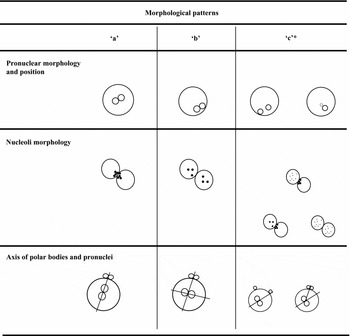
The pronuclear referring score used was the one developed by Gianaroli et al. (Reference Gianaroli, Magli, Ferraretti, Fortini and Greco2003). In particular, for zygotes with 2PN, three morphological patterns were defined for the localization of pronuclei inside the cytoplasm (a: juxtaposed and centralized pronuclei; b: peripheral pronuclei; c: centralized and separated or of unequal size and fragmented); three patterns for dimension and localization of nucleoli inside the pronuclei (a: nucleoli of large size and orientated; b: nucleoli of large size and scattered; c: large size and scattered in both nucleoli, large size aligned in one pronucleus and scattered in the other, small size in at least one pronucleus and scattered); and three patterns for the orientation of polar bodies in the axis of pronuclei (a: orientation of polar bodies in the longitudinal axis of pronuclei; b: orientation of polar bodies perpendicularly to the longitudinal axis; c: in different positions) as reported in Fig. 1.
Configurations of the three patterns ‘a’, ‘b’ and ‘c’ used for the zygotes classifications during statistical analysis. *, examples of the many possible ‘c’ patterns.

We examined 365 zygotes in 208 IVF cycles with the insemination of non-frozen oocytes: 144 zygotes from 85 cycles inseminated with conventional IVF (Group 1) and 221 from 123 cycles inseminated with ICSI (Group 3). Similarly, we examined 361 zygotes derived from frozen sibling oocytes always inseminated with ICSI: 164 zygotes from Group 1 patients’ sibling frozen oocytes (Group 2) and 197 zygotes from Group 3 patients’ sibling frozen oocytes (Group 4).
Sperm preparation
Sperm was collected by masturbation after 3–5 days of abstinence. The semen preparation for conventional IVF or ICSI was performed based on WHO Guidelines (World Health Organization, 1999), as reported in our previous study (Nicoli et al., Reference Nicoli, Valli, Di Girolamo, Di Tommaso, Gallinelli and La Sala2007).
IVF insemination was performed in cycles in which normal sperm morphology was >4% based on Kruger Strict Criteria (Kruger et al., Reference Kruger, Acosta, Simmons, Swanson, Matta and Oehninger1988) and sperm concentration after preparation was ≥1.5 × 10⁶/ml.
ICSI was performed in cycles in which normal sperm morphology was ≤4% based on Kruger Strict Criteria, sperm concentration was <1 × 10⁶/ml or sperm concentration after preparation was <1.5 × 10⁶/ml.
Statistical analysis
To evaluate the effect of the insemination techniques, semen quality and cryopreservation procedure on the zygote pronuclear morphology (position of pronuclei, shape and dimension of nucleoli and axis orientation) we chose to only compare sibling oocytes before and after treatments: this choice was done to limit the possible effects of confounding variables, such as individual indications to certain insemination techniques and individual characteristics within the pools of retrieved oocytes. Therefore comparisons made for the statistical analysis were Group 1 vs Group 2 and Group 3 vs Group 4. The correspondence analysis (CA) was performed for each zygote feature classified as ‘a’ pattern.
The CA is an explorative computational method for the study of associations between variables, in that the Pearson chi-squared statistic is decomposed into singular values by portioning the matrix of Pearson contingencies using the singular value decomposition.
Displaying a low dimensional projection of the data into a plane, the CA reveals associations between rows and columns. The graph consists of axes that reflect the variation in terms of the location (first principal axis) and dispersion (second principal axis) components, with the first two coordinates of which containing the bulk of the information.
The points significantly far from the origin indicate that they contribute to the dependency between row and column variables, while points close to the origin indicate they do not make such a contribution. Similar objects are clustered together, resulting in small distances, while dissimilar ones are far from each other (Greenacre, Reference Greenacre1993).
The contribute to the dependency between the row and column categories was estimated by means of the confidence circle of Lebart (CC) (Lebart et al., Reference Lebart1984). The CC is a method of observing the importance of the position of the profile and, if the correspondence plot consists of two dimensions, the radius of the confidence circle should be calculated with two degrees of freedom and at 5% level of significance for the i'th row/column co-ordinate.
Descriptive data analysis and CA were performed using the XL STAT PRO 6.0 (http://www.addinsoft.com).
Results
The present study included 190 patients who, between January 2005 and December 2006, underwent an oocyte pick-up with the recovery of more than three oocytes, at our public ART centre. All these patients underwent ET, but none of them obtained a pregnancy. In every oocyte pick-up, at least one oocyte was cryopreserved. We performed a total of 208 retrieval cycles (85 conventional IVF and 123 ICSI) with the insemination of three non-frozen oocytes (255 oocytes inseminated in conventional IVF and 369 in ICSI cycles). On the day of fertilization check we evaluated the pronuclear morphology of a total of 365 zygotes obtained from non-frozen oocytes (144 zygotes in Group 1 and 221 zygotes in Group 3).
From 3 to 10 months later, the patients came back to our centre to thaw the cryopreserved oocytes. We performed 208 cycles of oocyte thawing: we thawed 919 oocytes and 694 survived (75.5%) after the thawing protocol; after 4 h of post-thawing incubation, 635 oocytes resulted appropriate for ICSI; we inseminated, with ICSI, a total of 635 (2.5 ± 0.7) thawed oocytes and we obtained a total of 361 zygotes (164 zygotes in Group 2 and 197 zygotes in Group 4).
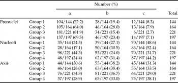
The rows of Table 1 show the four insemination treatments (Group 1 – non-frozen oocytes inseminated with conventional IVF; Group 2 – Group 1 patients’ sibling frozen oocytes inseminated with ICSI; Group 3 – non-frozen oocytes inseminated with ICSI; and Group 4 – Group 3 patients’ sibling frozen oocytes inseminated with ICSI) and their effects on the pronuclear morphological evaluation of 726 zygotes classified with an ordered three point scale (‘a’, ‘b’, ‘c’), as displayed in the columns.
Zygote morphology in non-frozen and frozen sibling oocytes.

Note: Group 1: zygotes from non-frozen oocytes inseminated with conventional IVF. Group 2: zygotes from Group 1 patients’ sibling frozen oocytes. Group 3: zygotes from non-frozen oocytes inseminated with ICSI. Group 4: zygotes from with Group 3 patients’ sibling frozen oocytes.
The correspondence plot for the row/column (groups and patterns) profile coordinates is shown in Fig. 2. The profile coordinates graphically depicted are accompanied by the 95% confidence circle for each technique. The two axes explain 100% of the variation in the groups and patterns (location = 94.6%, dispersion = 5.4%).
Graphic representation of the results obtained with the correspondance analysis. 1–4, pronuclei morphology and position (1: Group 1; 2: Group 2; 3: Group 3; 4: Group 4); 5–8: nucleoli morphology (5: Group 1; 6: Group 2; 7: Group 3; 8: Group 4); 9–12: axis orientation (9: Group 1; 10: Group 2; 11: Group 3; 12: Group 4).

Considering the position of the pronuclei inside the zygote cytoplasm, Groups 1 and 3 show the higher percentages of pattern ‘a’ (72.2% and 81.9%, respectively). In Fig. 2 the values related to the graphic representation of the position of pronuclei are depicted in the right side (points 1–4) of the plane; the statistical analysis evidenced a strict connection with the four points and the ‘a’ pattern, which represents the best morphological classification in zygotes. The 95% confidence circle, developed on Group 1 vs Group 2 and Group 3 vs Group 4, shows an elevated homogeneity between treatments with no statistically significant differences.
Insemination techniques, semen quality and oocyte cryopreservation did not seem to influence the position of pronuclei in zygotes obtained both from non-frozen oocytes and from frozen ones.
The analysis of nucleolar morphology shows a different distribution inside the three patterns. In Fig. 2 the graphic representation of the nucleoli inside the groups is shown by points 5 to 8 and it clearly shows the low connection with the ‘a’ pattern. Group 3 is statistically different from Group 4 (44.3% vs 24.4%, respectively), as graphically evidenced by the fact that the confidence circle developed on Group 3 does not intersect the circle developed on Group 4 (Fig. 2). Statistical analysis between Group 1 and Group 2 does not evidence a significant difference in relation to the position of the nucleoli.
Finally, considering the axis orientation, described by points 9 to 12 in Fig. 2, the four sets are the closest to the graphic origin and the most homogeneous when compared with points 1–4 and 5–8. No difference between the groups is evidenced in relation to the orientation of the axis.
Discussion
Oocyte cryopreservation is a technique that is still defined as ‘experimental’ because: first, the method is not yet standardized and reproducible; second, the results in terms of clinical pregnancies obtained by transferring embryos derived from frozen oocytes are still not satisfactory; and, finally, the effects of oocyte cryopreservation on the pediatric outcome of the children born by this technique must still be studied on a sufficiently wide sample (Fabbri et al., Reference Fabbri, Porcu, Marsella, Rocchetta, Venturoli and Flamigni2001; Jones et al., Reference Jones, Van Blerkom, Davis and Toledo2004; Gook, Reference Gook and Edgar2007; Oktay et al., Reference Oktay, Cil and Bang2006; Chian et al., Reference Chian, Huang, Tan, Lucena, Saa, Rojas, Ruvalcaba Castellón, García and Montoya Sarmiento2008).
Many biological aspects should be investigated to define exactly what damage could be caused by oocyte cryopreservation. Existing studies have shown anomalies in the frozen–thawed oocytes regarding the exocytosis of the cortical granules, the integrity of the mitochondrial membrane, the microtubular structure of the meiotic spindle and the speed of the embryonic cleavage (Fabbri et al., Reference Fabbri, Porcu, Marsella, Rocchetta, Venturoli and Flamigni2001; Jones et al., Reference Jones, Van Blerkom, Davis and Toledo2004; Gook, Reference Gook and Edgar2007). Most of these studies, however, focussed on the state of the thawed oocyte before the insemination.
The studies carried out by Payne et al. (Reference Payne, Flaherty, Barry and Matthews1997) on zygotes, obtained from non-frozen oocytes, have evidenced how the male and female pronuclei undergo a series of structural and spatial modifications so much so that 18–20 h after insemination the zygotes with a development considered ‘normal’ should have the pronuclei localized in the central zone of the cytoplasm, the nucleoli of large size and juxtaposed and the axis of pronuclei and polar globules parallel to each other.
More recently, Gianaroli et al. (Reference Gianaroli, Magli, Ferraretti, Fortini and Greco2003, Reference Gianaroli, Magli, Ferraretti, Lappi, Borghi and Ermini2007) have restated the concept, already expressed by their group, that the nuclear asset of the embryo is correlated with the pronuclear morphology of the zygote and that the use of this new score for the evaluation of the zygote viability can thus be hypothesized. In particular, they have shown how the configuration with centralized and juxtaposed pronuclei, large sized, aligned or scattered nucleoli and polar bodies located in the longitudinal or perpendicular axis of pronuclei corresponded to those with a higher proportion of chromosomally normal embryos and a higher proportion of implantation rate than all the others.
The aim of our study was to compare the pronuclear morphology of zygotes obtained from sibling frozen oocytes with the morphology of zygotes obtained from non-frozen oocytes inseminated with conventional IVF or ICSI (Group 1 vs Group 2 and Group 3 vs Group 4). At the same time, we verified if the insemination techniques and the presence or co-presence of a low semen quality could interfere with pronuclear morphology. We decided to use pronuclear morphology as it is a non-invasive instrument that also other recently published studies have put in correlation with the quality of the embryo and its potentiality of implantation (Scott, Reference Scott2003; Gianaroli et al., Reference Gianaroli, Magli, Ferraretti, Fortini and Greco2003, Reference Gianaroli, Magli, Ferraretti, Lappi, Borghi and Ermini2007).
The statistical analysis performed on pronuclei position inside the zygote cytoplasm in Group 1 vs Group 2 and Group 3 vs Group 4, did not evidence a significant statistical difference.
Similarly, no significant statistical differences were found regarding the axis of polar bodies and pronuclei comparing sibling oocytes, showing a highly homogeneous distribution of these values.
On the other hand, the statistical study of the morphology of the nucleoli evidenced an interesting distribution of pattern ‘a’ in the two comparisons. In the comparison between Group 1 (non-frozen oocytes and conventional IVF) and Group 2 (Group 1's sibling frozen oocytes and ICSI) no statistically significant difference emerged. This result seems to show that, in the presence of a good semen quality, the zygote pronuclear morphology is not affected by the association of the ICSI micromanipulation of oocytes and the oocyte cryopreservation procedure.
The statistical analysis performed on the comparison between Group 3 (non-frozen oocytes and ICSI) and Group 4 (Group 3's sibling frozen oocytes and ICSI) evidenced a high statistically significant difference. In fact, Group 4 presents a strongly inferior percentage of pattern ‘a’ compared with that of Group 3 (24.4% and 44.3%, respectively). In this case the two groups include sibling oocytes, non-frozen (Group 3) and frozen (Group 4), always inseminated with ICSI. Given the result of the comparison between Group 1 and Group 2, the deleterious effects of ICSI micromanipulation on zygote morphology could be excluded from the variables considered in the comparison between Group 3 and Group 4. Therefore, only the association between oocyte cryopreservation and an altered male component, present in Group 4, would seem to be unfavourable for the nucleolar morphology.
Resuming, the comparisons evidenced that: (i) the only parameter that showed the susceptibility to the examined variables (insemination technique, semen quality and oocyte cryopreservation) was the nucleolar morphology, while the position of pronuclei and the axis of pronuclei and polar bodies seem to be less affectable; (ii) the insemination technique itself (conventional IVF or ICSI), performed on non-frozen and frozen sibling oocytes, does not alter the morphological asset of the pronuclei in the zygote, as already reported by our group (Nicoli et al., Reference Nicoli, Valli, Di Girolamo, Di Tommaso, Gallinelli and La Sala2007); and (iii) the oocyte cryopreservation procedure negatively affects the nucleolar morphology on zygotes derived from frozen oocytes only if associated with a poor semen quality.
Our results show that oocyte cryopreservation does not induce on pronuclear morphology the same clear negative effects observed on other structures of the thawed oocyte, as reported in previous studies (Gook, Reference Gook and Edgar2007). These negative effects would only seem to be seen in the association between oocyte cryopreservation and a poor semen quality and this appears to be confirmed by recent studies in which the role of spermatozoa was reevaluated within the fertilization process (Seli, Reference Seli and Sakkas2005; Chatzimeletiou et al., Reference Chatzimeletiou, Morrison, Prapas and Handyside2008). Among the sperm structures that are probably more active during oocyte activation, polarization and embryogenesis, the centrosome was shown to be fundamental in oocyte fertilization, forming an aster comprising radially arrayed microtubules that brings the male and female pronuclei into close apposition and organize the first mitotic spindle in the zygote (Terada, Reference Terada2004; Sathananthan et al., Reference Sathananthan, Ratnasooirya, de Silva and Randeniya2006; Schatten, Reference Schatten2008). Starting from this knowledge, we hypothesized that the male centrosome could be one of the structures damaged in poor quality semen and that it could be involved in the alteration of the nucleolar morphology, amplifying the limited damage on the oocyte caused by cryopreservation, as observed in this study.
To confirm this hypothesis, future research should probably focus on the study of pathological semen parameters, centrosome structure and zygote morphology on a wide range of samples of non-frozen oocytes, also aiming to obtain information regarding the pregnancy outcome.
Acknowledgements
We sincerely thank Dr Leonardo Llewellyn Duncan De Pascalis for his contribution to the linguistic revision of the paper.