Congenital heart disease prevalence is reported at around 8–12 per 1000 live births globally.Reference Van Der Linde, Konings and Slager1 Screening with pulse oximetry to facilitate early diagnosis is recommended in all newborns to enable early diagnosis and management of children with critical congenital heart disease.Reference Mahle, Newburger and Matherne2 In resource limited settings however, children with congenital heart disease are not only often presenting acutely and being diagnosed late but heart disease is usually also not one of the main health priorities.Reference Chelo, Nguefack and Menanga3 Focussed cardiac ultrasound (also referred to as cardiac point of care ultrasound) is defined as a focussed examination of the cardiovascular system using ultrasound as an adjunct to the physical examination to recognise specific ultrasonic signs that represent a list of potential diagnoses in specific clinical settings.Reference Spencer, Kimura, Korcarz, Pellikka, Rahko and Siegel4 It is recommended in settings where echocardiography is either not available, unavailable in a timely fashion or impractical.Reference Spencer, Kimura, Korcarz, Pellikka, Rahko and Siegel4 In resource limited settings, one study in adults patients showed that focussed ultrasound performed by general practitioners after a brief training programme was accurate and could potentially influence acute patient assessment and management.Reference Shah, Shah and Fils-Aime5 Focussed studies in paediatrics have been used to assess for pericardial effusion, left ventricular size and function as well as features of rheumatic heart disease with promising resultsReference Van Der Linde, Konings and Slager1,Reference Spurney, Sable, Berger and Martin6,Reference Yacoub, Lang and Shebbe7 but focussed cardiac ultrasound has not been utilised to diagnose congenital heart disease.
Tele-echocardiography typically involves remote review of images acquired from a different geographic location via an established communications platform (e.g. an online platform). In one study, adult trained sonographers with additional training in paediatric echocardiography performed scans on newborns with suspected congenital heart disease that were then transmitted to a tertiary center and reviewed in real time by paediatric cardiologists.Reference Sable, Cummings and Pearson8 Tele-echocardiography was shown to be accurate, cost effective and improved patient care with significant impacts on referral patterns. The impact of point of care cardiac ultrasound in rural areas and community hospitals where access to trained paediatric echo sonographers and cardiologists is limited cannot be overstated. It has not only been shown to enable early diagnosis but also results in timely initiation of medical interventions while avoiding unnecessary patient transport and the associated expenditures which are known to be disproportionately severe in low income countries.Reference Sable, Cummings and Pearson8,Reference Sekar and Vilvanathan9
Médecins Sans Frontières has an established telemedicine programme that has previously been describedReference Martinez Garcia, Bonnardot and Olson10 with demonstrable improvements in the quality of care provided in often remote, humanitarian and resource limited settingsReference Zachariah, Bienvenue and Ayada11 however, the use of focussed cardiac ultrasound in patient management on this platform has not been previously assessed or described.
The aim of this study was to describe the use of remotely performed focussed cardiac ultrasound by non-cardiologists (with remote cardiologist involvement) to diagnose congenital and acquired heart disease and subsequently guide patient management using the Médecins Sans Frontières tele-medicine platform.
Methods
Training of Médecins Sans Frontières teams in cardiac point of care ultrasound commenced in South Sudan in 2016 and was then rolled out to multiple other sites in several countries including Niger, Nigeria, Guinea Bissau, Tanzania and Bangladesh over the next 2 years. Clinicians who were mostly ultrasound naïve underwent a rigorous 4 week face to face point of care ultrasound programme that included didactic lectures and 50 hours of ultrasound scanning time. This was held in the Médecins sans Frontières centers where the clinicians were based using portable point of care ultrasound machines.
Prior to being signed off, the clinicians needed to have completed at least 25 focussed cardiac exams that were graded by a point of care ultrasound qualified trainer. The cardiac views they were trained to acquire included parasternal long and short axes, apical four chamber and sub-xiphoid views. Additional coaching on specific views was provided in a non-real-time environment by the remotely based cardiologist as required to gain additional information. Follow up training was provided 3–9 months after the initial training to reinforce previously acquired skills. The indications for performing focussed cardiac ultrasound were mainly respiratory distress or hypoxia. The local trainers focussed the training on image acquisition as they were not trained to diagnose congenital heart disease. The clinicians were therefore trained to obtain the best possible images which were subsequently interpreted by a remotely placed paediatric cardiologist.
Clinical information on cases encountered in the field (humanitarian and resource limited settings) where cardiology consultation was sought were submitted to the Médecins Sans Frontières telemedicine platform by the clinicians on the ground. The cases were then reviewed by an Medecins sans Frontiers clinical case coordinator and assigned to one of two paediatric cardiologists (AD, LN) for review of clinical information and interpretation of imaging (including cardiac point of care ultrasound). Feedback on patient management was subsequently provided to the field teams via the telemedicine platform and was not real-time, but generally was within 24–48 hours. Each patient was assigned a number specific to that consultation and all information was entered under that consultation number on the Telemedicine platform.
We conducted a retrospective review of all the paediatric cardiology referrals for children below 18 years on the Médecins Sans Frontières Telemedicine platform from January 2016 to December 2018. The extracted information included information from the referring team and the reviewing cardiologist. This data included patient demographics, history and examination findings, test results and follow up clinical information where available. The final cardiac diagnosis was based on the cardiac point of care ultrasound interpretation. Whether or not management changed as a result of remote cardiologist involvement was determined from review of the follow up patient information. The outcome was based on the last available data entered on the patient condition in the telemedicine platform. The data was entered into a Microsoft Excel® (Version 16, Redmond, Washington, USA) spreadsheet.
Clinical characteristics were summarized using means and standard deviation or medians and interquartile ranges for continuous variables as appropriate; frequencies and proportions were reported for dichotomous and polytomous variables. Between-group differences in dichotomous/polytomous variables were evaluated with Fisher’s exact tests.
Cardiac point of care ultrasound images from 30 randomly selected patients were downloaded with a brief clinical summary and reviewed independently by the two paediatric cardiologists who were blinded to patient diagnoses or outcomes. Comparison of diagnoses reported by each cardiologist was done to determine the inter-observer agreement by calculating Kappa statistics, and the significance of divergence between raters was assessed using McNemar’s chi square tests.
Ethical review and approval for this study was provided by the Hospital for Sick Children Research Ethics Board and the Médecins Sans Frontières Independent Ethics Review Board.
Results
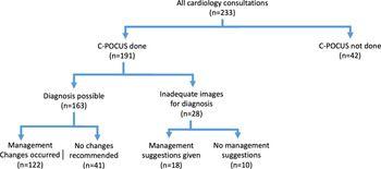
A total of 233 cases with suspected heart disease were referred for paediatric cardiology consultation on the platform from January 2016 to December 2018. Of the 233 referred cases, cardiac point of care ultrasound was performed in 82% (191) of the group (Fig 1). Males comprised 54% (126) of the referrals. The total group included neonates (12% - 29/233), infants (35% - 81/233), 1–5 years (23% - 53/233), 5–10 years (12% - 27/233) and >10–18 years (18% - 42/233). There was missing data on age for one patient.
Flowchart showing the proportions of study participants that underwent cardiac point of care ultrasound and the resultant changes in management.

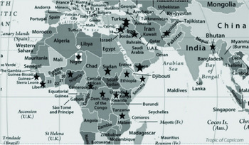
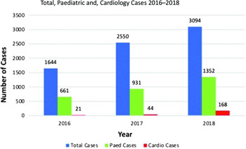
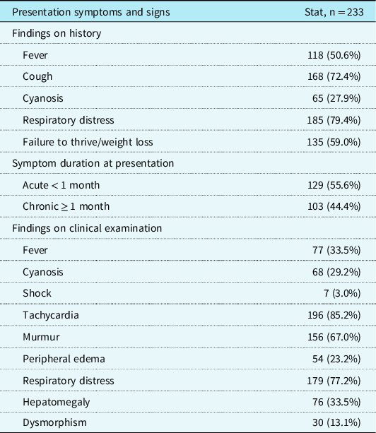
Cases originated mainly from sub-Saharan Africa, the Middle East, and South Asia (Fig 2). There was a year on year increase in overall, paediatric and paediatric cardiology cases referred for consultation on the telemedicine platform (Fig 3) with 21, 44 and 168 cases referred in 2016, 2017 and 2018 respectively. The majority of consultations were for in-patients (98%) of which the majority were in a general in-patient ward setting (94%). Presentation symptoms and signs are shown in Table 1 below. Weight, saturations and blood pressure were recorded in 65.7% (153), 51.1% (119), and 15.9% (37) patients respectively. Cardiac point of care ultrasound was the only investigation done in 55% (129) of all the referred cases. Other investigations included chest X-ray in 20% (45), lung ultrasound in 20% (46), and abdominal ultrasound in 15% (36).
Countries of origin for the cases referred to the Telemedicine platform marked by a star.

Year on year referrals through the Telemedicine platform.

Showing a summary of symptoms at presentation and signs at initial clinical examination in the study population

The most common indication to perform cardiac point of care ultrasound was to exclude congenital heart disease that was suspected in 61% (141) of the patients based on their clinical presentation. The most common differential diagnoses were pneumonia 43% (101) and rheumatic heart disease 15% (34). Signs and symptoms that prompted this concern included cyanosis, respiratory distress, murmur, and hepatomegaly as shown in Table 1.
Following cardiac point of care ultrasound, the diagnoses included septal defects (atrial and ventricular) in 11% (22/191), atrioventricular septal defect 7% (13/191), tetralogy of Fallot 9% (17/191) dilated cardiomyopathy/ myocarditis 8% (15/191), isolated pericardial effusion 6% (12/191), rheumatic heart disease features 8% (15/191), single ventricle/ complex congenital heart disease 4% (7/191), and other diagnoses (including double outlet right ventricle with malposed great arteries, transposition of the great arteries, aortic stenosis, persistent pulmonary hypertension of the newborn, pulmonary hypertension (no cause identified), mitral stenosis, ventricular masses, hypertrophic cardiomyopathy, pulmonary atresia, and decreased univentricular function with no cause identified) in 15% (29/191) of the cases. In 17% (33/191), the imaging showed no structural abnormality (presumed anatomically normal heart with normal function) while 15% (28/191) had imaging that was inadequate to make a diagnosis.
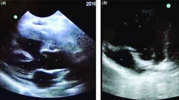
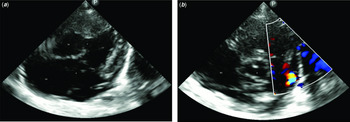
Despite the limited number of images obtained, the image quality was of sufficient quality to enable diagnosis as is shown in Figures 4 and 5 below. Further imaging is available in the supplementary material. The quality of images subjectively improved with increased use of imaging as user experience increased.
Images of Tetralogy of Fallot in a cyanosed 4-year-old child (a) shows the parasternal long axis view in Tetralogy of Fallot with the overriding aorta and sub-aortic ventricular septal defect. RV- right ventricle, LV – left ventricle, VSD – ventricular septal defect, Ao – aorta, LA – left atrium. The short axis image (b) shows the perimembranous ventricular septal defect and narrowed right ventricular outflow tract. RA – right atrium, RV – right ventricle, P – pulmonary valve/right ventricular outflow tract, A – aortic valve, VSD – ventricular septal defect.

Unbalanced atrioventricular septal defect with small patent arterial duct in a 7-week-old with clinical features of Trisomy 21 and congestive heart failure symptoms (a) shows the apical four chamber view of an unbalanced atrioventricular septal defect with large atrial and ventricular septal defect components. RA – right atrium, RV – right ventricle, LA – left atrium, LV – left ventricle, A – atrial septal defect component, V – ventricular septal defect component of the atrio-ventricular septal defect, AVV – atrial-ventricular valve leaflets. (b) Shows unobstructed outflow tracts with a patent arterial duct shunting left to right in the same patient. RA – right atrium, RV – right ventricle, P – pulmonary valve/ right ventricular outflow tract, A – aortic valve, PDA – patent arterial ductal flow in color.

Management recommendations after cardiologist involvement were implemented in 75% (122/163) cases in whom a diagnosis was possible on focussed cardiac ultrasound (Fig 1) including addition and removal of medications, changes to drug dosing, initiation of other supportive therapies or withdrawal of care. The latter was recommended in cases that were deemed nonsurvivable in their setting for example those with single ventricle physiology, transposition of the great arteries and one with a massive tricuspid valve mass (video of the echocardiograms in supplementary material). In the other 25% (41/163) cases in which a diagnosis was possible, no cardiac specific management was recommended due to a structurally normal heart on imaging or minor lesions identified on cardiac imaging (e.g. isolated pericardial effusion) and deemed non-contributory.
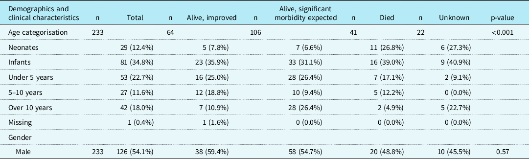
Considering the natural history of unrepaired cardiac lesions, in almost two thirds of the group in which a diagnosis was possible (65%, [106/163]), significant morbidity was anticipated despite the patients being alive at the time the consultation was concluded. Furthermore, among the entire group of referred patients, 38% (11/29) of all neonates and 20% (16/81) of all infants died (Table 2) compared to lower rates in other age groups.
Patient demographics summarised by patient outcome. The mortality was disproportionately higher in the neonatal and infant sub-groups of the study population

Follow up information was available for the majority of referred cases (62%–144/233) but in 37% (87/233), follow up information had not been entered in the Telemedicine system (by the managing team on the ground) at the time the cases were reviewed. There were two cases lost to follow up.
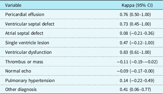
On independent echocardiography image review, significant divergence between reviewing cardiologists was found only for diagnoses of atrial septal defects with otherwise no difference noted for the other diagnoses as shown in Table 3 below.
Kappa statistics for inter-rater reliability were summarised for the different echocardiography diagnoses

Discussion
This study adds to a growing body of literature that supports the concept of clinician performed point of care ultrasound as a rapidly evolving and invaluable diagnostic tool in resource limited settings.Reference Shah, Shah and Fils-Aime5,Reference Sable, Cummings and Pearson8,Reference Hinton and Ware12
In our study, cardiac point of care ultrasound with remote paediatric cardiologist involvement improved health care access, supported health workers in isolated settings and altered patient care in settings where a paediatric cardiologist would otherwise have been unavailable. This transcendence of boundaries evidenced by the multiple countries from which cases originated is one of the hallmarks of telemedicine.Reference Martinez Garcia, Bonnardot and Olson10,Reference Wootton and Bonnardot13,Reference Graham, Zimmerman and Vassallo14
Our study differs from that by Shah et alReference Shah, Shah and Fils-Aime5 where non cardiologists were trained to identify specific pathology namely, left ventricular dysfunction, pericardial effusion, interstitial pulmonary edema and pleural effusion and that by Engelman et alReference Engelman, Kado and Reményi15 where nurses were trained to diagnose rheumatic heart disease features. Our study went further in involving a remotely based paediatric cardiologist to review the limited imaging. This significantly aided in the diagnosis of congenital and acquired heart disease that might have otherwise not been immediately clear to the non-cardiologist clinician. The image acquisition by largely previously ultrasound naïve clinical personnel in our study differed from the study by Sable et al,Reference Sable, Cummings and Pearson8 where a remotely placed paediatric cardiologist reviewed real time imaging obtained by a trained adult sonographer to diagnose congenital heart disease. We thereby demonstrated that it was possible on limited imaging to provide interpretation of paediatric echocardiograms from remote settings.
Similar to findings by Stachura et alReference Hinton and Ware12 where focussed ultrasound including cardiac ultrasound was performed in an emergency room setting, there were some studies that did not change clinical management. However, in these cases, there was still a benefit in narrowing the differential diagnosis, excluding a life threatening cardiac diagnosis or providing diagnostic certainty.
We found a high incidence of heart disease in our study, a finding that likely represents selection bias, with more severe congenital heart disease presentations in hospitalised patients with significant symptoms. This is due to the fact that the Médecins Sans Frontières sites are specifically acute care hospitals, with very limited outpatient care. It was not surprising in a study group where approximately half were infants below one year, almost all being inpatients who had presented with predominantly respiratory symptoms or hypoxia which are common manifestations of heart failure. It has been previously reported that up to 60% of heart failure cases in paediatric patients present in the first year of life.Reference Hinton and Ware16 In addition, there was a high proportion of patients with peripheral edema, which is uncommon in children with CHD. This was often seen in conjunction with severe malnutrition and an older cohort of children with these conditions.Reference Da Silva, De Oliveira Lopes and De Araujo17,Reference Batte, Lwabi and Lubega18 The spectrum of diagnoses encountered in this study was analogous to what has been described in population-based studies with sepal defects predominating.Reference Tantchou Tchoumi, Butera, Giamberti, Ambassa and Sadeu19,Reference Nkoke, Balti and Menanga20
The mortality noted in this study was higher than would be seen in settings where robust paediatric cardio-surgical programmes exist and is related to the natural history of unrepaired lesions. This was similar to findings by Hwang et alReference Hwang, Sisavanh and Billamay21 where a mortality of 11.7% was reported in their cohort. This was however in the setting of access to a cardiac surgery programme which was not the case in our study.
There was a trend to increased utilisation of cardiac point of care ultrasound year on year due to an increased number of sites with trained personnel and the necessary ultrasound equipment. Despite the limited experience of the operators with echocardiography and limited images available for review, this study demonstrated that it was possible to make a diagnosis of congenital or acquired heart disease across a telemedicine platform. The images obtained were sufficient to make a general diagnosis in the majority of cases which led to alterations in patient management in up to 75% of cases. However, the accuracy could not be assessed given that the diagnoses were not confirmed on a full echocardiogram interpreted by a paediatric cardiologist, at surgery or at autopsy given the setting in which the studies were done.
These are some of the key benefits of cardiac point of care ultrasound and Telecardiology realised in this study that enabled delivery of a cardiology service in otherwise remote and limited resource settings, as has been previously shown.Reference Sable, Cummings and Pearson8,Reference Sekar and Vilvanathan9,Reference Graham, Zimmerman and Vassallo14 This was not only advantageous for the clinical team who obtained advice to facilitate ongoing care but also to the families who would be made aware of the underlying diagnosis in their children thereby obviating the need to make potentially hazardous journeys to tertiary care centers with the prohibitive costs associated with travel.
Of note, despite the limited imaging available, the inter-observer variability in lesion diagnosis was only variable for atrial septal defects which are difficult to image in standard parasternal and apical imaging planes due to echo dropout of atrial septal regions.Reference Shub, Dimopoulos and Seward22
The implications of this work are such that in a few cases, referrals have been made to centres with cardiac surgery programmes and children have undergone repair for example, one with tetralogy of Fallot who was repaired in a Canadian center (full cardiology assessment confirmed the diagnosis). In one Médecins Sans Frontières site, plans are underway to establish a referral system to the cardiac surgical programme in that country using the cardiac point of care ultrasound findings as a screening test. This further shows the potential of cardiac point of care ultrasound to transform the care of children with congenital heart disease that is amenable to surgery.
There were limitations to our study including the fact that diagnoses could not be confirmed by a paediatric cardiologist, at surgery or autopsy (none of these services were available from the remote sites where the studies originated). In addition, there was limited clinical and follow up information on the study subjects (only information within the Telemedicine platform was reviewed and this varied widely depending on the site/ country of origin and likely clinical burden of the field team). We were also not able to access cases on the Médecins Sans Frontières telemedicine platform referred to other paediatric cardiologists.
In conclusion, cardiac point of care ultrasound performed by previously ultrasound naïve clinicians supported by remotely placed paediatric cardiologists over a Telemedicine platform was not only feasible, but also facilitated diagnosis and altered case management in the majority of children with heart disease in humanitarian and resource limited settings.
Acknowledgements
We acknowledge the work of the Médecins Sans Frontières field teams in various countries who submitted this information through the Médecins Sans Frontières telemedicine website.
Financial support
This work received no specific grant from any funding agency, commercial or not-for-profit sectors.
Conflicts of interest
None.
Ethical standards
The authors assert that all procedures contributing to this work comply with the ethical standards of Helsinki Declaration of 1975, as revised in 2008, and ethical review and approval for this study was provided by the Hospital for Sick Children Research Ethics Board and the Médecins Sans Frontières Independent Ethics Review Board.
Supplementary material
To view supplementary material for this article, please visit https://doi.org/10.1017/S1047951121000834