





Carotenoids constitute a diverse family of naturally occurring tetraterpenoid pigments, responsible for the vivid colouration of fruits and vegetables and play crucial physiological roles in human health. As essential precursors to vitamin A, carotenoids such as β-carotene, α-carotene and β-cryptoxanthin are critical for retinal function, cellular differentiation and immune regulation(Reference Yabuzaki1). In addition, non-provitamin A carotenoids including lutein, zeaxanthin and lycopene are implicated in antioxidant defense, modulation of gene expression and regulation of inflammatory pathways(Reference Yabuzaki1). These effects highlight the potential of dietary intake of carotenoids with decreased risk of various chronic diseases, including metabolic diseases such as type 2 diabetes mellitus (T2DM) and obesity(Reference Bohn, Bonet and Borel2–Reference Wu, Zhou and Hu5).
Meta-analyses and population studies consistently report inverse associations between dietary or serum carotenoid levels and measures of obesity, waist circumference and metabolic syndrome prevalence(Reference Bowen, Stacewicz-Sapuntzakis and Diwadkar-Navsariwala6–Reference Wang, Tam and Ng9). Supplementation trials further indicate that carotenoids can reduce BMI and markers of systemic inflammation, though their efficacy may depend on the specific compound, dose and food matrix involved(Reference Wu, Zhou and Hu5,Reference Shalini, Jose and Prasanthi8,Reference Wang, Tam and Ng9) . Mechanistically, carotenoids may influence adipogenesis, lipolysis and insulin signalling within the phosphatidylinositol 3-kinase/Protein kinase B pathway (PI3K/Akt) and adenosine monophosphate-activated protein kinase (AMPK) pathways, thereby regulating glucose metabolism, lipid transport and inflammation(Reference Kaulmann and Bohn4,Reference Shalini, Jose and Prasanthi8,Reference Ferreira, Jamar and Estadella10–Reference Zhuge, Ni and Wan14) . Despite these promising findings, the metabolism and bioavailability of carotenoids, especially in individuals with obesity or diabetes, remain incompletely understood. Factors influencing absorption and utilisation could also contribute bioavailability, which include the molecular structure of the carotenoid, the food matrix, dietary fat co-ingestion, host genetics (e.g. variation in β-carotene-15,15′-monooxygenase (BCO1), Scavenger Receptor class B type I (SR-BI) genes), inflammatory status and interactions with the gut microbiome(Reference Kaulmann and Bohn4,Reference Borel, Lietz and Goncalves15,Reference Rao and Rao16) . Individuals with metabolic disorders often exhibit lower circulating carotenoid levels, a phenomenon potentially attributable to altered lipid metabolism or impaired intestinal absorption(Reference Zia-Ul-Haq, Dewanjee and Riaz17).
In parallel, microgreens, particularly those from the Brassicaceae family (e.g., broccoli sprouts), are noteworthy as nutrient-dense dietary sources. They may serve as a promising functional food owing to their high carotenoid content combined with a short growth period and strong adaptability to controlled cultivation systems. Their carotenoid and phytochemical richness is relevant to metabolic health as these compounds exhibit antioxidant properties and may help modulate oxidative stress. Their short growth cycle enables rapid production, frequent harvests and efficient nutrient accumulation in a small space over a short period. In addition, Brassica microgreens are easy to manipulate under controlled environments such as hydroponic or indoor growing systems, where light, temperature, nutrients and other environmental conditions can be adjusted to enhance their phytochemical profile. Together, these traits make B. microgreens a practical and biologically attractive candidate for development as a functional food with potential benefits against metabolic disorders.
B. microgreens are also rich in bioactive phytochemicals, including glucosinolates, sulforaphane and carotenoids, which may act additively or synergistically to ameliorate metabolic dysregulation(Reference Favela-González, Hernández-Almanza and De la Fuente-Salcido18,Reference Le, Chiu and Hsieh19) . Experimental and clinical studies have shown that Brassicaceae microgreens can reduce oxidative stress, suppress proinflammatory cytokines and improve glycaemic parameters in diabetic models(Reference Favela-González, Hernández-Almanza and De la Fuente-Salcido18,Reference Le, Chiu and Hsieh19) . The precise contributions of microgreen-derived carotenoids to these effects, as well as their interplay with other phytochemicals, are areas of active investigation.
Although carotenoids and Brassicaceae vegetables have each been reviewed separately, the literature remains fragmented across broad carotenoid metabolism reviews, Brassica phytochemistry reviews and disease-specific summaries. A focused synthesis linking carotenoid structure, bioavailability, microgreen matrix effects and metabolic outcomes in obesity and T2DM is still lacking. This review attempts to fill that gap by integrating mechanistic, preclinical and clinical evidence to clarify both the potential value and the current limitations of Brassicaceae microgreens.
Scope of this review and methodology
This comprehensive review was conducted by synthesising available studies in PubMed, Scopus, Web of Science and Google Scholar from database inception to March 2026. Searches combined controlled vocabulary and free-text terms around three core concepts: carotenoids, Brassicaceae microgreens/sprouts and metabolic disease, using a representative string such as (‘carotenoid*’ OR ‘β-carotene’ OR ‘α-carotene’ OR ‘lutein’ OR ‘zeaxanthin’ OR ‘xanthophyll*’) AND (‘microgreen*’ ‘Brassicaceae*’ OR ‘Brassica microgreen*’ OR ‘broccoli sprout*’ OR ‘Brassica sprout*’) AND (‘obesity’ OR ‘metabolic syndrome’ OR ‘T2DM’ OR ‘insulin resistance’ OR ‘NAFLD’ OR ‘MASLD’). Additional searches omitted microgreen terms to capture broader carotenoid, metabolic disease literature and reference lists of key papers, and recent reviews were manually screened for further eligible studies. Given marked heterogeneity in carotenoid species, food matrices, doses, co-phytochemicals, populations and outcome measures, a narrative synthesis was adopted rather than meta-analysis, with emphasis on consistency of effects and critical appraisal of preclinical v. human evidence.
Included were peer-reviewed, original research articles investigating carotenoid-containing foods, extracts, supplements or Brassicaceae microgreens/sprouts; reporting at least one relevant metabolic outcome (body weight/adiposity, HOMA-IR, fasting glucose/HbA1c, serum lipids, oxidative stress/inflammation markers, liver enzymes or histological liver endpoints) and conducted in animal models (primarily rodent models of diet-induced obesity, T2DM or liver injury) or human populations (observational and interventional studies). All identified studies using Brassicaceae microgreens or sprouts as a primary intervention with metabolic outcomes were included in the Brassica-specific table.
Excluded were non-nutrition botanical studies (e.g. agronomic yield without physiological outcomes), articles lacking metabolic endpoints, commentaries, narrative reviews, conference abstracts and non-indexed theses. Studies of mature Brassica vegetables without a microgreen/sprout component were generally excluded from the microgreen synthesis but occasionally cited for background carotenoid or glucosinolate biology. Non-Brassica carotenoid-rich foods were retained only when providing mechanistic or clinical insight relevant to interpreting B. microgreen findings and are tabulated separately for clarity.
Study-level limitations, including small sample sizes, short intervention periods, heterogeneous diets and metabolic profiles and animal-to-human differences, are highlighted throughout to avoid overinterpretation and identify priorities for future mechanistic and clinical research.
Carotenoids
Carotenoids are a structurally diverse group of isoprenoid pigments biosynthesised predominantly via the 2-C-methyl-D-erythritol 4-phosphate pathway located within the plastids of plants, algae and photosynthetic bacteria(Reference Maiani, Periago Castón and Catasta12,Reference Rodriguez-Amaya20) . These pigments play indispensable roles in phototrophic organisms by facilitating light harvesting during photosynthesis and providing photoprotection through quenching of reactive oxygen species generated by excess light energy.
Chemically, carotenoids are tetraterpenoids composed of 40 carbon atoms (C40H64), constructed via the head-to-tail condensation of eight isoprene units (C5). To date, over 1100 distinct carotenoids have been characterised across various taxa, and ongoing advances in analytical chemistry continue to reveal novel variants with unique structural adaptations and biological functions(Reference Yabuzaki1). The extensive conjugated polyene chain along the carotenoid backbone enables absorption of visible light in the 400–500 nm range, producing vivid pigmentation spanning from pale yellow through orange to deep red. This conjugated system underlies their key biological functions, including energy transfer to chlorophyll during photosynthesis and dissipation of excess light energy for photoprotection(Reference Yabuzaki1).
Carotenoid structure and classification
Carotenoids can be broadly categorised into two major classes based on their chemical structure and degree of oxygenation (Figure 1): (i) Carotenes: are purely hydrocarbon carotenoids devoid of oxygen-containing functional groups, thus highly lipophilic molecules predominantly localised within lipid membranes and lipid droplets. Examples include α-carotene, β-carotene and lycopene. Among these, β-carotene serves as a predominate provitamin A compound capable of enzymatic cleavage to yield retinal (vitamin A aldehyde) in animals and humans(Reference Yabuzaki1,Reference Zia-Ul-Haq, Dewanjee and Riaz17) ; (ii) Xanthophylls are oxygenated carotenoids containing polar functional groups such as hydroxyl (–OH), keto (=O), epoxy or aldehyde moieties, conferring altered polarity and functional diversity. Prominent xanthophylls include lutein, zeaxanthin, astaxanthin, neoxanthin and violaxanthin(Reference Yabuzaki1,Reference Zia-Ul-Haq, Dewanjee and Riaz17) . The presence of oxygen atoms enables xanthophylls to interact more readily with membrane interfaces and participate in protective mechanisms like the xanthophyll cycle, which modulates non-photochemical quenching of chlorophyll fluorescence to prevent photo-oxidative damage in plants.
Common structures of major carotenoids.

Beyond these primary groups, several additional subclasses and derivatives exist that diversify carotenoid functionality: (iii) Apo-carotenoids (Apocarotenoids): arise from the oxidative cleavage of carotenoids catalysed by carotenoid cleavage dioxygenases and related dioxygenases(Reference Harrison21). Cleavage generates shorter carbon-chain molecules, which act as bioactive signalling molecules and precursors for important metabolites. Examples of apocarotenoids include retinal (visual pigment precursor), strigolactones (plant hormones regulating development and symbiosis), abscisic acid (stress hormone) and volatile compounds contributing to aroma and flavor. (iv) Epoxy-carotenoids and other oxygenated derivatives have modified structures with additional epoxy rings or other oxygen modifications that affect their chemical stability, antioxidant potential and cellular localisation(Reference Raj and Ramamoorthy22).
The structural diversity of carotenoids, driven by variations in cyclic end groups (e.g., β-ring, ϵ-ring), chain length, stereochemistry (cis-trans isomerism) and functional group placement, profoundly influences their biophysical properties and biological activities across kingdoms(Reference Harrison21).
Carotenoid absorption and metabolism
Carotenoid absorption in the human intestine is a multistep process involving both passive and active mechanisms, influenced by carotenoid concentration, structure and dietary context. At high luminal concentrations, carotenoids can permeate enterocyte phospholipid bilayers via passive diffusion owing to their hydrophobicity. When present at lower concentrations, facilitated transport becomes prominent, mediated by lipid transport proteins such as SR-BI, cluster of differentiation 36 (CD36) and Niemann-Pick C1-like 1 (NPC1L1)(Reference Geng, Bao and Sun23,Reference Hamed, Moradi and Ezati24) .This process is a regulated, multi-step physiological sequence contingent on lipid co-ingestion; because carotenoids are highly lipophilic, their release from the food matrix must be followed by incorporation into mixed micelles formed with bile salts and dietary lipids before intestinal uptake can proceed. Insufficient co-ingested lipids may limit micellar solubilisation and, consequently, restrict access to enterocyte transport pathways, including SR-BI, CD36 and NPC1L1. The transition from intestinal absorption to tissue distribution is presented in Figure 2.
Systemic bioavailability, metabolic bioconversion and tissue partitioning of dietary carotenoids. The schematic outlines the multi-stage physiological pathway of carotenoids from ingestion to target tissue accumulation. (Left Panel: Intestinal Uptake) Following mechanical release from the food matrix and solubilisation into mixed micelles, carotenoids enter the enterocyte through passive diffusion, at high luminal concentrations or facilitated active transport mediated by scavenger receptor class B type I (SR-BI), cluster determinant 36 (CD36) and NPC1L1, at low luminal concentrations. (Middle Panel: Enterocyte Metabolism) Inside the enterocyte, provitamin A carotenoids are cleaved by β-carotene-15,15′-monooxygenase (BCO1) into retinal, which is reduced to retinol and esterified to retinyl esters (via LRAT/DGAT1) for chylomicron packaging. Asymmetric cleavage by β-carotene 9′,10′-dioxygenase (BCO2) yields signalling metabolites (apo-carotenoids). Non-provitamin A carotenoids are largely absorbed intact. (Center: Transport) Nascent chylomicrons transporting retinyl esters and intact carotenoids bypass the portal vein, entering the systemic circulation via the lymphatic system. (Right Panel: Hepatic Fate & Distribution) The liver serves as the central metabolic hub; hepatic stellate cells act as the principal storage depot for vitamin A (as retinyl esters). Mobilized retinol (bound to RBP4) and other carotenoids are distributed via circulation to peripheral target tissues, including the retina, skin, adipose tissue and kidneys, to support antioxidant defenses and cellular signalling. BCO1, β-carotene-15,15′-monooxygenase; BCO2, β-carotene 9′,10′-dioxygenase; CD36, cluster determinant 36; DGAT1, diacylglycerol O-acyltransferase 1; FFA, free fatty acids; LRAT, lecithin retinol acyltransferase; NADPH, nicotinamide adenine dinucleotide phosphate; NPC1L1, Niemann-Pick C1-like 1; RBP4, retinol-binding protein 4; SR-BI, scavenger receptor class B type I.

Mechanistic flow of intestinal uptake
The intestinal uptake initiates with (i) release carotenoids from food matrix via mechanical processing and digestion; (ii) Incorporated into mixed micelles with bile salts and dietary lipids for solubilisation; (iii) Uptake by Enterocytes via passive diffusion, relatively at high luminal concentrations or active transport, involving SR-BI, CD36 and NPC1L1 receptors, at physiological (lower) concentrations(Reference Geng, Bao and Sun23); (iv) Intracellular Metabolism of provitamin A carotenoids, which are substrates for BCO1, yielding two molecules of retinal, further converted to retinol (vitamin A) or esterified to retinyl esters(Reference Geng, Bao and Sun23). Non-provitamin A carotenoids (e.g., lutein, lycopene) are generally absorbed intact and serve non-retinoid biological functions(Reference Harrison21,Reference Harrison and Bugg25) .
Metabolic fate and distribution
Only a fraction (∼3·5–30 %) of consumed β-carotene is bio-converted into active vitamin A, with efficiency tapering off at high intakes due to enzymatic saturation and feedback inhibition by retinoic acid(Reference Harrison21,Reference Harrison and Bugg25) . The remainder, as well as non-provitamin A carotenoids, are absorbed and distributed to tissues including the liver, adipose tissue, retina (macula lutea) and skin, where they contribute to antioxidant defenses and tissue-specific functions. Alternative cleavage pathways, such as asymmetric β-carotene cleavage by β-carotene 9′,10′-dioxygenase (BCO2), yield β-apo-carotenoic acids, with minor contribution to retinoid pools but potentially significant signalling or antioxidant roles(Reference Bohn, Bonet and Borel2) (Figure 2).
Transport, storage, excretion
Post-absorption, both retinyl esters and unmodified carotenoids are incorporated into nascent chylomicrons and transported to the systemic circulation via the lymphatics. The liver particularly hepatic stellate cells are the principal storage depot for vitamin A, mainly as retinyl esters, helping to buffer fluctuations in supply and maintain homeostasis (Figure 2) Carotenoids and retinoid compounds are subsequently trafficked to peripheral tissues (colon, pancreas, prostate, kidneys, skin), often in association with lipoproteins(Reference West and Castenmiller26–Reference Zeng, Furr and Olson29).Carotenoids not absorbed or utilised may be excreted in bile or, after further oxidative cleavage and conjugation (e.g., glucuronidation), eliminated via urine(Reference Bohn, Bonet and Borel2). The gut microbiota additionally catabolises unabsorbed carotenoids in the colon, producing a range of apo-carotenoids and other metabolites with potential bioactivity(Reference Bohn, Bonet and Borel2).
Determinants of bioavailability: the SLAMENGHI framework
Carotenoid and vitamin A bioavailability varies markedly between individuals and meals, shaped by numerous factors. Dietary factors, including the composition of the meal, the presence of dietary fat (which enhances absorption) and the food matrix (raw v. cooked or pureed foods), significantly impact how well carotenoids are absorbed. Host-related factors such as lifestyle, health status, gender, age and the presence of certain diseases can also modulate absorption and utilisation. Genetic variations, including single nucleotide polymorphisms in genes like BCO1 and SR-BI, can lead to significant inter-individual differences in carotenoid metabolism and vitamin A production. The acronym SLAMENGHI provides a comprehensive framework(Reference West and Castenmiller26): Species of carotenoid, physical and chemical properties, such as polarity and conjugation, determine micelle incorporation and uptake; Linkage to food matrix, plant cell wall structure, processing and co-ingestion with fats influence release and bio accessibility; Amount consumed, efficiency of absorption declines with increased intake due to transporter and enzyme saturation; Matrix of incorporation, raw v. cooked/pureed food, dietary fibre content and processing impact bioavailability; Effectors of absorption, dietary fats enhance solubilisation and transportation; inhibitors include excessive dietary fibre, phytates and certain medications; Nutrient status of the host, vitamin A sufficiency reduces β-carotene conversion via negative feedback; deficiencies enhance conversion efficiency; Genetic factors, polymorphisms in BCO1, BCO2 and SR-BI genes account for major inter-individual variability; Host-related aspects. age, health status, sex and lifestyle factors (e.g., smoking, alcohol) modulate absorption efficiency; Interactions, synergistic or antagonistic interactions with other dietary components can modulate bioavailability.
Obesity
Recent studies have provided compelling evidence that carotenoids and their derivatives play a significant role in lipid and energy metabolism(Reference Yu, Yuan and Song30). These bioactive compounds have been associated with reduced fat accumulation and modulation of adipocyte function(Reference Wu, Zhang and Li31), offering valuable insights into the prevention and management of obesity and related metabolic disorders. The liver and adipose tissue serve as major storage sites for carotenoids and retinoids, with these compounds predominantly accumulating in lipid droplets and cell membranes of adipocytes(Reference Saeed, Bartuzi and Heegsma13,Reference Yan, Chen and Liu32) . Several studies of carotenoids on obesity are listed in preclinical and clinical trials (Table 1).
Studies of carotenoid intervention on obesity

Table 1. Long description
The table compares various studies on carotenoid intervention in obesity, listing references, study details, carotenoid types and sources, duration and dose, and key findings. It includes four columns: Reference, Study, Carotenoid type and source, Duration and dose, and Key findings. The table has two main sections: Clinical studies and Pre-clinical studies. Each row provides specific details for each study, such as the type of subjects, the carotenoid used, the duration and dosage of the intervention, and the key findings from the study.
CRP, C-reactive protein; FFA, free fatty acids; KK-Ay mice, mice are a cross between diabetic KK and lethal yellow (Ay) mice; SREBP1, sterol regulatory element-binding protein-1; SREBP2, sterol regulatory element-binding protein-2.
Animal models of obesity
Because human and animal interventions using Brassicaceae microgreens are still relatively limited, representative trials with carotenoid–rich foods from other matrices are also (e.g. pumpkin, sweet potato, wolfberry, marine algae) are summarised here to illustrate generalisable carotenoid–mediated effects on adiposity, lipids and glycemic control. These studies provide mechanistic and translational context and are not intended as substitutes for Brassica–specific interventions, which are synthesised separately in the B. microgreens section.
Pre-clinical investigations of carotenoid interventions in obesity have demonstrated consistent improvements in lipid metabolism and protection against ectopic fat accumulation. Interventions utilising provitamin A carotenoids from whole-food sources have been particularly effective. In hyper triglyceridemic Wistar rats, supplementation with β-carotene derived from pumpkin and sweet potato significantly reduced triacylglyceride (TAG) levels across three dose ranges (4·8–14·5 mg per 100 g diet), establishing a dose-response relationship(Reference Sunarti, Santoso and Pramana33). This finding demonstrates that β-carotene retains lipid–lowering efficacy when consumed within complex food matrices, suggesting clinical applicability to dietary interventions.
Marine-derived xanthophylls have shown particularly robust effects on tissue-specific lipid dynamics. In diabetic/obese KK-A y mice, a model combining genetic obesity and insulin resistance, fucoxanthin supplementation (0·2 % diet) produced a paradoxically beneficial restructuring of hepatic cholesterol metabolism: serum high density lipoprotein (HDL) cholesterol increased while hepatic cholesterol content decreased, accompanied by upregulation of hepatic Sterol Regulatory Element-binding Proteins 1 and 2 (SREBP1 and SREBP2) and their target genes(Reference Beppu, Hosokawa and Niwano34). This molecular remodeling suggests that fucoxanthin promotes hepatic cholesterol efflux or clearance rather than storage. Similarly, astaxanthin derived from Hematococcus pluvialis (6 mg/kg/day for 60 days) significantly reduced total lipids, free fatty acids and cholesterol specifically within skeletal muscle of non-genetic insulin-resistant Swiss albino mice(Reference Arunkumar, Bhuvaneswari and Anuradha35). A more recent investigation using ob/ob mice (leptin-deficient genetic obesity model) demonstrated that fucoxanthin (0·05 % diet over 20 weeks) alleviated skeletal muscle lipid deposition, a critical mechanism for protecting against lipotoxicity-induced insulin resistance(Reference Wu, Zhang and Li31).
Human studies in obesity
Clinical trials investigating carotenoid interventions in obese populations have yielded substantially more modest outcomes compared to animal models, with particular limitations in inflammatory and oxidative biomarker modification. In a two-arm study of overweight and obese breast cancer survivors (BMI 25–45 kg/m2), supplementation with freshly juiced orange (Balero) and purple (beta-sweet) carrots rich in α-carotene, β-carotene, lutein and zeaxanthin (8 fluid ounces/day for 3 weeks) failed to produce significant changes in broad oxidative stress or inflammatory markers, with the isolated exception of a reduction in prostaglandin E2(Reference Butalla, Crane and Patil36). This selective effect on a single eicosanoid suggests mechanistically limited intervention rather than systemic antioxidant efficacy.
A study of obese patients (BMI 37·5 ± 2·5 kg/m²) measured plasma carotenoids after receiving tomato derived-lycopene supplementation (30 mg daily for 4 weeks) revealed a paradoxical metabolic phenotype(Reference Noa Markovits and Yishai Levy37). Despite supplementation, carotenoid levels were significantly reduced in obese subjects compared to controls, and C-reactive protein (CRP) was significantly elevated. Lycopene intervention produced no measurable changes in inflammation or oxidation products. These findings support the ‘oxidative sink’ hypothesis, wherein chronic low-grade inflammation and heightened metabolic stress in obesity accelerate the consumption of circulating antioxidants, thereby limiting the capacity of standard supplemental doses to achieve detectable systemic effects(Reference Carlson, True and Wilson38). Collectively, obesity-focused human trials demonstrate that while animal models show clear lipid-modulating benefits, translating these effects to obese human subjects requires either substantially higher doses, extended intervention durations or enhanced delivery strategies to overcome the metabolic inertia of established adiposity (Table 1). In addition, the background diets and metabolic phenotypes of participants differ substantially across obesity trials, ranging from overweight breast cancer survivors to individuals with morbid obesity and variable inflammatory burden, with limited control or reporting of habitual fat intake and overall dietary patterns. This heterogeneity in both diet and metabolic status likely contributes to the inconsistent magnitude and direction of carotenoid responses observed across studies.
Diabetes
Epidemiological and experimental studies have also demonstrated that higher dietary intake of carotenoids, such as β-carotene and α-carotene, is associated with a reduced risk of developing T2DM(Reference Qiu, Chen and Geng39). The mechanisms underlying these benefits include enhancement of insulin sensitivity, improvement of glucose metabolism and modulation of adipose tissue function(Reference Qiu, Chen and Geng39). Carotenoids exert protective effects against diabetes-related complications, including nephropathy, retinopathy and neurodegeneration, largely attributable to their ability to ameliorate hyperglycemia, reduce insulin resistance and attenuate dyslipidaemia. Moreover, an inverse association has been found between circulating carotenoid levels and the incidence of obesity, suggesting their role in maintaining healthy body weight and preventing metabolic disturbances. These conflicting results may be attributed to differences in carotenoid types, dosages, study populations or methodological variability as pointed SLAMENGHI. Several studies of carotenoids on diabetes are performed in preclinical model and clinical studies (Table 2).
Studies of carotenoid intervention on diabetes

Table 2. Long description
A table comparing studies on carotenoid intervention in diabetes. The table has 7 rows and 5 columns. Column headers are Reference, Experimental model, Carotenoid type and source, Duration and dose, and Key findings. Row 1: Reference: Upritchard et al.; Experimental model: T2DM patients, Parallel randomised controlled trial; Carotenoid type and source: Sugar free tomato juice (Campbells, Sydney, Australia), Lycopene as a major carotenoid; Duration and dose: 4 weeks, 250 ml/twice daily; Key findings: Threefold increase in plasma lycopene levels, Increased the resistance of isolated LDL to oxidation. Row 2: Reference: Lotfollahi et al.; Experimental model: T2DM patients (HbA1c≥ 6-5 percent), Powdered Green banana (Musa spp.) biomass; Carotenoid type and source: No particular carotenoid stated but focused on the total carotenoid content; Duration and dose: 6 months to any food preparation without heating (two tablespoons, 40 g/day); Key findings: Increased carotenoid content in the LDL-cholesterol particles, Enhanced prevention of LDL-cholesterol oxidation, reduced Total-C, Non-HDL-cholesterol, glucose and HbA1c. Row 3: Reference: Ramezani et al.; Experimental model: T2DM patients, A double-blind randomized clinical trial; Carotenoid type and source: Carrot juice (Sunich Company, Iran) enriched with β-carotene; Duration and dose: 8 weeks, 200 ml of carrot juice + 10 mg of β-carotene; Key findings: Significantly increased serum antioxidants with no significant changes in glycemic indices and inflammatory factors. Row 4: Reference: Hashimoto et al.; Experimental model: Male Wistar Rats; Carotenoid type and source: Lycopene-free tomato (negative control) v. lycopene rich tomato (LR), LR tomato variety, called PR-7; Duration and dose: 4 weeks, PR-7 homogenate at 1 ml/day via oral gavage; Key findings: Increased 25 percent insulin levels without statistical significance, Significantly increased plasma leptin. Row 5: Reference: Yu et al.; Experimental model: Male db/db leptin receptor-deficient T2DM mice; Carotenoid type and source: Wolfberry (Lycium barbarum L., Chinese name Goji berry) containing lutein and zeaxanthin in ester forms; Duration and dose: 8 weeks, Wolfberry diet 10.7 g/4057 kcal; Key findings: Induced accumulation of zeaxanthin and lutein in the liver, Upregulated carotenoid metabolic genes in the retina, Stimulates AMPK-α2 activation and nuclear enrichment in the retina. Row 6: Reference: Ali et al.; Experimental model: Male Wistar diabetes induced with STZ injection; Carotenoid type and source: Saffron (dried stigmas of the flowers of Crocus sativus L.), Active constituents: crocin and crocetin; Duration and dose: 6 weeks, Daily oral administration of an aqueous Saffron extract (100 mg/kg BWDT); Key findings: A significant decrease in serum insulin level and the liver enzyme activities and the hepatic glutathione content. Row 7: Reference: Wu et al.; Experimental model: Male ob/ob mice; Carotenoid type and source: Fucoxanthin supplement, No extraction source reported; Duration and dose: 20 weeks fucoxanthin (0.05 percent diet); Key findings: Improved insulin resistance accompanied by a significant reduction in the HOMA-IR index, Inhibited PKM1enzyme activity, disrupts glucose metabolism in skeletal muscle.
ALT, alanine aminotransferase; AMPK-α2, AMP-activated protein kinase alpha-2; BDWT, body weight; HOMA-IR, Homeostatic Model Assessment of Insulin Resistance; LR, lycopene-rich; Non-HDL-C, non-high-density lipoprotein cholesterol; PKM1, pyruvate kinase M1; STZ, streptozotocin; T2DM, type 2 diabetes mellitus; Total-C, total cholesterol; γ-GTP, gamma-glutamyl transpeptidase.
Animal models of diabetes
Pre-clinical studies of carotenoid interventions in diabetes have revealed multifaceted benefits ranging from insulin secretion modulation to glucose metabolic remodeling and protection against diabetic complications. In Wistar rats, lycopene-rich tomato homogenate (1 ml/day via oral gavage for 4 weeks) significantly increased plasma leptin levels, though it produced only a non-significant 25 % elevation in insulin levels, suggesting that tomato lycopene’s effects on glucose homeostasis may be mediated through altered adipokine signalling rather than direct pancreatic β-cell stimulation(Reference Hashimoto, Tominaga and Wakagi40). In contrast, more pronounced effects were observed with phytocompound-rich sources; saffron extract (crocin and crocetin, 100 mg/kg body weight daily for 6 weeks) administered to streptozotocin-induced diabetic Wistar rats significantly decreased serum insulin levels and hepatic enzyme activities, indicating improved insulin sensitivity and hepatoprotection(Reference Ali, Serag and Abdeen41).
Mechanistic investigations have elucidated specific pathways linking carotenoids to improved glucose metabolism and mitigation of diabetic complications. In db/db mice, leptin receptor-deficient model of T2DM, wolfberry-derived lutein and zeaxanthin (ester forms, 10·7 g/4057 kcal diet for 8 weeks) induced accumulation of zeaxanthin and lutein in the liver and upregulated carotenoid metabolic genes in the retina, while stimulating AMPK-α2 activation and nuclear enrichment(Reference Yu, Wark and Ji42). This finding is particularly significant as AMPK-α2 is a key energy sensor and metabolic regulator, and its activation promotes fatty acid oxidation and glucose utilisation. Additionally, in ob/ob mice, fucoxanthin (0·05 % diet for 20 weeks) improved insulin resistance as assessed by the Homeostatic Model Assessment of Insulin Resistance (HOMA-IR) index and inhibited Pyruvate Kinase M1 (PKM1) enzyme activity, thereby disrupting aberrant glucose metabolism in skeletal muscle(Reference Wu, Zhang and Li31). These findings collectively demonstrate that carotenoids, particularly xanthophylls and specific plant extracts, directly modulate key enzymes and signalling molecules involved in glucose homeostasis and metabolic regulation.
Human studies in diabetes
Clinical investigations in T2DM patients have confirmed carotenoid-mediated improvements in lipoprotein oxidation resistance and partial glycemic control, though effects remain modest compared to animal model efficacy. In a parallel randomised controlled trial, consumption of lycopene-rich tomato juice (250 ml twice daily for 4 weeks) produced a three-fold increase in plasma lycopene and significantly enhanced the resistance of isolated low-density lipoprotein (LDL) to oxidation(Reference Upritchard, Sutherland and Mann43), demonstrating that carotenoid bioavailability and incorporation into lipoproteins is achievable in diabetic populations. A longer-term intervention (6 months) involving T2DM patients with hemoglobin A1c level (HbA1c) ≥ 6·5 % consuming powdered green banana biomass (40 g/day without heating) increased carotenoid content in LDL particles, enhanced LDL oxidation resistance and significantly reduced total cholesterol, non-HDL cholesterol, fasting glucose and HbA1c(Reference Lotfollahi, Mello and Costa44). This study demonstrates that extended dietary interventions incorporating mixed carotenoids can achieve modest but statistically significant improvements in glycemic control.
However, the broader translational efficacy remains limited. In a double-blind randomised clinical trial, T2DM patients receiving carrot juice enriched with β-carotene (200 ml plus 10 mg β-carotene daily for 8 weeks) demonstrated significantly increased serum antioxidant status, yet this did not translate into improvements in glycemic indices or inflammatory factors(Reference Ramezani, Yousefinejad and Javanbakht45). This discrepancy between enhanced antioxidant capacity and stable glycemic control suggests that the pathophysiology of T2DM involves metabolic dysregulation that extends beyond simple oxidative stress. The differential efficacy between the green banana study(Reference Lotfollahi, Mello and Costa44) and the carrot juice study(Reference Ramezani, Yousefinejad and Javanbakht45) may reflect differences in carotenoid dosage, duration or the broader nutrient composition of the intervention. Green banana provides resistant starch and additional micronutrients alongside carotenoids, whereas isolated carrot juice offers primarily carotenoids in a purified form. Collectively, diabetic human trials indicate that carotenoid interventions can improve specific oxidative and lipoprotein parameters and, in extended interventions with whole-food sources, achieve modest glycemic improvements, but sustained glycemic control requires prolonged intervention and may benefit from multicomponent dietary approaches rather than isolated carotenoid supplementation. Furthermore, clinical studies in T2DM encompass diverse patient populations with differing baseline glycemic control, comorbidities and background medication use and often provide limited detail on habitual diet and dietary fat co–intake. Such variability in metabolic status and dietary context complicates direct comparison between trials and may mask carotenoid–related effects in some cohorts
It is important to acknowledge that the findings from animal studies may not translate directly to humans. However, these findings, including observations from both human studies and animal models, underscore the need for further research to elucidate the metabolic implications of altered carotenoid and retinol dynamics. As the relationship between dietary components, carotenoid metabolism and metabolic health continues to be explored, understanding these processes may provide new insights into therapeutic strategies for managing obesity, fatty liver conditions and related metabolic disorders. Furthermore, mechanistic studies are needed to fully elucidate the pathways by which carotenoids exert their beneficial effects on glucose metabolism, insulin sensitivity and inflammation in both animal models and human subjects. A deeper understanding of these mechanisms will enable the development of more targeted and effective dietary interventions for the prevention and management of T2DM and obesity.
Preclinical studies in rodent models consistently report substantial improvements in adiposity, insulin sensitivity and inflammatory signalling following carotenoid-rich or Brassicaceae microgreen interventions. Nevertheless, these data must be interpreted cautiously in terms of human relevance, as clinical trials in obesity and T2DM have generally yielded smaller and more variable effects, often confined to oxidative stress, lipoprotein oxidation or selected metabolic endpoints rather than sustained changes in body weight or glycemic control. The divergence between animal and human outcomes likely reflects species-specific differences in metabolism, variations in dose and intervention duration, heterogeneity in background diet, limitations in carotenoid bioavailability and the greater pathophysiological complexity of established obesity and T2DM in humans.
Vegetable carotenoids
Carotenoids are essential nutrients, which humans must obtain through dietary intake as they cannot synthesise them endogenously(Reference Yabuzaki1). Across diverse vegetable categories, carotenoid profiles vary substantially based on chromoplast v. chloroplast localisation and specific carotenoid composition. Orange and yellow vegetables including carrots, sweet potatoes and butternut squash are rich sources of β-carotene, containing approximately 8·2 mg/100 g, 11·5 mg/100 g and 4·5 mg/100 g, respectively(Reference Clevidence, Paetau and Smith46). Green leafy vegetables, including kale, spinach and romaine lettuce, are enriched sources of carotenoids, predominantly lutein and β-carotene(Reference Clevidence, Paetau and Smith46,Reference Sun, Tong and Guo47) , with kale standing as the richest cruciferous source of carotenoids and exceeding cabbage approximately 40-fold(Reference Hirakida, Nakamura and Inagaki3,Reference Pan, Cui and Gao48) . In these vegetables, carotenoids are localised in chloroplasts associated with thylakoid membranes, which impacts their bio-accessibility due to the complex interactions with plant cell structures and proteins(Reference Clevidence, Paetau and Smith46,Reference Sun, Tong and Guo47) . Despite the bioavailability challenges posed by chloroplast localisation, green leafy vegetables remain a valuable source of carotenoids, providing antioxidants essential for reducing oxidative stress and inflammation which are implicated in metabolic syndrome and neurodegenerative diseases.
This diversity of carotenoid sources and types across vegetable families underscores the nutritional importance of consuming a varied diet to obtain complementary carotenoid classes with distinct bioactivities and metabolic functions. Considering the low average carotenoid intake in North America of approximately 6–18 mg/day (with a mean of 9·7 mg/day according to NHANES data)(Reference Yu, Yuan and Song30,Reference Yan, Chen and Liu32,Reference Qiu, Chen and Geng39,Reference He, Yin and Chen49) , the daily intake of carotenoids through a balanced diet rich in diverse vegetables is critical to capitalise on these protective health effects(Reference Favela-González, Hernández-Almanza and De la Fuente-Salcido18,Reference Clevidence, Paetau and Smith46,Reference Kim, Koo and Park50) .
Among green leafy vegetables, Brassicaceae microgreens are noteworthy because some species(Reference Kyriacou, Rouphael and Di Gioia51) are promising functional foods with high concentrations of carotenoids and other bioactive compounds. Comparative studies show that some Brassicaceae microgreens contain higher carotenoid concentrations than mature leaves, although the magnitude of this difference is species, cultivational practices and genotype dependent. In addition, their glucosinolate and polyphenol profiles, together with antioxidant activity, may support mechanisms relevant to metabolic health(Reference Kyriacou, Rouphael and Di Gioia51–Reference Dereje, Jacquier and Elliott-Kingston53).
Factors affecting green leafy vegetable carotenoid bioavailability
To maximise the nutritional benefit of dietary carotenoids, it is essential to understand the distinction between bioavailability and bioaccessibility. Bioaccessibility refers to the fraction of a carotenoid that is released from the food matrix during digestion and becomes available for absorption in the gastrointestinal tract (i.e., solubilised in mixed micelles), whereas bioavailability encompasses the entire process of intestinal absorption, systemic circulation, tissue uptake and utilisation by target organs(Reference Molteni, La Motta and Valoppi54,Reference Reboul55) . In practical terms, a compound may be highly bioaccessible yet poorly bioavailable if subsequent steps in the absorption and utilisation pathway are rate-limiting(Reference Etcheverry, Grusak and Fleige56).
The bioavailability of carotenoids from green leafy vegetables is influenced by multiple factors including the food matrix, molecular structure, processing and dietary components. Carotenoids in leafy greens tend to have lower bio-accessibility compared to those in non-photosynthetic plant tissues (such as carrots and tomatoes) because they are tightly bound within chloroplast structures and associated with thylakoid membrane proteins(Reference Molteni, La Motta and Valoppi54). β-carotene bioaccessibility from raw leafy greens is approximately 14 %, substantially lower than from chromoplast-containing vegetables where it can exceed 50 %(Reference Molteni, La Motta and Valoppi54). Processing methods such as cooking and mechanical disruption can enhance carotenoid release by breaking down cell walls and protein complexes. Additionally, the co-consumption of dietary fats is vital to aid micelle formation in the gastrointestinal tract, facilitating carotenoid solubilisation and absorption; a minimum of 5 g of dietary fat per meal is generally required for adequate absorption(Reference Molteni, La Motta and Valoppi54,Reference Sánchez-Moreno, Plaza and De Ancos57) . This lipid requirement is not merely additive but rate-limiting; in the absence of adequate co-ingested fat, even carotenoid-rich vegetables and microgreens with favorable matrix properties may yield markedly reduced systemic absorption, regardless of their intrinsic carotenoid content. Advances in understanding carotenoid absorption mechanisms reveal that specific membrane transporters, SR-BI, CD36 and NPC1L1, regulate their cellular uptake, modulated by host genetic factors, age, BMI and nutrient interactions(Reference Reboul55). Novel nanostructured delivery systems are also emerging to improve carotenoid stability, solubility and bioavailability during digestion(Reference Molteni, La Motta and Valoppi54). Thus, to maximise carotenoid intake benefits, consumption of carotenoid-rich green leafy vegetables in conjunction with adequate dietary fat is recommended, alongside food preparation techniques that improve bio-accessibility(Reference Molteni, La Motta and Valoppi54).
Brassica microgreens
Concentrated source of carotenoids and bioactive compounds
B. microgreens are recognised as a rich source of carotenoids, ranging from 162 to 224 mg β-carotene equivalents per 100 g dry weight across different Brassicaceae species(Reference Moreira-Rodríguez, Nair and Benavides58), which is significantly higher than mature vegetables. The primary carotenoids identified in B. microgreens include β-carotene, lutein, zeaxanthin, violaxanthin and neoxanthin. Red cabbage microgreens contain more β-carotene than mature plants, and other species such as broccoli, mustard, kale, radish and cauliflower microgreens consistently exhibit higher carotenoid content relative to their adult forms(Reference Fabek Uher, Radman and Opačić59). Therefore, the majority of B. microgreens (around 83·3 %) are classified as very rich sources of carotenoids, with levels exceeding 10 mg per 100 g edible portion(Reference Xiao, Rausch and Luo52). Enriched but varying carotenoids in microgreens from broccoli, kale and kohlrabi depend on species, cultivation conditions and light exposure. These are attributable to their high metabolic activity during early growth stages, which leads to increased synthesis of bioactive phytochemicals, contributing to their health-promoting effects by quenching free radicals and supporting vision, immune function and cellular health(Reference Hamed, Moradi and Ezati24,Reference Xiao, Rausch and Luo52) .
Enhancement of carotenoid content through Controlled cultivation conditions
Controlled cultivation parameters, particularly light spectra and intensity, modulate carotenoid biosynthesis and can enhance their nutritional quality. Blue (447–470 nm) and white Light-Emitting Diode (LED), (containing a blue peak at ∼450 nm) light treatments have been shown to significantly increase β-carotene and other carotenoid accumulation in broccoli and kohlrabi microgreens, with carotenoid levels increased by 20–44 % through these specific light treatments, which stimulate carotenoid biosynthesis by regulating genes involved in its metabolism(Reference Alrifai, Mats and Liu60). Optimal light intensities of approximately 90 µmol·m⁻²·s⁻¹ for cabbage and 70 µmol·m⁻²·s⁻¹ for Chinese kale along with a 16-hour photoperiod have been identified to enhance carotenoid levels effectively(Reference Liu, Gao and Jiang61). Nutrient stress (such as nitrogen or water limitation) can also increase secondary metabolite production, elevating lutein by up to 110 % and β-carotene by 30 %(Reference Kumari, Banerjee and Verma62). Other factors including temperature, humidity, substrate composition and harvest timing also influence final carotenoid concentrations, underscoring the need for tightly controlled conditions to produce functional foods with consistent and enhanced bioactive profiles(Reference Liu, Gao and Jiang61,Reference Kumari, Banerjee and Verma62) . The capacity to manipulate growing environments for targeted carotenoid enrichment presents significant opportunities for commercial production and research.
Therapeutic potential in metabolic syndrome management
B. microgreens appear promising for metabolic syndrome prevention and management,(Reference Bhandari and Kwak63–Reference Hedges and Lister65). Preclinical studies reveal broccoli microgreens improve glucose homeostasis and reduce inflammatory markers in models of diet-induced obesity(Reference Bahadoran, Mirmiran and Azizi66–Reference Xu, Wang and Ji70). In vitro studies demonstrate anti-diabetic effects through enzymatic inhibition, and early human feasibility trials support the tolerability and nutritional benefits of daily microgreen consumption(Reference Mewis, Schreiner and Nguyen71).
The metabolic benefits observed in these studies most likely arise from complex, non-separable interactions among multiple bioactive compounds, such as glucosinolates, isothiocyanates and polyphenols and carotenoids present in B. microgreens. The use of whole Brassicaceae microgreens in the reviewed studies prevents isolation of individual compound effects; therefore, true synergy cannot be confirmed from the available evidence. Nonetheless enriched carotenoids may contribute through their antioxidant capacity to quench reactive oxygen species and modulate insulin signalling pathways, including phosphorylation of insulin receptor substrate-1 (IRS-1) and glucose transporter-4 (GLUT-4) translocation(Reference Bahadoran, Mirmiran and Azizi66,Reference Mirmiran, Bahadoran and Hosseinpanah69) . These interventions also suppress inflammatory cytokines such as IL-6 and TNF-α, which are characteristically elevated in metabolic syndrome. Similarly, glucosinolate-derived isothiocyanates, particularly sulforaphane, are equally plausible drivers of these effects through activation of the nuclear factor erythroid 2-related factor 2 (Nrf2) pathway and inhibition of nuclear factor-κB (NF-κB) -mediated inflammation(Reference Xu, Wang and Ji70,Reference Lei, Zhao and Pang72) . Agronomic strategies such as light spectrum and intensity manipulation have been used to experimentally alter carotenoid and glucosinolate/isothiocyanate content in B. microgreens, sometimes in divergent directions, and genotype screening studies have shown that these compound classes vary independently across microgreen varieties(Reference Bafumo, Alloggia and Ramirez73,Reference Kim and Park74) . To our knowledge, none of the animal or human metabolic intervention studies reviewed here used differently enriched microgreens; instead, all employed standard-grown preparations containing the full, natural phytochemical profile. Consequently, the relative contribution of carotenoids v. glucosinolates/isothiocyanates to the metabolic outcomes reported in whole Brassica microgreen interventions cannot be determined from the currently available evidence.
A key advantage of B. microgreens is their potentially improved bioaccessibility compared to mature vegetables. Their tender, immature tissue structure minimises cell wall lignification and reduces carotenoid-protein binding, facilitating better release during digestion(Reference Hamed, Moradi and Ezati24,Reference Donhowe and Kong75) . This enhanced release is further promoted when microgreens are co-ingested with dietary lipids, which improve micelle formation and subsequent transporter mediated absorption. This advantage may also extend to other phytochemicals, including glucosinolates and isothiocyanates, potentially amplifying their synergistic effects. However, the relative contributions of carotenoids v. glucosinolates to metabolic benefits in whole-food interventions remain to be systematically characterised.
Despite these promising findings, human clinical trials specifically investigating B. microgreens remain limited in number, sample size and duration, restricting definitive conclusions about therapeutic efficacy. B. microgreens could be a sustainable, nutrient-dense functional food with considerable potential to prevent and manage metabolic syndrome through their concentrated phytochemical profile, including carotenoids, glucosinolates, isothiocyanates and other bioactive compounds. Future well-controlled clinical studies with larger cohorts, extended intervention periods and co-ingested dietary fat are needed to confirm efficacy, isolate the contributions of specific phytochemical classes through targeted depletion or enrichment studies, elucidate precise molecular mechanisms and establish evidence-based dietary guidelines to harness their full therapeutic potential in diabetes and obesity management.
Brassica microgreen in obesity and diabetes
Human trials
Clinical studies demonstrate that B. microgreens provide meaningful metabolic benefits in patients with diabetes and obesity. Three related studies examined broccoli sprout powder supplementation (5–10 g/day or placebo) in 81 patients with T2DM over 4 weeks(Reference Bahadoran, Mirmiran and Azizi66,Reference Bahadoran, Mirmiran and Hosseinpanah67,Reference Mirmiran, Bahadoran and Hosseinpanah69) . The intervention significantly reduced serum insulin levels and HOMA-IR, indicating improved insulin sensitivity(Reference Bahadoran, Mirmiran and Azizi66). Oxidative stress markers also decreased substantially, including malondialdehyde (MDA) and oxidised LDL-cholesterol(Reference Bahadoran, Mirmiran and Hosseinpanah67). Importantly, inflammatory markers including CRP and IL-6 declined in a dose-dependent manner(Reference Mirmiran, Bahadoran and Hosseinpanah69), suggesting that broccoli sprouts address the chronic inflammation that drives diabetic complications.
Beyond glycemic control, B. microgreens show protective effects on liver health. A two-month intervention using broccoli sprout capsules in patients with fatty liver disease reduced liver enzyme markers Alanine Aminotransferase and Gamma-Glutamyl Transferase (ALT, γ-GTP, alkaline phosphatase) and significantly lowered urinary 8-hydroxy-2’-deoxyguanosine (8-OHdG), a marker of deoxyribonucleotides (DNA) damage from oxidative stress(Reference Kikuchi, Ushida and Shiozawa76). This is particularly relevant given the close links between fatty liver, oxidative damage and progression to more severe liver disease in obesity and diabetes.
For obesity prevention, a 10-week study in healthy overweight adults (n 40) using fresh broccoli sprouts at 30 g/day significantly reduced circulating IL-6 and CRP levels, demonstrating that even in people without diagnosed disease, B. microgreens can suppress low-grade inflammation that precedes metabolic dysfunction(Reference López-Chillón, Carazo-Díaz and Prieto-Merino68). These inflammatory markers returned toward baseline after the 10-week washout period, confirming a direct effect of broccoli sprout consumption (Table 3).
Clinical and pre-clinical studies of brassica Microgreen/sprouts intervention on obesity and diabetes

Table 3. Long description
A table comparing clinical and pre-clinical studies on the effects of brassica microgreens and sprouts on obesity and diabetes. The table has 10 rows and 4 columns. Column headers are Reference, Study, Carotenoid type and source, Duration and dose, and Key findings. Row 1: Reference: Bahadoran et al. Study: T2DM (n 81). Carotenoid type and source: Broccoli sprouts. Duration and dose: 4 weeks powder:10 g/d, 5 g/d or placebo. Key findings: Decreased MDA and oxidized LDL-cholesterol levels. Row 2: Reference: Bahadoran et al. Study: T2DM (n 81). Carotenoid type and source: Broccoli sprouts. Duration and dose: 4 weeks powder either 10 g/d, 5 g/d or placebo. Key findings: Decrease in serum insulin concentration and HOMA-IR. Row 3: Reference: Kikuchi et al. Study: Fatty liver patients (male n 52). Carotenoid type and source: Broccoli sprouts. Duration and dose: 2 months capsules (n 24) or placebo (n 28). Key findings: Decreased serum levels of liver function markers, ALT, and γ-GTP, and alkali phosphatase activity. Significantly reduced the urinary level of 8-OHdG, an established oxidative stress marker. Row 4: Reference: López-Chillón et al. Study: Healthy overweight subjects (n 40). Carotenoid type and source: Broccoli sprouts. Duration and dose: 10 weeks López fresh produce (30 g/day) followed by 10-week normal diet. Key findings: Significantly decreased IL-6 and C-Reactive protein levels. Row 5: Reference: Mirmiran et al. Study: T2DM (n 81). Carotenoid type and source: Broccoli sprouts. Duration and dose: 4 weeks powder: 10 g/d, 5 g/d or placebo. Key findings: Decreased serum C-reactive protein and IL-6 levels. Row 6: Reference: Lei et al. Study: Male C57BL/6J mice. Carotenoid type and source: Broccoli sprouts. Duration and dose: 2 weeks extract: 7-6, 25-2 and 50-4 mg/kg BW via oral administration twice a day. On the 13th day, alcohol challenge period (5 g/kg of BW) every 12 h for three times. Key findings: All the doses increased antioxidant capacity through enhancing glutathione, superoxide dismutase, glutathione peroxidase and glutathione S-transferase in the liver, by activating Nrf2 signalling pathway. Row 7: Reference: Li et al. Study: Male C57BL/6J mice fed high-fat and high-sugar diet. Carotenoid type and source: Broccoli sprouts. Duration and dose: 8 weeks juice 0-4 mL at 20 g/(kg/BW). Key findings: Reduced white adipose tissues mass, BDWT adipocyte size, improved glucose tolerance, reduced insulin level, HOMA-IR value and alleviated insulin resistance. Row 8: Reference: Huang et al. Study: Male C57BL/6NCrl mice, aged 5 weeks divided into six diet groups (n 10). Carotenoid type and source: Red cabbage microgreens. Duration and dose: 8 weeks Powder:10.9 g/kg of diet. Key findings: Decreased LDL-cholesterol, reduced liver inflammation markers (TNFα, CRP). Row 9: Reference: Lee et al. Study: Male C57BL/6J mice. Carotenoid type and source: Red radish coral sprouts. Duration and dose: 8 weeks Extract (RRSE) 1000 mg/kg BW orally administered daily. Key findings: Significantly reduced TAG. Row 10: Reference: Jia et al. Study: Male Wistar rats induced with liver injury. Carotenoid type and source: Red radish sprouts (selenium-enriched). Duration and dose: 6 weeks basal diet and Se-treatment radish sprouts were 1:1 in dry weight. Key findings: Alleviated liver damage, reduced inflammation. Row 11: Reference: Nakakaawa et al. Study: Male Wistar rats (n 50). Carotenoid type and source: Abyssinian cabbage microgreens (Brassica carinata). Duration and dose: 8 weeks Ethanol extract 100, 250 and 500 mg/kg BW. Key findings: Significant reduction in fasting blood glucose levels in diabetic rats. Significant reduction in the total cholesterol (TC) and TAG levels. Increased IRS-1 and GLUT-2 expression while reducing NF-κβ expression.
8-OHdG, 8-hydroxy-2′-deoxyguanosine; ALT, alanine aminotransferase; BW, body weight; BDWT, body weight; CRP, C-reactive protein; GLUT-2, glucose transporter-2; HOMA-IR, Homeostatic Model Assessment of Insulin Resistance; IRS-1, insulin receptor substrate-1; MDA, malondialdehyde; NFκβ, nuclear factor kappa-light-chain-enhancer of activated B cells; Nrf2, nuclear factor erythroid 2-related factor 2; T2DM, type 2 diabetes mellitus; TC, total cholesterol; γ-GTP, gamma-glutamyl transpeptidase.
Animal trials
Preclinical studies have revealed the molecular mechanisms underlying these clinical benefits. Broccoli sprout extract (7·6–50·4 mg/kg body weight) administered to mice for 2 weeks significantly increased liver antioxidant enzymes including glutathione, superoxide dismutase, glutathione peroxidase and glutathione S-transferase through activation of the Nrf2 signalling pathway, which regulates cellular antioxidant defenses(Reference Lei, Zhao and Pang72). This provides a mechanistic explanation for the reduced oxidative stress observed in human trials.
Anti-obesity effects were demonstrated in mice on a high-fat, high-sugar diet supplemented with broccoli sprout juice (0·4 mL at 20 g/kg body weight) for 8 weeks(Reference Li, Tian and Wang77). The intervention reduced white adipose tissue mass, body weight and adipocyte size, while improving glucose tolerance, reducing insulin levels and decreasing HOMA-IR. These results indicate that B. microgreens can limit fat accumulation while enhancing insulin sensitivity.
Lipid-lowering effects have been documented across different Brassica varieties. Red cabbage microgreen powder (10·9 g/kg diet) fed to mice for 8 weeks decreased LDL cholesterol and reduced liver inflammation markers (TNF-α, CRP)(Reference Huang, Jiang and Xiao78). Similarly, red radish coral sprout extract (1000 mg/kg body weight) administered daily for 8 weeks significantly reduced serum triglycerides in mice(Reference Lee, Cheon and Rhee79). Additionally, selenium-enriched red radish sprouts administered for 6 weeks in rats with induced liver injury alleviated liver damage and reduced inflammation(Reference Jia, Wang and Sun80), providing further evidence of hepatoprotective effects across different Brassica varieties.
The detailed mechanistic study examined Abyssinian cabbage microgreens (Brassica carinata) ethanol extract at doses of 100, 250 and 500 mg/kg body weight in diabetic rats for 8 weeks(Reference Nakakaawa, Gbala and Bargul81). The intervention produced significant reductions in fasting blood glucose levels and improvements in lipid profiles, including decreased total cholesterol and TAG. At the molecular level, the microgreen extract increased expression of IRS-1 and glucose transporter-2 while reducing NF-κB expression. IRS-1 is essential for insulin signalling through the PI3K/Akt pathway, glucose transporter-2 improves glucose uptake in liver cells and NF-κB suppression reduces inflammatory gene activation(Reference Bohn, Desmarchelier and El82,Reference Żółnowska, Gostyńska-Stawna and Stawny83) . Together, these changes enhance insulin action, improve glucose metabolism and dampen inflammation.
These findings collectively demonstrate that B. microgreens provide metabolic benefits through coordinated effects on glucose homeostasis, lipid metabolism and inflammatory status. However, human clinical trials specifically investigating B. microgreens remain limited in number, sample size and duration, restricting definitive conclusions about therapeutic efficacy. B. microgreens may represent a sustainable, nutrient-dense functional food with considerable potential to prevent and manage metabolic syndrome through their concentrated phytochemical profile, including carotenoids, glucosinolates, isothiocyanates and other bioactive compounds. Future well-controlled clinical studies with larger cohorts and extended intervention periods with provision of constant amount of lipids are needed to confirm efficacy, isolate the contributions of specific phytochemical classes through targeted depletion or enrichment studies, elucidate precise molecular mechanisms and establish evidence-based dietary guidelines to harness their full therapeutic potential in diabetes and obesity management (Table 3).
Proposed mechanisms underlying the metabolic benefits of Brassica microgreens; focusing on carotenoids
The metabolic efficacy of B. microgreens derives from their concentrated carotenoid profile comprising provitamin A carotenoids (β-carotene) and non-provitamin A xanthophylls (predominantly lutein), which mediate complementary actions realised only within the whole-food matrix(Reference Zia-Ul-Haq, Dewanjee and Riaz17,Reference Harrison84) . β-Carotene from B. microgreens (3·1–9·1 mg/100 g fresh weight) undergoes enzymatic cleavage by BCO1 to yield retinoic acid, which activates retinoic acid receptors (RAR/RXR) to regulate glucose metabolism, adipogenesis and inflammation(Reference Geng, Bao and Sun23). Retinoic acid upregulates IRS-1 and hepatic glucose transporter-2, enhancing insulin-dependent glucose uptake and hepatic glucose sensing(Reference Gudas27,Reference Bhaswant, Shanmugam and Miyazawa85) , with Abyssinian cabbage microgreen-treated diabetic rats showing significant reductions in fasting blood glucose levels(Reference Nakakaawa, Gbala and Bargul81). Retinoic acid simultaneously inhibits adipogenesis by suppressing Cytosine-Cytosine-Adenosine-Adenosine-Thymine/Enhancer-Binding Protein Alpha (C/EBPα) and PPAR-γ, reducing adipose tissue expansion and circulating leptin(Reference Zia-Ul-Haq, Dewanjee and Riaz17,Reference Shamarao and Chethankumar86) , while protecting pancreatic β-cells from endoplasmic reticulum stress through modulation of unfolded protein response pathways (Inositol-Requiring Enzyme 1 alpha, IRE1α) (also called ‘Inositol-Requiring Kinase 1α’, X-box Binding Protein 1, Activating Transcription Factor 6, Activating Transcription Factor 4 (IRE1α–XBP1, ATF6 and ATF4)), explaining sustained glycaemic control with reduced serum insulin levels(Reference Bahadoran, Mirmiran and Azizi66,Reference Li, Tian and Wang77,Reference Ahn and Kim87,Reference Chen, Chen and Chen88) (Figure 3).
Mechanistic network of metabolic regulation by Brassicaceae microgreen carotenoids. The schematic illustrates the divergent and convergent signalling pathways through which provitamin A (β-carotene) and non-provitamin A (e.g. lutein) carotenoids derived from the whole-food matrix mediate metabolic homeostasis. (Left Branch) Provitamin A carotenoids, such as β-carotene, undergo cleavage by β-carotene-15,15’-monooxygenase (BCO1) to generate retinoic acid. Retinoic acid activates nuclear retinoic acid receptors (RAR) and retinoid X receptors (RXR), which transcriptionally regulate downstream targets to: (i) enhance insulin-dependent glucose metabolism via upregulation of insulin receptor substrate-1 (IRS-1) and hepatic glucose transporter-2; (ii) inhibit adipogenesis by suppressing C/EBPα and PPAR-γ expression while reducing circulating leptin and (iii) mitigate endoplasmic reticulum (ER) stress in pancreatic β-cells through modulation of the unfolded protein response (IRE1α–XBP1, ATF6 and ATF4 pathways). (Right Branch) Non-provitamin A xanthophylls, primarily lutein, accumulate in metabolically active tissues (e.g. liver) and directly activate AMP-activated protein kinase (AMPK). Activated AMPK phosphorylates acetyl-CoA carboxylase (ACC), reducing malonyl-CoA levels to promote mitochondrial fatty acid β-oxidation and decrease hepatic TAG accumulation (indicated by reduced ALT and γ-GTP). Concurrently, AMPK phosphorylates glycogen synthase kinase-3 (GSK-3), promoting insulin-independent GLUT-4 translocation. (Center Integration) Both pathways, amplified by the synergistic food matrix effect (including glucosinolates/sulforaphane), converge to activate the Nrf2-dependent antioxidant response (upregulating SOD, GPx, GST) and suppress the NF-κB proinflammatory pathway (reducing TNF-α, IL-6). These integrated mechanisms collectively confer hepatoprotection, ameliorate oxidative stress markers (MDA, oxidised LDL and 8-OHdG) and improve systemic insulin sensitivity. ACC, acetyl-CoA carboxylase; ALT, alanine aminotransferase; AMPK, AMP-activated protein kinase; ATF, activating transcription factor; BCO1, β-carotene-15,15’-monooxygenase; C/EBPα, CCAAT/enhancer-binding protein alpha; GLUT, glucose transporter; GPx, glutathione peroxidase; GSK-3, glycogen synthase kinase-3; GST, glutathione S-transferase; γ-GTP, gamma-glutamyl transpeptidase; IRE1α, inositol-requiring enzyme 1 alpha; IRS-1, insulin receptor substrate-1; MDA, malondialdehyde; NF-κB, nuclear factor kappa-light-chain-enhancer of activated B cells; Nrf2, nuclear factor erythroid 2-related factor 2; RAR, retinoic acid receptor; RXR, retinoid X receptor; SOD, superoxide dismutase; XBP1, X-box binding protein 1.

Figure 3. Long description
A flowchart illustrating the metabolic regulation by Brassicaceae microgreen carotenoids. The diagram is divided into two main branches: provitamin A (β-carotene) and non-provitamin A (lutein) carotenoids. The left branch shows provitamin A carotenoids being cleaved by BCO1 to generate retinoic acid, which activates RAR and RXR receptors. This regulation enhances glucose metabolism, inhibits adipogenesis, and mitigates ER stress in pancreatic β-cells. The right branch shows non-provitamin A xanthophylls accumulating in metabolically active tissues and activating AMPK. Activated AMPK phosphorylates ACC and GSK-3, promoting fatty acid β-oxidation and insulin-independent GLUT-4 translocation. The center integration shows both pathways converging to activate the Nrf2-dependent antioxidant response and suppress the NF-κB proinflammatory pathway, conferring hepatoprotection and improving insulin sensitivity.
In contrast, lutein is absorbed intact via SR-BI, CD36 and NPC1L1 transporters and accumulates in metabolically active tissues where it directly activates AMPK(Reference Żółnowska, Gostyńska-Stawna and Stawny83,Reference Kijlstra, Tian and Kelly89) . AMPK phosphorylates acetyl-CoA carboxylase, reducing malonyl-CoA and promoting mitochondrial fatty acid β-oxidation, thereby reducing hepatic TAG accumulation reflected in clinical reductions in liver enzyme markers (ALT and γ-GTP)(Reference Pan, Cui and Gao48,Reference Kikuchi, Ushida and Shiozawa76,Reference Żółnowska, Gostyńska-Stawna and Stawny83) . Serum lutein serves as a promising biomarker for T2DM and diabetic kidney disease, underscoring its metabolic relevance(Reference Pan, Cui and Gao48) (Figure 3). AMPK also phosphorylates glycogen synthase kinase-3, promoting GLUT-4 translocation in an insulin-independent manner, creating metabolic redundancy with the retinoic acid-dependent pathway and explaining robust glucose-lowering effects across broccoli sprout interventions in both diabetic and healthy overweight populations(Reference Bahadoran, Mirmiran and Azizi66,Reference López-Chillón, Carazo-Díaz and Prieto-Merino68,Reference Mirmiran, Bahadoran and Hosseinpanah69,Reference Chen, Chen and Chen88) .
Both carotenoid classes converge on shared Nrf2-dependent antioxidant mechanisms. Broccoli sprout supplementation and red cabbage microgreens activate Nrf2, upregulating antioxidant enzymes (superoxide dismutase), glutathione peroxidase, glutathione S-transferase (SOD, GPx, GST) that counteract oxidative damage from hyperglycaemia, explaining clinical reductions in MDA, oxidised LDL-cholesterol and urinary 8-OHdG(Reference Bahadoran, Mirmiran and Hosseinpanah67,Reference Xu, Wang and Ji70,Reference Lei, Zhao and Pang72,Reference Kikuchi, Ushida and Shiozawa76) . Carotenoids simultaneously suppress NF-κB pathway activation, reducing proinflammatory cytokines (TNF-α and IL-6) that impair insulin signalling(Reference Bahadoran, Mirmiran and Azizi66,Reference Bahadoran, Mirmiran and Hosseinpanah67,Reference Huang, Jiang and Xiao78) . Broccoli sprout interventions (5–10 g/day for 4 weeks) in T2DM patients produced dose-dependent reductions in serum C-reactive protein and IL-6, while fresh broccoli sprouts (30 g/day) in healthy overweight subjects significantly suppressed circulating inflammatory markers(Reference Bahadoran, Mirmiran and Azizi66,Reference López-Chillón, Carazo-Díaz and Prieto-Merino68,Reference Mirmiran, Bahadoran and Hosseinpanah69) . Red cabbage microgreens specifically reduced circulating proinflammatory cytokines by inhibiting NF-κB signalling(Reference Huang, Jiang and Xiao78). Carotenoid-mediated PPAR-γ enhancement integrates antioxidant and insulin-sensitising effects(Reference Zia-Ul-Haq, Dewanjee and Riaz17,Reference Li, Tian and Wang77) , while broccoli microgreens juice promoted apoptosis of hypertrophic adipocytes, reducing adipose mass and body weight in high-fat diet-induced obese mice(Reference Li, Tian and Wang77).
Hepatoprotection exemplifies multi-mechanism convergence through three synergistic pathways. AMPK-mediated fatty acid oxidation reduces hepatic steatosis(Reference Kikuchi, Ushida and Shiozawa76,Reference Żółnowska, Gostyńska-Stawna and Stawny83,Reference Pan, Sun and Zhang90) , Nrf2-dependent phase II detoxification enzyme upregulation enhances hepatic lipid peroxide neutralisation(Reference Xu, Wang and Ji70,Reference Lei, Zhao and Pang72,Reference Kikuchi, Ushida and Shiozawa76) and NF-κB suppression limits macrophage infiltration and hepatocellular injury(Reference Bahadoran, Mirmiran and Hosseinpanah67,Reference Huang, Jiang and Xiao78,Reference Jia, Wang and Sun80) . Selenium-enriched red radish sprouts significantly alleviated liver damage and reduced inflammation in liver injury models(Reference Jia, Wang and Sun80), while sulforaphane-rich broccoli sprout extract improved hepatic abnormalities by suppressing inflammatory cascades(Reference Kikuchi, Ushida and Shiozawa76).
The superiority of whole Brassica microgreen interventions over isolated carotenoid supplementation reflects plausible synergistic amplification by glucosinolates and their hydrolysis products, particularly sulforaphane(Reference Mirmiran, Bahadoran and Hosseinpanah69,Reference Lei, Zhao and Pang72,Reference Kikuchi, Ushida and Shiozawa76) . Sulforaphane activates Nrf2 by covalently modifying Keap1, producing sustained Nrf2 activation that exceeds carotenoid-mediated mechanisms alone and amplifies antioxidant enzyme induction(Reference Mirmiran, Bahadoran and Hosseinpanah69,Reference Lei, Zhao and Pang72,Reference Kikuchi, Ushida and Shiozawa76) . Broccoli sprouts with high sulforaphane concentration suppressed inflammatory markers through combined Nrf2 and NF-κB inhibition in T2DM patients(Reference Bahadoran, Mirmiran and Azizi66,Reference Mirmiran, Bahadoran and Hosseinpanah69) (Fig. 3). Isothiocyanates also induce phase II detoxification enzymes, complementing carotenoid-driven anti-inflammatory effects(Reference López-Chillón, Carazo-Díaz and Prieto-Merino68,Reference Huang, Jiang and Xiao78) . The microgreen’s immature tissue architecture enhances bioaccessibility and enables simultaneous delivery of carotenoids and glucosinolates, facilitating coordinated cellular absorption and interaction(Reference Lei, Zhao and Pang72,Reference Donhowe and Kong75) . Consequently, whole Brassica microgreen interventions produce superior metabolic improvements, more robust reductions in inflammatory and oxidative stress biomarkers and improvements in insulin sensitivity, compared with isolated carotenoid supplementation(Reference Bahadoran, Mirmiran and Azizi66–Reference Mirmiran, Bahadoran and Hosseinpanah69,Reference Li, Tian and Wang77,Reference Nakakaawa, Gbala and Bargul81) . This evidence supports a (carotenoids, glucosinolates and isothiocyanates) converge on overlapping metabolic targets (Nrf2, AMPK, NF-κB, PPAR-γ and IRS-1), producing effects exceeding individual compound contributions. A key limitation of the available evidence is that most studies did not isolate carotenoids from other coexisting bioactive constituents. As a result, it is challenging to attribute the observed metabolic benefits to individual bioactive compounds or to synergistic interactions.
Summary and future directions
This review summarises current evidence regarding the therapeutic potential of dietary carotenoids and Brassicaceae microgreens in the prevention and management of T2DM and obesity(Reference Bohn, Bonet and Borel2,Reference Kaulmann and Bohn4,Reference Wu, Zhou and Hu5,Reference Bahadoran, Mirmiran and Azizi66–Reference Mirmiran, Bahadoran and Hosseinpanah69) . Carotenoids, including provitamin A compounds (β-carotene and α-carotene) and non-provitamin A carotenoids (lutein, zeaxanthin, lycopene and fucoxanthin), modulate metabolic homeostasis through multiple convergent mechanisms involving activation of AMPK and Nrf2 signalling, suppression of NF-κB-mediated inflammation, enhancement of IRS-1 phosphorylation and glucose GLUT-4 translocation, modulation of PPAR-γ activity and regulation of adipocyte differentiation and lipolysis(Reference Kaulmann and Bohn4,Reference Shalini, Jose and Prasanthi8,Reference Ferreira, Jamar and Estadella10,Reference Zhuge, Ni and Wan14) .
Preclinical investigations consistently demonstrate metabolic improvements across multiple animal models of diabetes and obesity(Reference Saeed, Bartuzi and Heegsma13,Reference Yu, Yuan and Song30,Reference Yuan, Ren and Liu91) . Fucoxanthin supplementation (0·2 % diet) in diabetic KK-Ay mice restructured hepatic cholesterol metabolism through upregulation of SREBP1 and SREBP2(Reference Beppu, Hosokawa and Niwano34), while astaxanthin (6 mg/kg/day) significantly reduced lipid accumulation in skeletal muscle of insulin-resistant mice(Reference Arunkumar, Bhuvaneswari and Anuradha35). In db/db mice, wolfberry-derived lutein and zeaxanthin (10·7 g/4057 kcal diet) induced hepatic accumulation and stimulated AMPK-α2 activation, a critical energy sensor regulating fatty acid oxidation and glucose utilisation(Reference Yu, Wark and Ji42). However, translation to human clinical outcomes reveals considerably more modest and variable effects(Reference Butalla, Crane and Patil36,Reference Noa Markovits and Yishai Levy37,Reference Ramezani, Yousefinejad and Javanbakht45) . While some trials report improvements in oxidative stress biomarkers and inflammatory markers (CRP and IL-6), glycaemic control benefits remain inconsistent(Reference Ramezani, Yousefinejad and Javanbakht45). This translational gap appears attributable to inadequate dosing relative to animal studies, short intervention durations (typically 4–8 weeks) and the ‘oxidative sink’ hypothesis, wherein chronic low-grade inflammation accelerates circulating antioxidant consumption(Reference Noa Markovits and Yishai Levy37). Individuals with obesity exhibit significantly reduced plasma carotenoid levels despite supplementation, suggesting impaired intestinal absorption or accelerated metabolic utilisation(Reference Zia-Ul-Haq, Dewanjee and Riaz17,Reference Noa Markovits and Yishai Levy37) .
Brassicaceae microgreens emerge as promising functional foods, providing concentrated carotenoid content (162–224 mg β-carotene equivalents per 100 g dry weight) substantially exceeding mature vegetables, alongside synergistic phytochemicals including glucosinolates and isothiocyanates(Reference Favela-González, Hernández-Almanza and De la Fuente-Salcido18,Reference Le, Chiu and Hsieh19,Reference Moreira-Rodríguez, Nair and Benavides58) . Their immature tissue structure enhances carotenoid bio-accessibility compared with mature vegetables due to reduced cell wall lignification and decreased protein–carotenoid complex stability(Reference Hamed, Moradi and Ezati24,Reference Donhowe and Kong75) . Clinical trials demonstrate that broccoli microgreen supplementation (5–10 g/day for 4 weeks to 3 months) significantly reduces insulin levels and HOMA-IR in diabetic patients, indicating improved insulin sensitivity(Reference Bahadoran, Mirmiran and Azizi66). Additional benefits include reductions in inflammatory cytokines (IL-6 and TNF-α), oxidative stress biomarkers (malondialdehyde and 8-hydroxy-2’-deoxyguanosine) and hepatic enzyme markers (ALT, γ-GTP)(Reference Bahadoran, Mirmiran and Hosseinpanah67–Reference Mirmiran, Bahadoran and Hosseinpanah69,Reference Kikuchi, Ushida and Shiozawa76) . These metabolic improvements arise from coordinated actions of multiple phytochemical classes: carotenoids contribute through Nrf2-mediated antioxidant enzyme induction and AMPK activation, while glucosinolate-derived isothiocyanates activate Nrf2 via Keap1 cysteine modification and inhibit NF-κB signalling(Reference Xu, Wang and Ji70,Reference Lei, Zhao and Pang72) . However, the relative contributions of individual phytochemical classes cannot be definitively isolated in whole-food interventions(Reference Bahadoran, Mirmiran and Azizi66,Reference Mirmiran, Bahadoran and Hosseinpanah69) .
Despite substantial mechanistic and preclinical evidence supporting the metabolic benefits of Brassicaceae microgreens, the current evidence base is limited by small human sample sizes, short intervention durations, heterogeneous study populations and the inability to distinguish carotenoid-specific effects from those of the broader phytochemical matrix. Consequently, it remains unclear whether the observed metabolic benefits are attributable to carotenoids alone or to synergistic interactions among multiple bioactive compounds. Another challenge is the weaker translation of findings from animal models to humans, which may reflect the chronic inflammatory and oxidative burden associated with obesity and type 2 diabetes. This ‘oxidative sink’ may increase the utilisation and degradation of dietary carotenoids, thereby reducing their accumulation in target tissues and attenuating their physiological effects. Together with differences in baseline metabolic status, dietary patterns and carotenoid bioavailability, these factors likely contribute to the inconsistent clinical outcomes. Future studies should employ larger, longer and well-controlled intervention designs, standardise dietary factors, comprehensively characterise phytochemical profiles and compare whole microgreens with carotenoid-enriched or carotenoid-depleted preparations to better distinguish matrix effects from carotenoid-specific mechanisms and improve translational relevance.
Therefore, critical knowledge gaps require systematic investigation. First, large-scale randomised controlled trials with extended follow-up periods (≥6–12 months) and adequate statistical power are essential to establish definitive therapeutic efficacy, dose–response relationships and long-term safety profiles. Current human trials are limited by small sample sizes (n 40–81) and short durations(Reference Bahadoran, Mirmiran and Azizi66–Reference Mirmiran, Bahadoran and Hosseinpanah69). Second, mechanistic studies employing targeted phytochemical depletion or selective enrichment approaches are needed to quantify synergistic v. additive interactions and determine optimal carotenoid-to-glucosinolate ratios for metabolic benefit. Third, investigations must elucidate mechanisms underlying paradoxically low circulating carotenoid concentrations in individuals with metabolic disorders despite supplementation(Reference Zia-Ul-Haq, Dewanjee and Riaz17). This phenomenon may reflect dysregulated expression of intestinal carotenoid transporters (SR-BI, CD36 and NPC1L1), altered gut microbiota composition affecting micelle formation and carotenoid biotransformation or accelerated oxidative degradation in chronic inflammatory states(Reference Borel, Lietz and Goncalves15,Reference Geng, Bao and Sun23,Reference Hamed, Moradi and Ezati24) . Development of enhanced delivery systems, including lipid-based nanoencapsulation and optimised dietary fat co-administration strategies, may overcome bioavailability barriers(Reference Molteni, La Motta and Valoppi54,Reference Sánchez-Moreno, Plaza and De Ancos57) .
Pharmacogenomic investigations examining polymorphisms in BCO1, SR-BI and apolipoprotein genes would enable identification of genetic determinants of therapeutic response and facilitate precision nutrition approaches(Reference Borel, Lietz and Goncalves15,Reference Hamed, Moradi and Ezati24) . Integration of multi-omics platforms, including untargeted metabolomics, lipidomics and gut microbiome metagenomics, would provide comprehensive mechanistic insights and identify predictive biomarkers of therapeutic efficacy(Reference Bohn, Bonet and Borel2). Such platforms, applied to microgreen preparations with experimentally varied carotenoid-to-glucosinolate ratios, would directly address whether carotenoids act independently or require the presence of the wider phytochemical matrix to achieve metabolic benefit. Agricultural research should establish standardised cultivation protocols optimising light spectra (blue and white LED at 447–470 nm), intensity (70–90 µmol·m⁻²·s⁻¹), photoperiod (16-h cycles) and nutrient stress conditions to maximise phytochemical content while maintaining commercial viability(Reference Alrifai, Mats and Liu60–Reference Kumari, Banerjee and Verma62). Post-harvest studies examining storage stability, processing effects and culinary preparation methods would inform evidence-based consumption recommendations.
Future clinical trials should prioritise diverse populations stratified by ethnicity, metabolic phenotype and socio-economic status to assess generalisability. Implementation science research is needed to evaluate cost-effectiveness, cultural acceptability and strategies for integrating microgreens into institutional food systems and underserved communities with elevated metabolic disease burden. Long-term intervention studies (≥12 months) should assess whether sustained carotenoid-rich microgreen consumption prevents or delays progression of diabetic complications including retinopathy, nephropathy and CVD(Reference Abdul Basith Khan, Hashim and King92,Reference Bhupathiraju and Hu93) . Mechanistic investigations employing tissue-specific models and advanced imaging techniques should elucidate organ-specific effects, particularly regarding pancreatic β-cell preservation, adipose tissue remodeling, hypothalamic insulin and leptin sensitivity and intestinal barrier function(Reference Hamed, Moradi and Ezati24,Reference West and Castenmiller26–Reference O’Connor, Varshosaz and Moise28) . Addressing these research priorities through rigorously designed clinical trials and mechanistic studies will determine whether carotenoid-rich Brassicaceae microgreens represent viable, evidence-based dietary interventions for metabolic disease prevention and management.
Data availability statement
This study does not involve the novel creation, generation or analysis of primary research materials. All relevant literature is cited in the reference.
Acknowledgement
The authors would like to sincerely acknowledge Shanella Senadhiraja for a figure visulization support using BioRender images.
Author contributions
RS, conceptualisation, literature review and manuscript preparation; RN, manuscript preparation and editing; BS manuscript review; MS, conceptualisation, supervision, manuscript review, editing and responsible for the final content and financial support.
Financial support
Financial support was provided under the Sustainable Canadian Agricultural Partnership, a federal-provincial-territorial initiative (ID: T02658) and a partial fund from NSERC-Discovery Horizons (ID: DH-2024-00076).
Competing interests
All authors read, edited and approved the final manuscript, and authors declare there are no conflicts of interest. The funders had no role in the design of the study of interpretation of data in the writing of the manuscript.
Declaration of generative AI and AI-assisted technologies in the writing process
The author(s) declare that no generative AI or AI-assisted technologies were used in the writing of this manuscript.


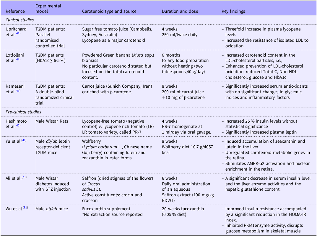
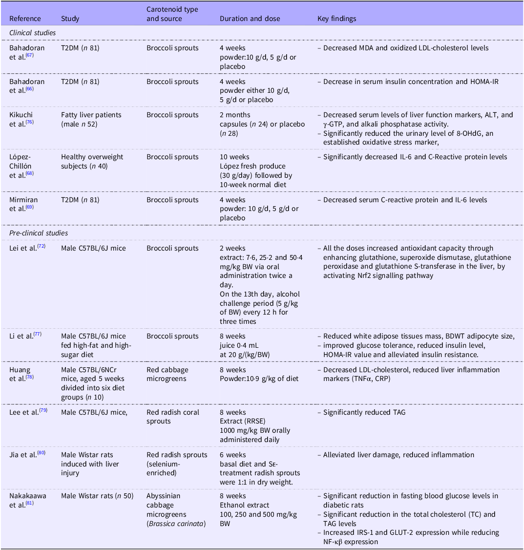


 Open access

