Introduction
It is well recognised that high noise intensity can lead to permanent hearing loss (i.e. noise-induced hearing loss). Much research has focused on the effects of occupational exposure to high noise intensity, leading the Occupational Safety and Health Administration to issue noise standards to protect workers from noise-induced hearing loss. The Occupational Safety and Health Administration has suggested that exposure to an impulse noise should not exceed 140 dB, which is the threshold of pain.1 It has also indicated that the levels of noise associated with aural suctioning are sufficient for noise-induced hearing loss to occur.2 Previous studies have utilised the Occupational Safety and Health Administration guidelines and the National Institute for Occupational Safety and Health guidelines to address the potential danger of aural suctioning on hearing sensitivity.Reference Wetmore, Henry and Konkle3, Reference Nelson, Giraud, Walsh and Mortelliti4
Surgical intervention for chronic otitis media with effusion often involves the use of suction by the otolaryngologist. The frequent utilisation of suction instruments for many otological procedures (e.g. removal of cerumen, myringotomies) may have effects on patients' hearing sensitivity. During aspiration of middle-ear effusion following myringotomies, suction noise has previously been recorded to range from 74 to 117 dB.Reference Wetmore, Henry and Konkle3 Because these mean intensities are sufficient to pose a potential risk for hearing loss, further investigation on the possible auditory consequences of routine ear suctioning is necessary. Tos et al. implied that there is a cause and effect relationship between suction-induced noise and acoustic trauma, although there is considerable controversy regarding this topic.Reference Tos, Lau and Plate5 Both MillsReference Mills6 and HumesReference Humes7 have reported that children are more susceptible to noise-induced hearing loss than adults. This is of particular concern as most patients undergoing myringotomies are children.
Several studies on suction-induced noise exist; however, many of these studies used older technology that has since been superseded by more sensitive instruments. Additionally, most studies have not reported the effects of differing viscosities of middle-ear effusion on peak noise intensities. This study was designed to test the following hypotheses: firstly, peak sound intensity during suctioning of middle-ear effusion reaches potentially harmful levels; and secondly, mucoid effusion results in increased sound pressure levels (SPLs) compared with serous effusion, and both are greater than SPLs in ears without effusion.
Materials and methods
Patient enrolment
This prospective, observational study was approved by the Texas Tech University Health Sciences Center Institutional Review Board. Subjects were recruited for the study if they were aged between 6 months and 18 years, had been diagnosed with otitis media with effusion (OME), and were scheduled to undergo myringotomy and tympanostomy tube placement. All subjects were initially examined by an otolaryngologist using otoscopy and tympanometry to confirm the diagnosis of chronic OME and to exclude any other concurrent pathology. Subjects were excluded if the external auditory canal was too narrow to fit the probe microphone attached to the suction tip. Informed consent was obtained from the parents of all subjects. Data on age, sex and type of effusion (classified as mucoid, serous or no effusion) were collected.
Clinical procedure
A single otolaryngologist performed all procedures. Subjects underwent bilateral myringotomies for OME that was refractory to standard medical management. Operations were performed under general anaesthesia (using a mask). After induction, cerumen was cleared from the external auditory canal using a curette; this was followed by irrigation with 70 per cent alcohol. An aural speculum was used to enhance surgical visualisation and the placement of the suction tip. A radial incision was then made in the anterior inferior quadrant of the tympanic membrane. Middle-ear effusion was aspirated utilising a Frazier 5 French gauge suction tip, connected to either a Neptune 2 Ultra waste management system (Stryker, Kalamazoo, Michigan, USA) or a wall suction unit.
Sound measurement
The SPLs generated during suctioning were recorded using an ER-7C Series B clinical probe microphone (Etymotic Research, Elk Grove Village, Illinois, USA). This was attached and stabilised to the suction tip using Steri-strips (3M, St Paul, Minnesota, USA) (Figure 1). The probe microphone was calibrated (for each patient) in the operating theatre, prior to surgery, using the built-in 1 kHz calibrator. The probe microphone was oriented at a 90° angle to the tympanic membrane. Sound was recorded during the entire fluid aspiration procedure using a custom sound level meter coupled to the Matlab program (MathWorks, Natick, Massachusetts, USA). In cases where noise intensity saturated the recording equipment, a programmable attenuator (PA5; Tucker-Davis Technologies, Alachua, Florida, USA) was used to decrease the output by −20 to −70 dB SPL. This adjustment was subsequently corrected for in all calculations. The following measurements were obtained for each ear: range and peak intensity of sound at the tympanic membrane, decibels in voltage at each frequency, and suction duration.
Etymotic Research ER-7C Probe Microphone System Series B connected to a Frazier 5 French gauge suction tip.

Sound pressure levels and spectral data analysis
The maximum intensity (in dB SPL), the peak frequency (in Hz) and the duration of suctioning (in seconds) were calculated utilising the Matlab acquisition toolbox (MathWorks). Frequency domain analysis was performed to explore the relationship between decibels in voltage and frequency (Hz). In order to conduct this analysis, the frequency domain data were smoothed to remove noise by calculating mean and maximum intensities over frequency ranges of 200 Hz. Analysis of the frequency and duration data was performed utilising Adobe Audition CS5.5 software (Adobe Systems, San Jose, California, USA). A normality test revealed marginal evidence against the assumption of normality within the mucoid effusion group (Kolmogorov–Smirnov test, p = 0.021). However, the mucoid effusion group was the largest of the three groups (n = 20), and as the t-test is robust against the assumption of normality, we proceeded with parametric analysis. Welch's analysis of variance (ANOVA) and pairwise Welch's t-test were used in light of heterogeneous variance between the three groups (Brown–Forsythe test, p > 0.038). Bivariate analysis using the Students t-test was employed to compare means, and an ANOVA was used to compare all three groups (via GraphPad Prism 5; GraphPad Software, La Jolla, California, USA). Significance was set at p < 0.05.
Results
Thirteen boys and 12 girls were enrolled, with an average age of 2 years (range, 10 months–6 years). On examination with a handheld otoscope, no abnormalities were visualised in the external auditory canal or the tympanic membrane. Of the 50 ears, 20 had mucoid effusion, 11 had serous effusion, and 13 had no effusion at the time of surgery. Six ears were not analysed because of missing SPL data from the intra-operative recording.
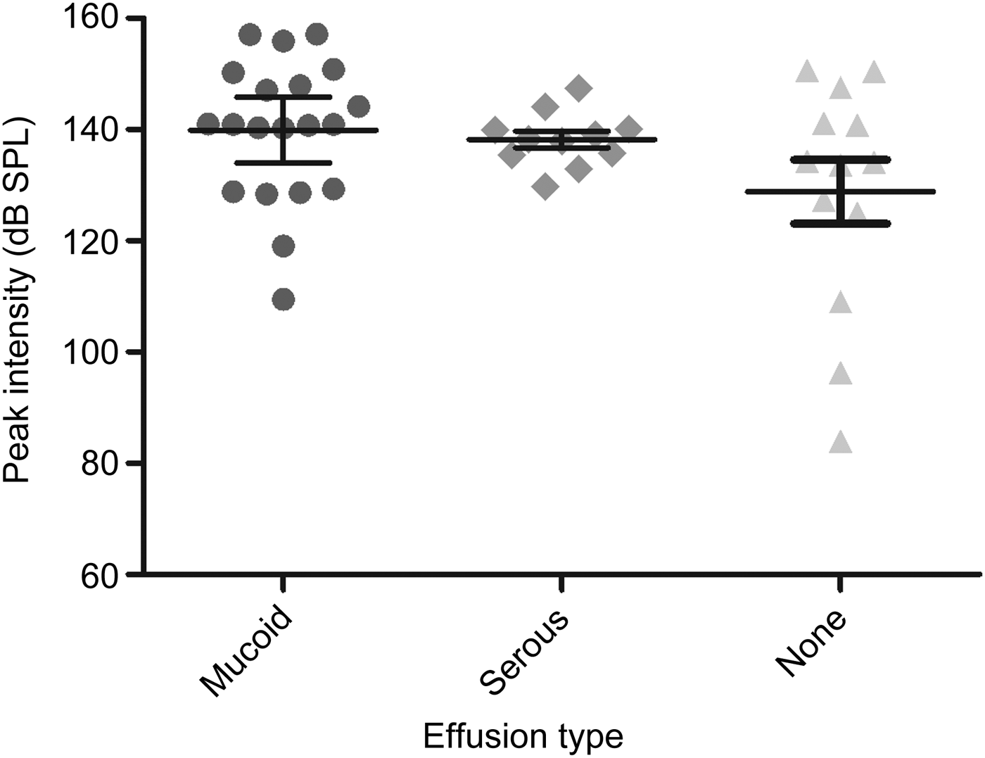
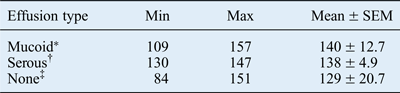
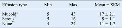
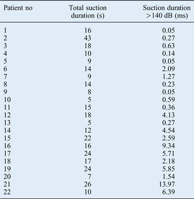
Maximum peak intensity ranged from 84 to 157 dB SPL, and suction duration ranged from 4 to 43 seconds; the means and ranges for each group are shown in Tables I and II. A scatterplot of the distribution of peak intensities is shown in Figure 2. There was no significant difference in mean peak intensity between groups. The lowest peak intensity of 84 dB SPL was recorded from a subject with no effusion and the highest peak intensity of 157 dB SPL was from a subject with mucoid effusion. Half of the ears (50 per cent) were exposed to greater than 140 dB SPL; of these, 82 per cent were exposed for longer than 0.2 ms (range, 0.05–13.97 ms) (Table III). Ears with mucoid effusion required significantly longer suction time than those with serous effusion (p = 0.0030) or no effusion (p = 0.0385).
Scatterplot of peak intensities in ears with mucoid, serous and no effusion (bars represent 95 per cent confidence intervals of the mean).
Peak intensities
Data values represent decibels. *n = 20; †n = 11; ‡n = 13. SEM = standard error of the mean
Duration of suction
Data values represent seconds. *n = 20; †n = 11; ‡n = 13. SEM = standard error of the mean
Total suction duration and suction duration above 140 dB
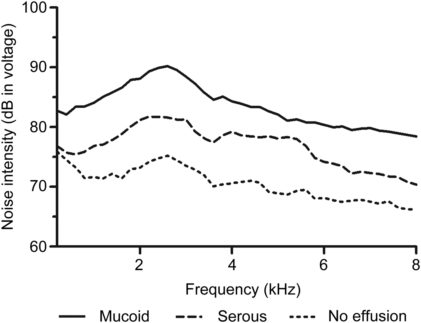
Analysis of the frequency domain data showed that the peak intensities occurred between 2 and 4 kHz for all three groups (Figure 3). However, the decibels in voltage were significantly different between all three groups (p < 0.0001).
Frequency domain analysis showing differences in noise intensity between ears with mucoid, serous and no effusion as a function of frequency. The resonance frequency of the external auditory canal in children less than four years old is approximately 3 kHz, which coincides with the peak decibel voltage in each group.Reference Dempster and Mackenzie8 (Graph constructed using bins of maximum intensities over a frequency range of 0.2 kHz).
Discussion
This study revealed that the peak noise intensities encountered during suctioning as part of middle-ear effusion aspiration were above the pain threshold and within the range deemed harmful for occupational exposure. While the peak intensities did not differ between ears with mucoid, serous and no effusion, the duration of suctioning was significantly longer in ears with mucoid effusion; thus exposure times to harmful noise were longer in these patients.
It is well known that noise-induced hearing loss is a slow, progressive process, primarily dependent on the intensity and duration of noise exposure. The damage is usually seen in the 3–5 kHz range with the characteristic 4 kHz downward notch related to the resonance of the external auditory canal.Reference Dempster and Mackenzie8 Temporary threshold shifts are reversible hearing losses; the outer hair cells have the ability to repair the damage, with resolution in 24 to 48 hours.Reference Nelson, Giraud, Walsh and Mortelliti4 Permanent threshold shifts are more severe, with irreversible damage within the cochlea.Reference Lonsbury-Martin, Martin, Flint, Haughey, Lund, Niparko, Richardson and Robbins9 Aspiration of mucoid effusion has previously been documented to cause temporary threshold shifts, as measured by auditory brainstem response (ABR) testing after aural suctioning.Reference Mason, Mason and Gibbin10
Impulse noise has been linked to noise-induced hearing loss and acoustic trauma. Impulses in the range of 0.2 ms in duration have peak energy at 2 to 3 kHz and are extremely detrimental to human hearing.Reference Dobie, Bailey, Johnson and Newlands11 Such impulses greater than 140 dB are regarded as potentially harmful to human hearing.Reference McRobert and Ward12 The Occupational Safety and Health Administration (OSHA) suggests that 140 dBA impulse noise is the threshold for pain, while levels above 115 dB may be harmful.Reference Dobie, Bailey, Johnson and Newlands11, Reference McRobert and Ward12 A comparison of commonly encountered noise sources is shown in Figure 4. In the current study, 50 per cent of ears were exposed to greater than 140 dB, and 82 per cent of these were exposed for greater than 0.2 ms.
National Institute for Occupational Safety and Health noise meter, showing examples of some common sources of noise and their expected noise levels. Permission to reprint granted from the Occupational Safety and Health Administration, 2013.
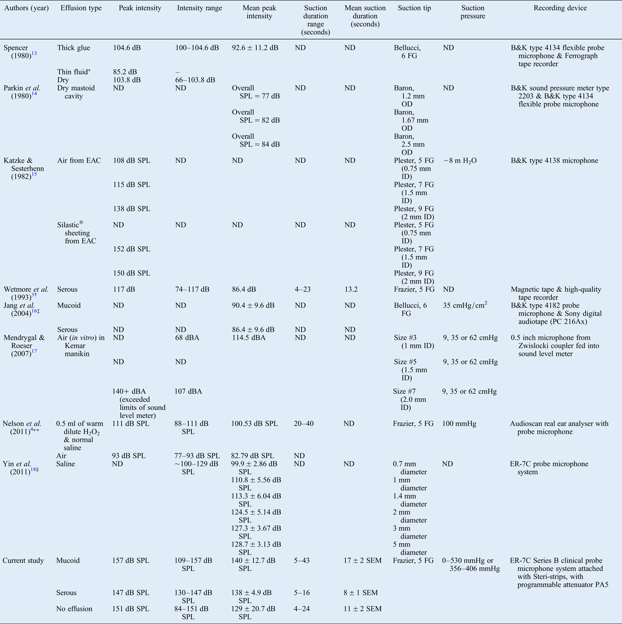
There is considerable controversy regarding whether ear suctioning causes noise-induced hearing loss or acoustic trauma. There is wide variation in the methodology of studies (conducted from 1980 to the present) that have examined this issue, as highlighted in Table IV.Reference Wetmore, Henry and Konkle3, Reference Nelson, Giraud, Walsh and Mortelliti4, Reference Spencer13–Reference Yin, Stromberg and Duan18 Three studies suggest that even though high peak intensities are recorded during myringotomies, it is not likely that the noise from the suction causes noise-induced hearing loss.Reference Nelson, Giraud, Walsh and Mortelliti4, Reference Spencer13, Reference Jang, Song, Kim and Wang16
Summary of suction studies
*n = 1. †Median intensity data reported. ‡Mucoid effusion mean intensity reported as 96.4 dB (abstract) and 90.4 dB (main text). **Study in healthy adult volunteers. §Study using fresh human temporal bones. ND = not documented; FG = French gauge; B&K = Brüel & Kjær; SPL = sound pressure level; OD = outer diameter; EAC = external auditory canal; H2O = water; ID = inner diameter; H2O2 = hydrogen peroxide; ~ = approximately; SEM = standard error of the mean
In a series of 24 ears studied, Spencer reported that the maximum intensities recorded were produced by suctioning of thick ‘glue’ ear.Reference Spencer13 Mean intensity in that study was 92.6 dB, with a standard deviation of 11.2 dB. Our lowest peak intensity measured during the suctioning of mucoid effusion (of 109 dB) exceeds their mean intensity; this discrepancy probably reflects differences in recording technology over the past 30 years.
In a study of 60 ears, Jang et al. showed no noise-induced sensorineural hearing loss associated with suctioning.Reference Jang, Song, Kim and Wang16 The mean intensities of the suction noise for ears with serous and mucoid effusion were 86.4 ± 9.6 dB and 90.4 ± 9.6 dB, respectively. Audiological evaluations performed prior to tube placement and after surgery were indistinguishable (paired t-test, p = 0.942).
Nelson et al. studied the effects of suctioning 0.5 ml of warm hydrogen peroxide diluted with normal saline using a Frazier 5 French gauge suction instrument in 21 healthy adults.Reference Nelson, Giraud, Walsh and Mortelliti4 Prior to suctioning, subjects underwent tympanometry, pure tone air conduction audiometry, and distortion product otoacoustic emission (DPOAE) testing. Five minutes post-suction, pure tone audiometry and DPOAE testing were repeated. No significant changes were seen in DPOAEs before and after suctioning. The lowest peak intensity recorded from suctioning with irrigation was 88 dB SPL with a maximum peak intensity of 111 dB SPL. In regards to suctioning a dry canal (e.g. air), the SPLs ranged from 77 to 93 dB SPL. In that study, suctioning was brief (20–40 seconds), controlled and unaggressive. The authors did not find any evidence of hearing detriment associated with ear canal suctioning; however, they did suggest that such hearing loss may be possible during office and surgical intervention.
The aspiration of mucoid effusion has previously been documented to cause temporary threshold shifts, as measured by ABR testing before and after aural suctioning.Reference Mason, Mason and Gibbin10 Mason et al. reported that 6 out of 14 ears aspirated showed clear evidence of a threshold shift greater than or equal to 15 dB, as measured by ABR testing.Reference Mason, Mason and Gibbin10 In addition, three of these six cases demonstrated a hearing threshold change from 15 to 50 dB nHL as measured by click-stimulus ABR testing, and a shift from 5 to 60 dB nHL for a 1 kHz tone-pip threshold ABR test.
Other studies recommend caution when utilising suction instruments. Parkin et al. states that noise due to suctioning and its effects on hearing loss should be considered.Reference Parkin, Wood, Wood and McCandless14 Katzke and Sesterhenn investigated the SPLs of one aural suction unit for removal of cerumen impaction.Reference Katzke and Sesterhenn15 In that study, they showed that noise intensity varied as a function of tip size and finger cut-off control being open or closed, with a larger tip size and closed finger cut-off increasing the noise intensity. Peak intensities ranged from 108 dB SPL for 5 French gauge tubes to 138 dB SPL for 9 French gauge tubes. Greater than 150 dB SPL was measured during actual debris extraction.
On the basis of similar outcomes for aspirated and non-aspirated middle ears, Youngs and Gatland suggested that middle-ear effusion evacuation should not be conducted, in order to avoid acoustic trauma.Reference Youngs and Gatland19 In that study, there was no statistically significant improvement in audiometric thresholds between cases of aspirated and non-aspirated effusion 24 hours following ventilation tube placement. In addition, the incidence of lumen obstruction was no higher for non-aspirated effusion cases than for aspirated effusion cases.
Wetmore et al. studied the suctioning of patients with only serous effusions, utilising a high-fidelity tape recorder with magnetic tapes.Reference Wetmore, Henry and Konkle3 The median intensities measured ranged from 74 to 117 dB, with a mean of 86.4 dB. It was suggested that the potential risk to hearing included the possibility of a temporary threshold shift.
Egeli and Kiris measured pre-operative and post-operative pure tone audiometry air–bone gaps, and concluded that the aspiration (vs non-aspiration) of middle-ear effusion resulted in no acoustic trauma.Reference Egeli and Kiris20 That study advocated ventilation tube insertion without aspiration of middle-ear effusion, to avoid noise exposure. However, the aspiration of middle-ear fluid prior to tube placement is so routinely performed that it is standard procedure.
Mendrygal and Roeser examined the effects of noise intensity related to suctioning air based on several variables, including suction pressure, suction tip size and depth of suction tip insertion.Reference Mendrygal and Roeser17 The findings showed that greater suction pressure was associated with higher noise intensity. In addition, larger suction tip diameters resulted in stronger intensity, regardless of pressure. The authors also examined the effects of finger valve closure, and found that it caused variability in noise intensity, for metal and plastic suction tips of varying sizes. Yin et al. showed a positive significant relationship between noise and increased inner diameter of the suction tip in a study wherein a fresh cadaveric temporal bone was suctioned.Reference Yin, Stromberg and Duan18 Noise levels recorded ranged from 100 to 129 dB SPL. The study findings implied that the utilisation of smaller diameter suction tips may protect against hearing loss.
There are many factors that play a role in SPL intensity and which need to be considered, such as suction pressure, tip diameter and tip material (e.g. plastic, metal).Reference Mendrygal and Roeser17 We speculate that suction noise may potentially cause temporary threshold shifts as a result of the proximity of the suction tip to the tympanic membrane and middle-ear space. Suction technique may also affect the SPLs; for instance, the clinician's movements while suctioning, and an open or closed cut-off valve during suctioning.
• High noise intensity can lead to permanent hearing loss
• The Occupational Safety and Health Administration suggests that impulse noise should not exceed 140 dB, which is the threshold of pain
• Impulses longer than 0.2 ms have peak energy at 2–3 kHz and are detrimental to human hearing
• In this study, maximum peak intensities ranged from 84 to 157 dB SPL, and 50 per cent of ears were exposed to greater than 140 dB SPL; of these, 82 per cent were exposed for longer than 0.2 ms
• Ears with mucoid effusion required longer suction times than those with serous effusion (p = 0.0030) or no effusion (p = 0.0385)
• The peak noise intensities encountered during suctioning were above the pain threshold and within the range deemed harmful for occupational exposure
The current study has several limitations. There was variability in the vacuum pressure, which may have caused inconsistency in the noise intensities measured. Some of the operating theatres in the previous studies used portable machines (0–530 mmHg) while others used a direct line from the wall suction unit (356–406 mmHg), and these pressures were not recorded. Because of the age group of our subjects, both pre and post bone conduction ABR testing should have been conducted. Post-operative audiometry was not conducted because of the young age of the subjects, and so temporary threshold shifts were not measured. We did not measure pre- and post-ABRs because there would most likely be an enhancement of the bone conduction results due to the occlusion effect caused by the middle-ear effusion. In this study, only a Frazier 5 French gauge suction tip was utilised. A larger suction tube is often required to aspirate mucoid effusions compared with serous effusions. All procedures were performed by a single surgeon; however, it is possible that individual variation may have caused undetected differing noise exposures. Future studies are necessary to examine whether these levels of sound intensity do indeed cause a shift in hearing threshold.
Conclusion
We ascertain that the noise intensity measured during the suctioning of middle-ear effusion is at levels conducive to hearing loss. Clinicians working in the ear with suction should be cognisant of the potential damage to patient hearing.
Acknowledgements
The authors would like to acknowledge Melissa Leiker (clinical research co-ordinator) and Phillip Watkins (biostatistician) from the Clinical Research Institute at the Texas Tech University Health Sciences Center for their time and dedication in this study.