Non-technical Summary
It is rare to find the ventral structure of the trilobite family Lichakephalidae. Recently, a nearly complete internal mold of Archikainella vomerinus, one species of that family, was collected from the Sandu Formation (Cambrian, Furongian) of South China. The new specimen shows its ventral structure. It is the first time allowing identification of the ventral structure in Lichakephalidae. The newly found ventral structure is similar to that of the more derived Lichidae and supports the hypothesis that lichakephalid trilobites are a primitive group of lichids.
Introduction
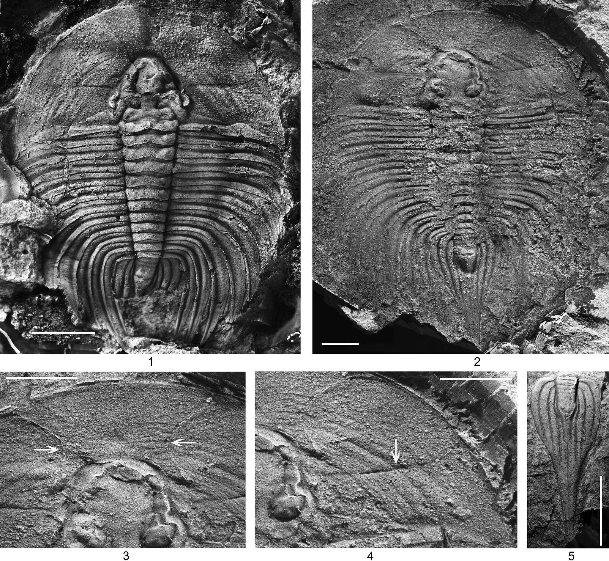
The phylogenetic relationships between major post-Cambrian trilobite groups and their Cambrian sister taxa is still very little known. This is known as the cryptogenesis problem (Stubblefield, Reference Stubblefield1959; Whittington, Reference Whittington1981; Edgecombe, Reference Edgecombe, Novacek and Wheeler1992; Lieberman and Karim, Reference Lieberman and Karim2010; Paterson, Reference Paterson2019). Lichakephalidae Tripp, Reference Tripp1957, which crosses the Cambrian-Ordovician boundary, plays an important role in understanding the Cambrian root of Lichida Moore, Reference Moore1959 (Fortey, Reference Fortey1990; Adrain, Reference Adrain, Harper and Servais2013). However, knowledge of trilobites in the family Lichakephalidae is still relatively sketchy. Only two species have been previously described from articulated specimens—Lichakephalus stubbsi Fortey, Reference Fortey2011 from the Ordovician and Archikainella vomerinus Liu, Reference Liu and Li1982 (Zhu et al., Reference Zhu, Peng, Pegel and Zuo2021; Fig. 2.1) from the Cambrian. Other species are known from fragmentary material and consist mainly of cranidia. The rostral plate and hypostomal attachment conditions of lichakephalids were hitherto unknown, even from the articulated specimens of Lichakephalus stubbsi and Archikainella vomerinus. The hypostome of lichakephalids has been illustrated in only one species, Paraacidaspis ultima (Shergold, Feist, and Vizcaino, Reference Shergold, Feist and Vizcaino2000). In most lichakephalids (i.e., Acidaspidella Pokrovskaya in Rozova, Reference Rozova1963; Acidaspidina Lazarenko, Reference Lazarenko1960; Archaeopleura Ramsköld, Reference Ramsköld1991; Belovia Poletaeva in Chernysheva et al., Reference Chernysheva, Egorova, Ogienko, Poletaeva and Repina1956; Bestjubella Ivshin, Reference Ivshin1962; Brutonia Thomas and Holloway, Reference Thomas and Holloway1988; Colossaspis Zhang in Qiu et al., Reference Qiu, Liu, Zhu, Bi and Lin1983; Eoacidaspis Poletaeva in Chernysheva et al., Reference Chernysheva, Egorova, Ogienko, Poletaeva and Repina1956; Lichokephalina Antsygin, Reference Antsygin, Varganov, Antsygin, Nasedkina, Militsina and Shurygina1973; Lichakephalus Sdzuy, Reference Sdzuy1955), there is no preglabellar field, therefore their hypostomal attachment conditions are reasonably inferred as conterminant. So far as we know, there is no substantive evidence to show that these lichakephalids (i.e., Usoviana Poletaeva, Reference Poletaeva1977; Paraacidaspis Poletaeva in Egorova et al., Reference Egorova, Xiang, Li, Nan and Guo1963; Archikainella Liu, Reference Liu and Li1982), each bearing a preglabellar field, have a conterminant hypostomal attachment condition.
Recently, a nearly complete internal mold of Archikainella vomerinus indicates that, in these lichakephalids, the hypostomal attachment condition is conterminant. This specimen was collected from the Sandu Formation (Cambrian, Furongian), Guole Town, Jingxi County, Guangxi Zhuang Autonomous Region, South China (Fig. 2.2). It is the second nearly complete specimen of this rare species found in this area. A complete exoskeleton was previously described by Zhu et al. (Reference Zhu, Peng, Pegel and Zuo2021; Fig. 2.1). The newly found internal mold is dorsoventrally compacted such that the glabella is compressed upon the hypostome. The rostral plate is long (exs.) and its inner margin extends medially as far as the front of the glabella; its hypostomal attachment condition is conterminant. Because more comprehensive morphological knowledge of lichakephalids is necessary for any future phylogenetic analysis of this important group of trilobites, this new specimen merits description. Based on the new material, the relationship between the family Lichakephalidae and the order Lichida is discussed.
Geological setting
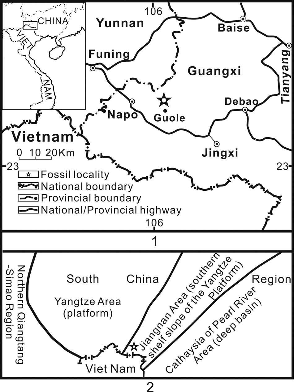
The new specimen is from the Sandu Formation (Cambrian, Furongian), near Guole Town, Jingxi County, southwestern Guangxi Zhuang Autonomous Region, South China (Fig. 1.1). Recently, a new Furongian Lagerstätte, the Guole Lagerstätte, was discovered in this region (Zhu et al., Reference Zhu, Peng, Zamora, Lefebvre and Chen2016).
Map of Jingxi County, Guangxi Zhuang Autonomous Region, China, showing location of the fossil-bearing site near Guole (1) and general geologic setting (2). Modified from Zhu et al. (Reference Zhu, Peng, Zamora, Lefebvre and Chen2016).

Figure 1. Long description
Panel 1 is a geographical map of the border region between China and Vietnam. An inset in the top-left shows a broad view of China and Vietnam with a rectangle highlighting the study area. The main map shows Yunnan to the Northwest and Guangxi to the Northeast. Cities labeled include Funing, Baise, Tianyang, Debao, Napo, Guole, and Jingxi. A star icon near Guole indicates the Fossil locality. A scale bar shows 0, 10, and 20 K m. A legend defines symbols for Fossil locality, National boundary (thick dashed line), Provincial boundary (thin dashed line), and National or Provincial highway (solid line). The 106 degree longitude and 23 degree latitude lines are marked.
Panel 2 is a geological map of the same region. From Northwest to Southeast, the zones are labeled: Northern Qiangtang-Simao Region, South China Yangtze Area (platform), Jiangnan Area (southern shelf slope of the Yangtze Platform), and Cathaysia of Pearl River Area (deep basin). The fossil star icon is located on the boundary between the Yangtze Area and the Jiangnan Area. Viet Nam is labeled at the bottom-left corner.
Archikainella vomerinus Liu, Reference Liu and Li1982: (1) latex cast from the external mold of a nearly complete exoskeleton, NIGP 170489; (2) internal mold of a nearly complete exoskeleton, NIGP 201820; (3) enlargement of the rostral plate of (2), showing the course of the rostral sutures (arrows); (4) enlargement of the right gena of specimen in (2) showing impression of the facial suture (arrow); (5) partially exfoliated pygidium, NIGP 201821. Specimens are from the Sandu Formation (Cambrian, Furongian), Guole Town, Jingxi County, Guangxi Zhuang Autonomous Region, South China, which is equivalent to the Probinacunaspis nasalis-Peichiashania hunanensis Zone of western Hunan. Scale bars = 10 mm.

Figure 2. Long description
A multi-panel figure containing five grayscale photographs of trilobite fossils.
Panel 1 at the top-left shows a latex cast of a nearly complete exoskeleton (N I G P 170489). It features a broad, rounded cephalon, a segmented thorax with long pleural spines curving backward, and a small pygidium. A white scale bar is in the bottom-left.
Panel 2 at the top-right shows an internal mold of a nearly complete exoskeleton (N I G P 201820). The preservation is flatter than panel 1, showing the central axis and the radiating pleural ribs. A white scale bar is in the bottom-left.
Panel 3 at the bottom-left is a high-magnification view of the rostral plate from the specimen in panel 2. Two horizontal white arrows point inward toward the rostral sutures located above the glabella.
Panel 4 in the bottom-center is an enlargement of the right gena (cheek) of the specimen in panel 2. A vertical white arrow points downward to the impression of the facial suture running across the genal field.
Panel 5 at the bottom-right shows a partially exfoliated pygidium (N I G P 201821). It is elongated with a distinct central axis and long, trailing marginal spines. A vertical white scale bar is on the right side.
In Jingxi County and neighboring areas, the Sandu Formation consists of siltstone and carbonate rocks with its lower stratigraphic levels not exposed. It is unconformably overlain by dolostones of the Huangqiongshan Formation (Lower Devonian). Associated trilobites (Zhu et al., Reference Zhu, Hughes and Peng2007, Reference Zhu, Hughes and Peng2010) indicate that the formation is equivalent to the Probinacunaspis nasalis-Peichiashania hunanensis Biozone (middle Furongian, upper Jiangshanian Stage) of northwestern Hunan (Peng, Reference Peng1992; Peng et al., Reference Peng, Babcock, Cooper, Gradstein, Ogg, Schmitz and Ogg2012).
Because outcrops in the Guole Town area are poor, there is a lack of consensus about the Cambrian paleoenvironmental setting of the Guole area. Interpretations range from outer shelf (Han et al., Reference Han, Tang, Wei and Wang2000) to basinal (Feng et al., Reference Feng, Peng, Jin, Jiang, Bao, Luo, Ju, Tian and Wang2001). Traditionally, Guole and its neighboring regions have been regarded as part of the transitional region between the Yangtze Platform and Jiangnan Basin (Fig. 1.2), based on typical slope facies trilobites. The trilobite fauna representative of slope facies includes the key Furongian agnostoids Lotagnostus Whitehouse, Reference Whitehouse1936 and Glyptagnostus Whitehouse, Reference Whitehouse1936 (Guangxi Geological Bureau, 1985; Peng et al., Reference Peng, Babcock and Lin2004). Brachiopods of the Guole Biota were interpreted as nearshore, shallow water organisms by Zhan et al. (Reference Zhan, Jin, Rong, Zhu and Han2010). The paleoenvironmental setting is important for understanding the ecology of the biota but cannot presently be interpreted unambiguously.
Implications for phylogenetic relationship between Lichida, Lichakephalidae, and Dameselloidea
The lichakephalid glabella is constricted at S1, and this feature is a key synapomorphy for the Lichakephalidae (Thomas and Holloway, Reference Thomas and Holloway1988). As such, the convex area opposite S1 in Lichakephalus and Archikainella is part of the fixigena (Fortey, Reference Fortey2011; Zhu et al., Reference Zhu, Peng, Pegel and Zuo2021); a similar structure in Paraacidaspis, regarded as a discreet baccula (Shergold et al., Reference Shergold, Feist and Vizcaino2000), might also be part of the fixigena. The degree of constriction at S1 is variable. It is strong in Belovia, Eoacidaspis, Paraacidaspis, Archikainella, Lichakephalus, and Lichakephalina; weak in Acidaspidella, Acidaspidina, and Usoviana; and obscure in Archaeopleura.
The family Lichakephalidae was recognized as a senior synonym of Eoacidaspididae by Thomas and Holloway (Reference Thomas and Holloway1988). Zhang (Reference Zhang1990), Shergold et al. (Reference Shergold, Feist and Vizcaino2000), and Peng et al. (Reference Peng, Babcock and Lin2004) rejected this synonymy, as discussed at length by Adrain (Reference Adrain, Harper and Servais2013). The synonymy was accepted by Ramsköld (Reference Ramsköld1991), Jell and Adrain (Reference Jell and Adrain2003), Pollitt et al. (Reference Pollitt, Fortey and Wills2005), Fortey (Reference Fortey2011), Adrain (Reference Adrain, Harper and Servais2013), and Zhu et al. (Reference Zhu, Peng, Pegel and Zuo2021). The cranidium of Eoacidaspis, Belovia, Paraacidaspis, and Archikaninella (classified as Eoacidaspididae by Zhang, Reference Zhang1990; Shergold et al., Reference Shergold, Feist and Vizcaino2000; and Peng et al., Reference Peng, Babcock and Lin2004) is strikingly similar to that of Lichakephalus stubbsi (see Fortey, Reference Fortey2011, figs. 1, 2), which apparently belongs to the eponymous family Lichakephalidae, in its strong constriction at S1 as mentioned above, and its complex glabellar lobation. The new specimen described in this paper indicates that both Archikaninella and Lichakephalus (see Fortey, Reference Fortey2011, figs. 1, 2) have 11 thoracic segments. These resemblances support Thomas and Holloway’s (Reference Thomas and Holloway1988) recognition of Eoacidaspididae as a synonym of Lichakephalidae.
The Lichakephalidae is considered to be a paraphyletic grade from which the Lichidae was derived, and both of them are included in the Lichida (see also Thomas and Holloway, Reference Thomas and Holloway1988; Fortey, Reference Fortey2011; Adrain, Reference Adrain, Harper and Servais2013). The newly collected specimen reveals more cogent evidence to support this argument. The rostral plate, found for the first time in lichakephalids, is wide (tr.) and long (sag.). Its posterior margin extends medially backward to the anterior margin of the glabella. Because the front margin of the hypostome and that of the glabella correspond closely (Whittington, Reference Whittington1988), the hypostomal attachment condition in Archikainella is conterminant. Most lichakephalids are similar to derived lichids in terms of the absence of a preglabellar field and narrow anterior border. The hypostomal attachment condition in these lichakephalids is reasonably inferred to be conterminant. The new material indicates that in lichakephalids, e.g., Paraacidaspis and Archikainella, which have long (sag.) preglabellar fields, the mode of hypostomal attachment is also conterminant. This feature is consistent with that of derived lichids. Archikainella has 11 thoracic segments, similar to most lichids (Thomas and Holloway, Reference Thomas and Holloway1988; Whittington, Reference Whittington2002). The pygidium of Archikainella is distinguished from that of lichids in being narrower (tr.). This could have resulted from the strongly backward curvature in the thoracic pleural region. The pygidium of Archikainella has four pleural ribs, which is less than those of other lichakephalids, but more than those of most lichids; most lichids have three pleural ribs (Thomas and Holloway, Reference Thomas and Holloway1988; Whittington, Reference Whittington2002). It seems likely that the pygidium of Archikainella is transitional between those of lichakephalids and lichids. The close similarities between the new material of Archikainella and derived lichids support the assignment of Lichakephalidae to the order Lichida.
The origin of lichids is controversial. It is inferred that lichids root within dameselloids and the families should be assigned to the order Lichida based on the striking similarities between Cambrian dameselloids and lichakephalids, and the assertion that dameselloids have conterminant hypostomal attachment condition (Fortey, Reference Fortey1990, Reference Fortey and Kaesler1997, Reference Fortey2001). This idea was followed by subsequent authors (Peng et al., Reference Peng, Babcock and Lin2004; Adrain, Reference Adrain and Zhang2011; Yuan et al., Reference Yuan, Li, Mu, Lin and Zhu2012, Reference Yuan, Zhang and Zhu2016; Ren et al., Reference Ren, Wei and Yuan2017). Recent studies demonstrate that Fortey’s (Reference Fortey1990) assertion that dameselloids have conterminant hypostomal attachment condition might possibly not be completely correct if dameselloid form a monophyletic group, because some dameselloids (ascribed to Dorypygellidae Kobayashi, Reference Kobayashi1935) have natant hypostomal attachment condition and the others (ascribed to Damesellidae Kobayashi, Reference Kobayashi1935) have conterminant hypostomal attachment condition (Peng et al., Reference Peng, Babcock, Yang and Zhu2024a, Reference Peng, Babcock, Yang, Zhu and Liub). The co-occurrence of these two types of hypostomal attachment condition indicates that some dameselloid trilobites possibly developed the conterminant condition secondarily from ancestors having the natant condition. Subsequently, Dameselloidea was transferred to the order Corynexochida (Peng et al., Reference Peng, Babcock, Yang, Zhu and Liu2024b). In fact, both Corynexochida and Lichida have been regarded as primarily (rather than secondarily) conterminant groups (Fortey, Reference Fortey1990, Reference Fortey and Kaesler1997); if so, then it is not acceptable to transfer Dameselloidea from Lichida to Corynexochida. In view of the striking similarities between conterminant Cambrian dameselloids (e.g., Palaeadotes Öpik, Reference Öpik1967) and lichakephalids, the assignment of dameselloids and lichakephalids to the order Lichida is tentatively supported herein, although Adrain (Reference Adrain and Zhang2011) noted that there has been almost no detailed evidence to support this view.
Materials and methods
Illustrated specimens were photographed under a Zeiss stereomicroscope (Model Axio Zoom V16) using a digital head (AxioCamMrM) and a circle light around the lens to provide uniform lighting. Directional light was added from the left-anterior side. A black latex mold was used to illustrate one specimen. The other specimens were coated with black ink prior to photography. Both the specimen and the latex mold were coated with magnesium oxide before photography.
Terminology, repository, and institutional abbreviation
Terminology for description in this study follows that of Whittington and Kelly (Reference Whittington, Kelly and Kaesler1997). All illustrated specimens are deposited in Nanjing Institute of Geology and Paleontology, Chinese Academy of Sciences, Nanjing, China (NIGPAS), and prefixed NIGP.
Systematic paleontology
Class Trilobita Walch, Reference Walch1771
Order Lichida Moore, Reference Moore1959
Family Lichakephalidae Tripp, Reference Tripp1957
Genus Archikainella Liu, Reference Liu and Li1982
Type species
Archikainella vomerinus Liu, Reference Liu and Li1982, p. 323, pl. 234, fig. 5, from the Shenjiawan Formation at Qiangongping, Fenghuang, western Hunan, China, by original designation.
Emended diagnosis
Exoskeleton ovoid in outline. Cephalon, exclusive of genal spines, subsemicircular. Glabella tapering forward, constricted at S1, acutely rounded anteriorly. Axial furrows sinuous, deep. S1 short, deep, proximally connected to S2; S3 and S4 short, faint. Frontal lobe rounded triangular. Occipital ring long (sag.) with median node, arching forward medially. Eye ridge absent; palpebral lobe short, situated slightly posterior to the midlength of glabella. Anterior branch of facial suture divergent forward and anterolaterally; posterior branch extending posteriolaterally. Fixigena narrow (tr.) between palpebral lobe and glabella. Frontal area long. Lateral border furrow shallow, lateral border wide (tr.) and convex; genal spine short. Doublure wide. Genal field covered with coarse granules.
Rostral suture marginal or submarginal. Connective suture smoothly extending posteromedially from anterior margin of anterior border, extending across 5/6 of preglabellar length, then gradually curving posterolaterally, terminating at the level of S4; rostral plate extending below entire frontal area, terminating below anterior glabellar margin. Rostral plate covered with terrace ridges. Anterior hypostomal margin coincident with anterior cranidial margin; attachment condition conterminant.
Thorax of 11 segments. Axis narrow (tr.), equivalent to approximately one-half width of pleural region at first segment. Inner portion of pleurae transverse and outer portion progressively curving gently rearward; curvature gentle in first pleura, progressively becoming stronger in succeeding pleurae; curvature posteromedially directed; pleural furrow deep; pleural tip pointed.
Pygidium triangular in outline. Axis short, consisting of three rings and long terminal piece, extending rearward as stout postaxial ridge. Pleural region consisting of four pleural ribs. Pleural furrows deep; interpleural furrows absent. Doublure long (tr.), extending into axis, covered with terrace ridges.
Remarks
Archikainella is easily distinguished from other Cambrian lichakephalids by its unique pygidium, with only four pleural ribs. This number of pleural ribs is less than that in other Cambrian lichakephalids, e.g., Paraacidaspis (see Peng et al., Reference Peng, Babcock and Lin2004, pl. 46) and Eoacidaspis (see Thomas and Holloway, Reference Thomas and Holloway1988, fig. 348), which have at least six pleural ribs. In other Cambrian lichakephalids, the pygidia are wider than long, but in Archikainella, the pygidium is much longer than wide. The cranidium of Archikainella is reminiscent of that of Paraacidaspis, but differs in having more divergent anterior branches of the facial sutures.
Acknowledgments
I am grateful to M. Gao and Y. Du who generously donated specimens to Xuejian. S.J. Wernette (Texas State University, San Marcos, Texas, USA) is thanked for helping to polish the English in this paper. L.E. Babcock (The Ohio State University) and an anonymous reviewer provided thorough reviews that greatly improved the paper. This work was supported by grants from the National Key Research and Development Program of China (2023YFF0803602).
Competing interests
The author declares none.