Implications
The achieving of adequate piglet performance during the weaning process is a primary economic target in pork production. As a result of the European ban of in-feed growth promoting antibiotics, the adoption of new systems for pig production involving immune competent, healthy and fast growing animals is a fundamental step to adjust the current production system to the legislative system. In this case, a dietary nucleotide supplementation could help piglets to cope with this transition phase.
Introduction
Social and nutritional stressors associated with the weaning process are important factors that affect digestive physiology, neuroendocrine and immune status and increase energy expenditure in piglets. One of these critical factors is the shift from milk to a dry feed, which induces a period of fasting. This results in alterations in gut architecture (Pluske et al., Reference Pluske, Hampson and Williams1997b). The presence and absorption of nutrients are essential for growth and function of the epithelial lining of the gut (Brooks and Tsourgiannis, Reference Brooks and Tsourgiannis2003). The intestinal epithelium provides an extensive and complex interface between the piglet's immune system and its environment. Diets formulated to maximize performance of weaning pigs need to support the development of intestinal tissue. To this aim, nucleotide-supplemented diets have been proposed in weaned piglets (Di Giancamillo et al., Reference Di Giancamillo, Domeneghini, Paratte, Dell'Orto and Bontempo2003; Martinez-Puig et al., Reference Martinez-Puig, Manzanilla, Morales, Borda, Pérez, Piñeiro and Chetrit2007; Godlewski et al., Reference Godlewski, Bierla, Strzalkowski, Martinez-Puig, Pajak, Kotunia, Chetrit and Zabielski2009).
Nucleotides are low-molecular-weight, intracellular compounds that are functional to numerous biochemical processes. They have important effects on the growth and development of rapid turnover cells, such as those in the immune system and in the gastrointestinal tract (Wang et al., Reference Wang, Gong, Le and Shi2008). In healthy conditions dietary nucleotides are probably not essential, however, during stressful or limited nutrient intake periods, dietary nucleotides may become essential (Dancey et al., Reference Dancey, Attree and Brown2006). They are continuously degraded and salvaged by all cells and they are naturally present in foods and feeds of animal and vegetable origin (Mateo and Stein, Reference Mateo and Stein2004).
The aim of the study was to evaluate the effect of a dietary nucleotide supplementation during lactation and post-weaning on the performance of piglets and on the neuroendocrine and immune systems, both involved during the response to stress and disease insults (Sapolsky et al., Reference Sapolsky, Romero and Munck2000).
Material and methods
Experimental procedures for this study were approved by the Ethics on Animal Experimentation Committee of Parma University, in accordance with the EC Directive 86/609/EEC.
Animals, housing and diets
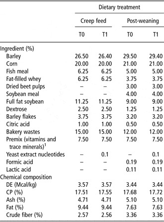
Ten [HBI Fomeva11 × (Large White × Landrace)] litters (n = 108 piglets) were reared in the same farrowing room (environmental temperature: 26°C; temperature of piglets zone: 30°C) from birth to 21 days, and in the same environmentally controlled nursery facility (temperature: 27°C in the 1st week after weaning, then reduced by 1°C per week) from 22 to 55 days (1 litter/pen). During each period litters had ad libitum access, respectively, to creep feed and to post-weaning standard diets supplemented with 0% (T0; n = 53 piglets) or 0.1% (T1; n = 55 piglets) of yeast extract nucleotides (Nucleoforce Piglets™ Bioiberica, Palafolls, Spain). The ingredients and chemical composition of the diets are presented in Table 1. In both T1 diets barley was replaced with yeast extract nucleotides. The allocation of litters to the dietary treatments (5 litters/treatment) was random. Yeast extract nucleotides were designed to provide the same amount of nucleotides per day than sow's milk at 21 days of lactation (Mateo et al., Reference Mateo, Peters and Stein2004), considering an estimated daily milk intake of 1 kg/day (Pluske et al., Reference Pluske, Fenton, Lorschy, Pettigrew, Sower and Aherne1997a) and a post-weaning feed intake of 250 g/day. Piglets had free access to drinking water. The diets did not contain antimicrobial growth promoters (AGPs). Piglets were vaccinated twice (4 and 21 days of age) against Mycoplasma hyopneumoniae (Suvaxyn™M Hyo, Pfizer Animal Health, Madison, NJ, USA).
Ingredients and chemical composition, as-fed basis, of T0 and T1 diets for piglets from 9 to 21 (creep feed) and from 22 to 55 (post-weaning) days of age

DE = digestible energy.
1The premix provides for kilogram: vitamin A, 160 000 IU; vitamin D3, 26 660 IU; vitamin E, 1330 mg; vitamin K, 27 mg; vitamin B1, 20 mg; vitamin B2, 80 mg; pantothenic acid, 200 mg; vitamin B6, 27 mg; vitamin B12, 0.4 mg; vitamin PP, 400 mg; biotin, 1.33 mg; folic acid, 13.3 mg; vitamin C, 533 mg; betaine, 2000 mg; I, 20 mg; Co, 7 mg; Se, 5.6 mg; Cu, 2000 mg; Mn, 666.6 mg; Zn, 1400 mg; Fe, 2133 mg; 6-phytase EC 3.1.3.26 8000 FTU.
Measurements
Piglets were identified by ear tags and they were individually weighed at 9 days of age, at weaning (day 21), at day 35 and at the end of the post-weaning period (55 days of age). The average daily feed intakes (days 9 to 21 and days 22 to 55) were evaluated per litter. Fecal consistency was recorded daily at litter level and assessed by a subjective four-point scale where 1 = firm and 4 = watery (Wellock et al., Reference Wellock, Fortomaris, Houdijk and Kyriazakis2007). At day 9, at weaning (day 21), and after the second vaccination against Mycoplasma hyopneumoniae (days 28 and 35), blood samples were collected (0800 h) using jugular venopuncture in 10 ml vacutainer tubes with lithium heparin. Whole blood was refrigerated and then analyzed, as plasma was obtained by centrifugation at 1800 × g for 10 min at room temperature and then stored at −20°C until analysis.
Hormone analysis
Cortisol plasma content was determined by a validated radioimmunoassay (Tamanini et al., Reference Tamanini, Giordano, Chiesa and Seren1983). Cortisol, H3-labelled cortisol and anti-cortisol antiserum were purchased from Sigma-Aldrich (St Louis, MO, USA). The intra- and interassay coefficients of variation were 7.4% and 9.5%, respectively. The minimal detection limit was 500 pg/ml.
Plasma samples were assayed for growth hormone (GH) by ELISA (Baratta et al., Reference Baratta, Saleri, Mainardi, Valle, Giustina and Tamanini2002). Effective dose (ED)90, ED50 and ED10 were 0.039, 0.166 and 5 ng/well, respectively. The intra- and interassay coefficients of variation were 5.3% and 7.8%, respectively. The minimal detection limit was 100 pg/ml. Porcine GH (pGH; AFP10864B) and anti-pGH antiserum (AFP5672099) were provided by Dr A. F. Parlow (National Hormone and Pituitary Program, Harbor-University of California-Los Angeles Medical Center, La Jolla, CA, USA).
Plasma IGF-1 content was evaluated using a multispecies IGF-1 ELISA (Alpco Diagnostic, Salem, NH, USA), according to manufacturer's instructions, previously applied by Rogan et al. (Reference Rogan, Reznikov, Pezzullo, Gansemer, Samuel, Prather, Zabner, Fredericks, McCray, Welsh and Stoltz2010). The intra- and interassay coefficients of variation were 7.8% and 5.3%, respectively. The minimal detection limit was 30 pg/ml.
Peripheral blood lymphocytes
The phenotypic characterization of T lymphocyte subpopulations in the whole blood was performed using flow cytometry (Borghetti et al., Reference Borghetti, De Angelis, Saleri, Cavalli, Cacchioli, Corradi, Mocchegiani and Martelli2006a). Briefly, 50 μl of heparinized blood were mixed with 5 μl of the specific antibody for each surface antigen in a plastic tube. A direct approach for CD4α and CD8α staining was performed by incubation with a phycoerithrin (PE)- and fluorescein isothiocyanate (FITC)-conjugated primary antibody (anti-pig CD4-PE and anti-pig CD8-FITC; Southern Biotech, Birmingham, AL, USA) for 15 min in the dark, at room temperature. After incubation, cells were washed with PBS (phosphate buffered saline) + 1% FBS (fetal bovine serum) and centrifuged for 5 min at 400 × g at room temperature. The contaminating red cells were lysed by treatment with an NH4Cl solution, pH 7.2, for 15 min at room temperature in the dark. The cell suspension was then washed twice with PBS + 1% FBS, centrifuged for 5 min at 400 × g at room temperature, suspended in 0.5 ml of PBS + 1% FBS and finally analyzed by flow cytometry (Epics XL-MCL, Beckman-Coulter, Brea, CA, USA). The results are based on lymphocyte gating on a forward scatter v. side scatter graph after acquisition of at least 10 000 cell events.
Cytokine gene expression
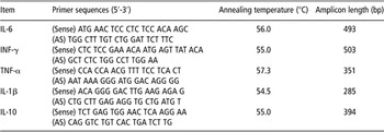
Interleukin (IL)-6, IL-10, interferon(IFN)-γ, IL-1β and tumor necrosis factor (TNF)-α gene expression was evaluated on peripheral blood mononuclear cells (PBMC) as previously described (Borghetti et al., Reference Borghetti, Saleri, Ferrari, Morganti, De Angelis, Franceschi, Bottarelli and Martelli2011). PBMC were separated by density gradient using Histopaque-1077 (Sigma-Aldrich, St Louis, MO, USA) and washed twice with sterile PBS. Ribonucleic acid was extracted with a TRI Reagent solution (Applied Biosystems, Monza, Italy). Briefly, 1 ml TRI Reagent solution was added to 1 × 10⁷ PBMC and RNA was extracted according to the manufacturer's instructions. The RNA quantification was carried out with a spectrophotometer (Genequant pro, Amersham Pharmacia, St Albans, UK). Ribonucleic acid retrotranscription (RT) was carried out using a Ready-to-go You-Prime First-Strand Beads kit (Amersham Pharmacia Biotech, Uppsala, Sweden) as described by the manufacturer. Two micrograms of total RNA were used in the RT. Aliquots (5 μl) of the cDNA obtained were used for the subsequent PCR amplification in a reaction buffer containing 1.5 μl MgCl2 (50 mM), 1 μl deoxynucleotide triphosphates (12.5 mM) and 1 μl Taq DNA polymerase (1 μg/μl), to a final volume of 50 μl. Amplification was carried out for 27 cycles, when the reaction was in the middle of the linear range (before reaching the amplification plateau). Each cycle consisted of denaturation at 94°C for 1 min, annealing at a specific temperature for each primer set for 1 min, extension at 72°C for 1 min; at the end of the 27th cycle, an additional extension step was carried out for 5 min. In Table 2 the specific primer sets used were summarized. QuantumRNA 18S was used (no. 1717, Ambion, Inc., Applied Biosystems, Monza, Italy) as an internal positive control for a relative quantitative RT-PCR. The 18S fragment (323 bp) was run in parallel with cytokine genes by using a 18S PCR alternate primer pair (5 μM) and 18S PCR competimers (5 μM; ratio 2 : 8) according to the manufacturer's instructions. PCR products were visualized after electrophoresis on a 2% agarose gel by SYBR Safe DNA gel stain (Invitrogen S.R.L., San Giuliano Milanese, Italy). The values are shown as the ratio of the band intensity of each cytokine normalized with the corresponding ribosomal 18S and expressed as relative arbitrary units.
Oligonucleotide sequences designed for the detection of different porcine cytokines

IL = interleukin; AS = antisense; INF = interferon; TNF tumor necrosis factor.
Statistical analysis
The data were subjected to ANOVA analysis using the GLM procedure (Statistical Analysis System, 2008) with the animal as the experimental unit for growth and hematological data and the litter as the experimental unit for feed intake and feed efficiency. Growth performance data were analyzed using a mixed model with group, sex and group × sex interaction as fixed factors, and sow nested within group as a random factor. Feed intakes and feed efficiency data were analyzed using a model with group as a fixed factor. For hematological data, group, sex, day of sampling and group × sex and group × day of sampling interactions were used as fixed factors and sow nested within group was considered as a random factor. Piglets performance and hematological data recorded at day 9 were used as a covariate. The results were expressed as least squares means ± standard error of the mean. A correlation analysis was performed on cortisol, IGF-1 and GH plasma levels. Statistically significant differences were assumed using P ≤ 0.05, as 0.10 > P > 0.05 was considered as a trend.
Results
Growth performance
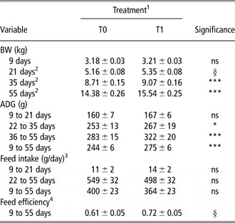
In both groups the fecal consistency was the same and it was always below three during the study. The effects of nucleotide supplementation on piglets’ growth performance are reported in Table 3. Despite an overall feed intake similarity between groups (P > 0.05), BW of piglets at days 21 (P < 0.10), 35 and 55 (P < 0.05) was greater in T1 compared with T0. Average daily gain (days 9 to 55) was higher (P < 0.05) and feed efficiency tended to improve (P = 0.10) in T1 than in T0.
Least squares means (±s.e.) of piglets growth performance as affected by nucleotide supplementation

ADG = average daily gain.
Probability of significance: *P < 0.05, ***P < 0.001, ns = non-significant (P > 0.05).
§Approaching significance (0.10 > P > 0.05).
1T0 = control diets (0% nucleotides added); T1 = diets supplemented with 0.1% yeast extract nucleotides.
2Least squares means covariated with initial BW (day 9).
3Feed intake was recorded at litter level.
4Feed efficiency = g of ADG/g of feed intake.
Hormones
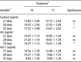
Mean plasma cortisol, GH and IGF-1 levels are shown in Table 4. Cortisol content was lower at days 28 and 35 and GH at day 35 in T1 compared with T0 (P < 0.05). The levels of IGF-1 were similar across groups (P > 0.05). A positive correlation was found between cortisol and GH (r = 0.326; P = 0.002).
Least squares means (±s.e.) of piglets plasma cortisol, GH and IGF-1 levels at different ages as affected by nucleotide supplementation

GH = growth hormone.
Probability of significance: *P < 0.05, ns = non-significant (P > 0.05).
1T0 = control diets (0% nucleotides added); T1 = diets supplemented with 0.1% yeast extract nucleotides.
2Least squares means covariated with initial values (day 9).
Immune response
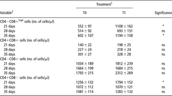
The CD4−CD8+high expressing cells comprehend the majority of cytotoxic T lymphocytes within the PBMC population, whereas total CD4−CD8+ cells comprehend CD3−CD8+ natural killer cells, cytotoxic T lymphocytes and a CD8+ γ/δ T lymphocytes.
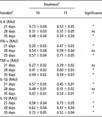
As shown in Table 5, nucleotide-supplemented diets increased CD4−CD8+high cells at days 21 and 35 (P < 0.05), but did not influence lymphocyte subpopulation CD4−CD8+ (P > 0.05). At 21 days of age CD4+CD8− cells were higher in T1 than in T0 (P < 0.05). No differences were observed between groups on helper double positive lymphocytes (CD4+CD8+; P > 0.05). PBMC cytokine expression was influenced by nucleotide supplementation as summarized in Table 6. At weaning, IL-6 and IL-1β expression was lower in T1 than in T0 (P < 0.05), whereas no effect was observed on TNF-α expression (P > 0.05). At the same time the expression of IFN-γ and IL-10 was higher in T1 than in T0 (P < 0.05). At day 28, higher values of TNF-α expression and lower values of IL-10 expression were showed in T1 than in T0 (P < 0.05). Dietary nucleotide supplementation had a suppressive effect on IL-6 and IL-10 at day 35 (P < 0.05). On the contrary, the expression of IFN-γ, TNF-α and IL-1β was enhanced (P < 0.05).
Least squares means (±s.e.) of piglets immunological parameters at different ages as affected by nucleotide supplementation

Probability of significance: *P < 0.05, ns = non-significant (P > 0.05).
1T0 = control diets (0% nucleotides added); T1 = diets supplemented with 0.1% yeast extract nucleotides.
2Least squares means covariated with initial values (day 9).
Least squares means (±s.e.) of piglets peripheral blood mononuclear cells cytokine expression at different ages as affected by nucleotide supplementation.

IL = interleukin; RAU = relative arbitrary units; INF = interferon; TNF = tumor necrosis factor.
Probability of significance: *P < 0.05, ns = non-significant (P > 0.05).
1T0 = control diet (no nucleotides added); T1 = diet supplemented with 0.1% of yeast extract nucleotides.
2Least squares means covariated with initial values (day 9).
Discussion
As in the past no effect on growth performance of piglets has been observed when nucleotide supplementation diet occurred after weaning (Di Giancamillo et al., Reference Di Giancamillo, Domeneghini, Paratte, Dell'Orto and Bontempo2003; Domeneghini et al., Reference Domeneghini, Di Giancamillo, Savoini, Paratte, Bontempo and Dall'Orto2004; Andrés-Elias et al., Reference Andrés-Elias, Pujols, Badiola and Torrallardona2007), the results of this study demonstrated that a nucleotide supplementation both in pre- and post-weaning diets (from 9 to 55 days of age) not containing AGPs, improved the overall growth performance of piglets without affecting the average daily feed intake. Mean feed intake during lactation was poor in T0 and T1 piglets, but at weaning T1 showed a BW higher than T0. The daily feed intake pre-weaning was expressed as a mean value between days 9 and 21 and feed was not weighted every day. So it is possible that feed intake was negligible at the beginning and higher at the end of the period. Nucleotide supplementation may have contributed to spare, during the critical transition phase of weaning, the cost of de novo nucleotide synthesis and/or salvage pathway (Burrin and Stoll, Reference Burrin and Stoll2002). Voluntary feed intake in post-weaning has a deep effect on the functioning of several neuroendocrine systems (Carroll et al., Reference Carroll, Veum and Matteri1998; Hay et al., Reference Hay, Orgeur, Levy, Le Dividich, Concordet, Nowak, Schaal and Mormède2001). Although we observed the same voluntary feed intake between groups, a different hormonal response occurred during the first 2 weeks after weaning. In particular, mean plasma cortisol levels in T0 were consistent with data previously reported (Carroll et al., Reference Carroll, Veum and Matteri1998; Wu et al., Reference Wu, Meininger, Kelly, Watford and Morris2000; Borghetti et al., Reference Borghetti, De Angelis, Saleri, Cavalli, Cacchioli, Corradi, Mocchegiani and Martelli2006a), whereas T1 mean plasma cortisol levels were markedly lower over time. Cortisol affects the regulation of mucosal amino acids uptake, and facilitates an appropriate amino acids metabolism by the small intestine post-weaning (Wu et al., Reference Wu, Meininger, Kelly, Watford and Morris2000). It was observed that blocking of glucocorticoid receptors abolished weaning-enhanced enterocyte utilization of glutamine (Flynn and Wu, Reference Flynn and Wu1997). Glutamine, a precursor for the synthesis of purine and pyrimidine nucleotides (Ardawi and Newsholme, Reference Ardawi and Newsholme1983; Wu, Reference Wu1998), stimulates cell proliferation and protein synthesis (Burrin and Stoll, Reference Burrin and Stoll2002). It has been shown that glutamine reduces cortisol synthesis in the adrenal cortex due to reductions in the activities of key enzymes (Wu et al., Reference Wu, Bazer, Johnson, Knabe, Burghardt, Spencer, Li and Wang2011). On this basis, we may hypothesize a negative feedback on cortisol levels in T1 as the nucleotides, which consist of a pyrimidine or purine base, themselves are the building blocks for protein synthesis. So a nucleotide-supplemented diet during the pre-weaning phase can exert a direct control on metabolic resources of piglets. The greater BW shown by T1 than T0 was not coupled with a higher GH plasma content. GH is the postnatal master regulator of growth, and its effects are mediated mainly by IGF-1 (Møller and Jørgensen, Reference Møller and Jørgensen2009). Under conditions of stress, such as fasting, GH secretion is amplified (Vendelbo et al., Reference Vendelbo, Jørgensen, Pedersen, Gormsen, Lund, Schmitz, Jessen and Møller2010). In this study, an overall positive correlation was found between cortisol and GH but not between GH and IGF-1 plasma levels; the mean GH plasma level was lower in T1 at day 35. These observations furtherly sustain an uncoupling between the plasmatic levels of GH and IGF-1 and that the level of plasmatic GH could be mainly involved to counteract the levels of cortisol and their negative effect on metabolism and on immune efficiency in weaned piglets (Borghetti et al., Reference Borghetti, De Angelis, Saleri, Cavalli, Cacchioli, Corradi, Mocchegiani and Martelli2006a, Reference Borghetti, Ferrari, Cavalli, De Angelis, Saleri, Corradi and Martelli2006b and Reference Borghetti, Saleri, Mocchegiani, Corradi and Martelli2009). In this view, the different trend in mean cortisol and GH plasma levels between groups could testify a reduction of the negative effect of stress on growth in T1. Our data take into account the well-demonstrated existence of a crosslink between endocrine and immune systems (Sapolsky et al., Reference Sapolsky, Romero and Munck2000). The immune system is ordinarily brought back to a baseline response level after immune challenge through homeostatic processes, in part regulated by the hypothalamic–pituitary axis (Salak-Johnson and McGlone, Reference Salak-Johnson and McGlone2007). Lymphocytic cytokines such as IFN-γ and IL-10 are preferentially responsible for the immune response T-helper (Th)1 or 2 mediated, whereas inflammatory cytokines such as IL-1β, IL-6 and TNF-α are quickly produced when the inflammation is induced in the organism (Zelnickova et al., Reference Zelnickova, Leva, Stepanova, Kovaru and Faldyna2008). At weaning, T1 compared with T0 showed higher IFN-γ and IL-10 and lower IL-1 β and IL-6 expression levels, whereas TNF-α expression levels did not differ between groups. On these bases, we assume that supplemented piglets showed a better fitting to their environment. The increased number of both total CD8+ and especially CD8+high observed in treated piglets may be related to the possible activation by Th1 cells of IFN-γ, a cytokine with a fundamental role for the activation of cytotoxic T cells (Hayday and Tigelaar, Reference Hayday and Tigelaar2003). Stress hormone-induced suppression or enhancement of cytokine expression may affect disease susceptibility and then an unbalance between Th1 and Th2 cells may represent a mechanism to achieve homeostasis (Elenkov and Chrousos, Reference Elenkov and Chrousos2002; Wiegers et al., Reference Wiegers, Stec, Sterzer and Reul2005). The general effect of glucocorticoids is to inhibit synthesis, release or efficacy of cytokines. However, not all cytokines are suppressed by glucocorticoids. IL-10 secretion is increased by glucocorticoids (Blotta et al., Reference Blotta, DeKruyff and Umestsu1997; Richards et al., Reference Richards, Fernandez, Caulfield and Hawrylowicz2001), whereas others (i.e. IL-6) act synergistically with glucocorticoids (Wiegers et al., Reference Wiegers, Stec, Sterzer and Reul2005). Our results were consistent with these data at 35 days of age, when we observed a higher expression of IFN-γ, TNF-α and IL-1β in the nucleotide-supplemented group, associated with a lower expression of IL-6 and IL-10. At 28 days of age we noted a lack of significance between groups, probably because of a residual weaning-associated negative effect in T1 that flattened the differences for some cytokines, such as IFN-γ, IL-1β and IL-6.
In conclusion, our data confirmed that nucleotides could be considered as essential micronutrients during piglets’ weaning. Nucleotide supplementation of creep feed and post-weaning diets has improved the growth performance of piglets, by positively modulating (lower cortisol plasma levels) the adaptive response of piglets to weaning stress factors and overcoming their possible negative effects.
Acknowledgment
The authors thank DR. P. Benaglia (Mantova, Italy) for veterinary assistance and Bompieri farm (Pavone Mella Brescia, Italy) for logistic support.