Introduction
Although lithium is effective and principally safe in the treatment and prophylaxis of bipolar disorders, it has a narrow therapeutic ratio. It is well known that overdoses are neurotoxic, potentially leading to severe and sometimes irreversible disabilities (Kores and Lader, Reference Kores and Lader1997; Grandjean and Aubry, Reference Grandjean and Aubry2009). Lithium is known to cause drug-induced Parkinsonism (Bondon-Guitton et al., Reference Bondon-Guitton, Perez-Lloret, Bagheri, Brefel, Rascol and Montastruc2011), depending on dose. Drug administration, however, is complicated as lithium is known to accumulate over time and to interact with factors such as age, concurrent medication, and comorbidity (e.g. renal insufficiency, diabetes, or infections) (Adityanjee et al., Reference Adityanjee Munshi and Thampy2005). This may be a reason for the finding that lithium can lead to adverse effects even at therapeutic serum levels (Holroyd and Smith, Reference Holroyd and Smith1995).
Acute lithium intoxication cannot only lead to a variety of neurological symptoms, including Parkinson's syndrome, but also to cognitive and behavioral abnormalities (Kores and Lader, Reference Kores and Lader1997; Bartha et al., Reference Bartha, Marksteiner, Bauer and Benke2002). Cognitive deficits have been mentioned many times in the literature, but studies with a comprehensive neuropsychological assessment are rare (Bartha et al., Reference Bartha, Marksteiner, Bauer and Benke2002). Bartha et al. (Reference Bartha, Marksteiner, Bauer and Benke2002) present a patient with a bipolar disorder and an excess lithium serum level, who suffered severe cognitive deficits in different domains, esp. long-term memory, visuo-spatial functions, and praxis. Most of these deficits could still be found more than two years after lithium had been stopped, suggestive of Alzheimer's dementia. Tonn et al. (Reference Tonn, Bartenstein and Dahmen2005) report an elderly patient with an excess lithium serum level and with a temporo-parietal FDG–PET hypometabolism, typical of Alzheimer's disease (AD). Dementia screenings revealed abnormal results, but deficits were not further qualified by means of a comprehensive neuropsychological assessment. Here, we report a case with probable lithium intoxication at normal serum levels where both an extensive neuropsychological assessment and an FDG–PET examination were performed.
Case report
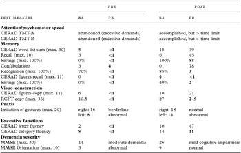
The patient was a 70-year old male, retired engineer who presented in 2012 with quickly progressing problems in movement, suggestive of Parkinson's disease. The patient's wife additionally reported increasing cognitive problems in temporal association with the motor symptoms. There was no family history of psychiatric or neurological diseases. Clinical examination revealed hypokinesia, rigor, and tremor (all non-lateralized). The patient was oriented for person, place, and situation, but not for time. Affect was a little bit flattened, but unremarkable in other respects. An extensive assessment disconfirmed idiopathic or atypical Parkinson's syndrome (L-DOPA-test negative, bilateral rigor). The patient had been implanted a shunt in 2009 due to suspected normal pressure hydrocephalus (NPH), but a shunt dysfunction could be excluded and several releases of cerebrospinal fluid (CSF) did not lead to any improvement. A comprehensive neuropsychological assessment applying the Consortium to establish a registry for Alzheimer's disease (CERAD) test battery, the Rey Complex Figure Test (RCFT), and a gesture imitation task (apraxia) revealed a severe dementia with deficits in all domains under investigation (verbal and visual long-term memory, executive, and visuospatial functions, praxis; see Table 1 and Figure 1), suggestive of typical AD (Dubois et al., Reference Dubois2007). The patient was hardly oriented and disease awareness was clearly impaired. A depression was found neither in the clinical examination nor in a standardized questionnaire (Geriatric Depression Scale/GDS).
Neuropsychological test results (raw scores/RS and percentile ranks/PR, PRs below average in bold) before (pre) and after (post) stopping lithium treatment in the patient. Abbreviations: TMT: Trail Making Test; RCFT: Rey Complex Figure Test; MMSE: Mini-Mental Status Examination; GDS: Geriatric depression scale. For further information see text

Results in copying a complex figure (Rey Complex Figure Test/RCFT), before (left side) and after (right side) cessation of lithium treatment. In addition to the profound problems of the patient in the spatial arrangement of the figure parts in the drawing on the left-hand side, time needed to perform the task was much greater for the figure on the left (17 minutes) compared to the one on the right-hand side (three and a half minutes).

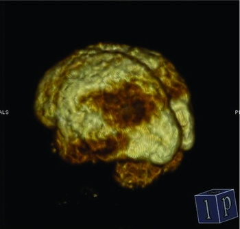
Analysis of CSF revealed elevated Tau protein (876 pg/mL (normal < 445)) and phosphorylated Tau (129 pg/mL (normal < 61)) whereas beta-amyloid 1–42 was in the normal range (869 pg/mL (normal > 375)). Protein 14-3-3 was negative. In the electroencephalogram (EEG), an irregular pattern of alpha was found with intermitting episodes of theta and no sign for epileptic activity. Magnetic resonance MRI was unremarkable (neither signs of relevant microangiopathy nor of AD- or FTD-typical regional cerebral atrophy). Cerebral glucose metabolism was measured by Positron–Emission–Tomography (PET/CT). Data acquisition with a Siemens Biograph 6 PET/CT-Scanner started 30 minutes after injection of 195 MBq of 2-deoxy-2-18F-fluoro-β-D glucose (18F-FDG). Data analysis revealed parietal and temporal hypometabolism bilaterally (see Figure 2), typically associated with AD (Hoffman et al., Reference Hoffman2000; Schroeter et al., Reference Schroeter, Stein, Maslowski and Neumann2009). Furthermore, hypometabolism was found in the striatum bilaterally. This was neither typical for Alzheimer nor for Parkinson's disease (Berti et al., Reference Berti, Pupi and Mosconi2011), but for Huntington's disease or multisystem atrophy (Berti et al., Reference Berti, Pupi and Mosconi2011), for which, however, clinical neurological evidence was absent and temporo-parietal hypometabolism was untypical.
Regional metabolism of 2-deoxy-2-18F-fluoro-β-D glucose (18F-FDG) in Positron Emission Tomography (PET) before stopping lithium treatment in the patient. Bilateral glucose hypometabolism in the parietal and temporal cortices bilaterally was highly suggestive of Alzheimer's disease.

The patient had been medicated with lithium for bipolar disorder for five years. When he presented at our clinic in 2012, serum level was elevated (1.1 mmol/L). Although the level was still within the therapeutic range (0.6–1.2 mmol/L), it was higher than recommended for prophylaxis (0.6–0.8 mmol/L). Lithium had been administered as lithium carbonate 450 mg twice daily (= 24.4 mmol) at least since 2011. According to an external medical record, serum level had been 0.49 mmol/L in 2009. At our clinic, thyroid-stimulating hormone (TSH) in the patient was 2.1 mU/L (no value available from 2009) and renal function was unrestricted (creatinine 1.05 mg/dL, glomerular filtration rate (GFR) 69.8 mL/min; comparable in 2009: creatinine 1.05 mg/dL, GFR 77.0 mL/min). Blood pressure was in the (upper) normal range (131/78). Besides lithium, the patient was taking Tamsolusin (0.4 mg/d) due to benign prostatic hyperplasia on a regular basis and Memantine (20 mg/d). Memantine had already been documented in the external medical file (2009), but the reason for prescription was left unclear (no diagnosis of AD; most probable reason were cognitive deficits due to NPH).
Detailed anamnesis as well as psychiatric consultation cast doubt on the diagnosis of bipolar disorder (atypical age, no unambiguously manic episodes, no depressive symptoms other than reduced drive and irritability according to the patient's wife). Thus, lithium was stopped and Valproate (1200 mg/d) was applied instead (as precaution). Tamsolusin and Memantine were continued as before.
In the following days, Parkinsonian symptoms markedly improved. The patient presented again three months later for a neuro-(psycho-)logical examination. Orientation and disease awareness were intact and Parkinson's symptoms were absent. Both the patient and his wife reported dramatic improvements in everyday life, with respect to both motor and cognitive functioning. Neuropsychological assessment revealed dramatic improvements across domains and even average results in several tests (esp. with respect to verbal long-term memory and visuo-construction; see Table 1 and Figure 1). The Mini-Mental Status Test result also increased dramatically, but was still in the range of a mild cognitive impairment. The score was the same (26) as in 2009 when the same test had been applied after shunt implantation (for treatment of the suspected NPH). According to the Naranjo ADR (adverse drug reaction, see Naranjo et al., Reference Naranjo1981) probability scale, the total ADR score was 6 (probable ADR).
However, we also found a slight increase in depressive symptoms in a standardized questionnaire, which we interpreted with improvements in awareness. Clinically, criteria for affective disorder were not fulfilled. Unfortunately, a re-examination one year later could not take place as the patient had died from an intracerebral hemorrhage (loco typico). The patient's wife provided informed consent to publish the case.
Discussion
Although there are numerous cases reported in the literature on lithium-induced motor and cognitive deficits, only one study (Bartha et al., Reference Bartha, Marksteiner, Bauer and Benke2002) provides a comprehensive neuropsychological assessment and demonstrates a range of cognitive deficits, including visuo-construction, and praxis, which indicate an affection of temporo-parietal cortex, such as in AD. Likewise, there is only one FDG–PET study (Tonn et al., Reference Tonn, Bartenstein and Dahmen2005) so far demonstrating a temporo-parietal hypometabolism after lithium intoxication, but without qualifying the neuropsychological deficits. Our study is the first in which both neuropsychology and FDG–PET were indicative of AD as a consequence of lithium intoxication. However, there are several issues that need to be discussed.
First, the fact that lithium serum level was within the upper therapeutic range (though was too high for prophylaxis) does not disprove intoxication, as lithium may accumulate over time and interact with other factors, especially in the elderly (Adityanjee et al., Reference Adityanjee Munshi and Thampy2005). In our case, the additional brain disease (NPH) may have increased the brain's sensitivity for lithium (Kores and Lader, Reference Kores and Lader1997). Furthermore, the finding of pronounced initial extrapyramidal symptoms which completely disappeared after lithium had been stopped provides convincing evidence for our interpretation. Second, although the cognitive abilities had greatly improved after cessation of lithium treatment, remaining deficits may be explained in several ways, namely (i) as a syndrome of irreversible lithium effectuated neurotoxicity (Kores and Lader, Reference Kores and Lader1997) or (ii) as a result of the fact that the NPH had probably not been treated early enough (cf. McGirt et al., Reference McGirt, Woodworth, Coon, Thomas, Williams and Rigamonti2005). Furthermore, we cannot exclude an additional neurodegenerative disease, but the evidence was inconclusive: Cognitive abilities had been stable over a period of at least three years, as indicated by identical Mini-Mental Status Test results after the NPH surgery (three years before our first contact with the patient) and our second assessment after lithium had been stopped. Although the CSF results were suggestive for AD, the stable (and relatively high) MMST scores over a period of three years do not support this interpretation. Furthermore, the fact that the patient died from cerebral hemorrhage is suggestive of vascular pathology, but clear evidence for this was also absent (MRI was unremarkable and blood pressure was in the (upper) normal range). We may conclude that a multimodally damaged brain (possible AD, possible vascular pathology, NPH) is particularly vulnerable to lithium intoxication. To conclude, our case study provides evidence that lithium intoxication may lead to a clinical picture very suggestive of AD, both neuropsychologically and with respect to brain metabolism, even at high therapeutic serum levels. This may lead to misdiagnosis and should be taken into account in the assessment of dementia.
Conflicts of interest
None.
Description of authors’ roles
The patient was examined neuropsychologically by Dr Frisch and neurologically as well as psychiatrically by Dr Friedrichs. Prof. Grünwald analyzed and interpreted the PET data.
Acknowledgments
We would like to thank Thanuja Victor for assistance in PET data analysis.