Introduction
Congenital Heart Disease (CHD) children present with structural abnormalities of the heart and major blood vessels, which induce abnormal hemodynamics, elevated energy requirements, inadequate energy intake, and impaired nutrient absorption. These abnormalities subsequently lead to feeding difficulties, recurrent respiratory tract infections, cardiac dysfunction, growth and developmental delay, severe malnutrition, and even life-threatening heart failure in severe cases. Researchers have found in a meta-analysis that CHD patients, even after receiving surgical treatment, have a higher risk of cardiovascular disease in later life than normal.Reference Wang, Chen and Yang1
Ghrelin, also known as growth hormone-releasing peptide, is mainly secreted by gastric mucosal cells, cardiomyocytes, and blood vessels, which is distributed throughout the body via the bloodstream. Ghrelin mainly binds to the growth hormone secretagogue receptor-1a to exert its biological effects. Growth hormone secretagogue receptor-1a receptors are also expressed in the heart and blood vessels.Reference Iglesias, Pineiro and Blanco2 Exogenous ghrelin and its synthetic analogue hexarelin can also exert cardioprotective effects by specific heart receptors CD36.Reference Mao, Tokudome and Kishimoto3 WangReference Wang, Liu and Lü4 confirmed that ghrelin plays an important role in the functional imbalance and compensation mechanism of CHD through animal experiments. Similarly, later studies foundReference Wang, Fang and Wang5,Reference Zhang, Guo, Yang and etal6 that the level of ghrelin in CHD children was higher than that of normal age-matched healthy children.
Currently, numerous domestic and overseas studies have confirmed that ghrelin regulates the stability of the cardiovascular system and plays a physiological and pathological role in the cardiovascular system. Ghrelin has a protective effect on the heart, but there has been less research on the structural and functional heart of CHD, so this study aims to detect the preoperative serum ghrelin level of CHD children and analyse its correlation with cardiac structure and function, providing a certain guidance for evaluating the early changes in the cardiac structure and function of CHD children.
Data and methods
Objects of study
Prospective analysis of 93 left-to-right shunt CHD patients (including 58 cases of ventricular septal defect and 35 cases of patent ductus arteriosus) who underwent interventional closure and surgical repair at the cardiovascular department of Children’s Hospital of Soochow University from September 2021 to February 2023 as the CHD group and 57 children who underwent elective surgery at our hospital during the same period were included as the control group(mainly for inguinal hernia). The inclusion criteria for the CHD group were as follows: (1) the child was diagnosed with CHD through medical history, clinical examination, physical examination, and auxiliary examination (including echocardiography, electrocardiography, three-dimensional heart film, cardiac catheterisation, and cardiovascular angiography), without severe aortic and ventricular valve regurgitation, and the operation was successful; (2) auxiliary examination such as chest X-ray, blood routine, and liver and kidney function was normal. The inclusion criteria for the control group were as follows: (1) no history of CHD or congenital malformation; (2) auxiliary examination such as chest X-ray, blood routine, and liver and kidney function was normal. Exclusion criteria for both groups were children with infectious diseases, diagnosable chromosomal and inherited metabolic diseases, haematological and immune system diseases, and incomplete clinical data.
This study was approved by the Board of Trustees of the Children’s Hospital of Soochow University (ethics approval number: 2022CS084), and all clinical samples and clinical data were collected with the informed consent of the children’s families.
Data collection and processing
Basic clinical data, such as date of birth, date of admission, gender, age in months, height, weight, and BMI of the children in the two groups, were collected, and body surface area was calculated using Haycock formula. The Z score of weight for age and Z score of height for age were calculated using the software WHO Anthro and AnthroPlus
Ultrasonic electrocardiogram results were collected before operation in the CHD group, such as left ventricular end-diastolic diameter (LVEDd), left ventricular end-systolic diameter (LVESd), ventricular septal diastolic thickness interventricular septum diastolic thickness (IVSd), left ventricular posterior wall thickness (LVPWd), left ventricular ejection fraction (LVEF), left ventricular fractional shortening (LVFS), etc. Relative wall thickness (RWT) = (LVPWd+ IVSd)/LVEDd; according to Devereux correction formula, left ventricular mass (LVM) = 1.04 × 0.8 × [(LVEDd+IVSd+LVPWd)3-LVEDd3] + 0.6; left ventricular mass index (LVMI) = left ventricular mass/body surface area; the left ventricular end-diastolic volume was calculated by Teichholz formula (LVEDV) = 7 × LVEDd3/ (2.4+LVEDd). Left ventricular remodelling index (LVRI) = LVM/LVEDV. Z-scores of LVEDd、 LVESd、IVSd、LVWPd、LVM、LVEDV were calculated by Z-scores calculator to evaluate the atrioventricular structure and function of the heart.
Collection and preservation of serum samples on the day of admission, 2 ml of fasting peripheral venous blood was placed in yellow blood collection tubes containing inert separation glue, centrifuged whole blood samples in a centrifuge at 3000rpm/ min for 10 minutes, and the upper serum was transferred to a sterile labelled Eppendorf (EP) tube and stored in a refrigerator at −80°C for future use. Repeated freeze–thaw cycles were avoided.
Determination of serum ghrelin Serum ghrelin was detected by enzyme-linked immunosorbent assay. The kits were purchased from Shanghai Enzyme-linked Biotechnology Co., LTD., and were performed strictly in accordance with the instructions.
Statistical methods
SPSS26.0 software was used for data analysis. The measurement data were tested for normality and homogeneity of variance. Those meeting normal distribution were represented by mean ± standard deviation (
 $\overline{X}$
±S). T-test was used for comparison between two groups, and analysis of variance test was used for comparison between multiple groups. Those who did not conform to the normal distribution were represented by the quartile method [M(P
25,P
75)], and the comparison between the two groups was performed by the Mann-Whithey U test in the rank sum test. The correlation between serum ghrelin and each index was analysed by Spearman. P < 0.05 was considered statistically significant.
Results
Basic information
A total of 150 children were enrolled in this study, including 93 cases in the CHD group (36 males and 57 females) and 57 cases in the control group with no statistical significance (P > 0.05). The age of the two groups was 35 (15,50.5) months and 41 (23,57.5) months, and BMI was 15.81 (14.87,17) and 16 (14.76,16.95) kg/m2, respectively, with no statistical significance (P > 0.05). Weight-for-age Z-score and height-for-age Z-score of CHD group and control group were 0.05 ± 1.27 and 0.54 ± 0.96, respectively, and height-for-age Z-score were 0.08 (−1.08,0.78) and 0.55 (−0.31,1.54), respectively, and there were statistical differences between the two groups (P < 0.05).
Comparison of serum ghrelin
The results showed that the serum ghrelin concentration in CHD group was significantly higher than control group, and the difference between the two groups was statistically significant (P < 0.05)(Table 1).
Comparison of serum ghrelin M (P 25, P 75)

Spearman correlation analysis of serum ghrelin and clinical information
There was no correlation between serum ghrelin and BMI, weight-for-age Z-score, and height-for-age Z-score(P > 0.05).
Comparison of serum ghrelin in children with different cardiac load
CHD with cardiac enlargement was divided into two groups according to the Z-scores of left ventricular end-diastolic diameter. The Z-scores of left ventricular end-diastolic diameter ≥ 2 were the enlarged heart group, and the Z-scores of left ventricular end-diastolic diameter < 2 were the non-enlarged heart group. There were 47 cases in the cardiomegaly group, including 18 cases of patent ductus arteriosus and 29 cases of ventricular septal defect. There were 46 patients in the non-cardiomegaly group, including 17 patients with patent ductus arteriosus and 29 patients with ventricular septal defect. Ghrelin in the cardiomegaly group was lower than the non-cardiomegaly group, and there were statistical significance of among those groups (P < 0.05) (Table 2).
Comparison of cardiac indexes of different cardiac loads in children with patent ductus arteriosus and ventricular septal defect
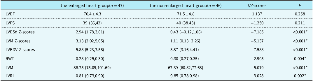
There were no significant differences in left ventricular ejection fraction and LVFE among different cardiac load groups (P > 0.05). Left ventricular end-systolic diameter Z value, left ventricular mass Z value, left ventricular mass index, and left ventricular end-diastolic volume Z value the cardiomegaly group were higher than those in the non-cardiomegaly group, and those two groups were significantly different (P < 0.05). The relative wall thickness and left ventricular remodelling index in the cardiomegaly group were lower than those in the non-cardiomegaly group, and the differences between the groups were statistically significant (P < 0.05) (Table 3).
Correlation analysis of serum ghrelin and cardiac indexes in children in patent ductus arteriosus and ventricular septal defect groups
Spearman correlation analysis: Serum Ghrelin in patent ductus arteriosus and ventricular septal defect groups was negatively correlated with the left ventricular end-diastolic diameter Z-scores, left ventricular end-systolic diameter Z-scores and left ventricular end-diastolic volume Z-scores of cardiac indexes, was positively correlated with left ventricular remodelling index, but had no correlation with relative wall thickness, left ventricular mass Z-scores and left ventricular mass index (P > 0.05).
Discussion
CHD is one of the prevalent congenital malformations, with a persistently high morbidity rate. Surgical intervention remains the primary therapeutic modality for CHD, and significant improvements have been achieved in terms of postoperative quality of life and survival rate among patients. However, with the increase of age, the risk of cardiovascular comorbidities gradually increases such as atherosclerosis, stroke, myocardial infarction etc. Ghrelin exerts a crucial protective effect on the cardiovascular system, but there are few studies on the clinical value of ghrelin in CHD.
Ghrelin is a small molecule polypeptide chain composed of 28 amino acid sequences, which exerts physiological effects by binding to corresponding receptors directly or by activating the growth hormone (GH)/insulin-like growth factor (IGF) axis. Ghrelin promotes GH secretion through GH/IGF-1 axis, which not only improves the growth, development, and nutritional status of CHD children but also modulates the growth, development, and function of cardiomyocytes to a certain extent. Haemodynamic alterations can locally regulate the cardiac production and secretion of ghrelin and GH. Evidence from experimental animal models of cardiac stress and volume overload demonstrates that the expression levels of IGF-1, GH, and IGF-1 receptors in cardiac tissue are upregulated, whereas ghrelin expression is downregulated.Reference Wang, Liu and Lü4,Reference Pecker7
The biological role of ghrelin encompasses a diverse array of signalling pathways. Within the cardiovascular system, its primary function is to regulate cardiovascular homeostasis. Previous research reports have consistently shown that serum ghrelin levels are higher in children with CHD compared to those without CHDReference Wang, Liu and Lü4,Reference Wang, Fang and Wang5 In the present study, we measured serum ghrelin levels in 93 children with CHD and 57 children without CHD, yielding results consistent with these prior findings. Kitamura et al.Reference Kitamura, Shimizu, Ohkawa, Oda, Ohtsuki and Kishiro8 investigated the association between serum ghrelin and growth, development, and cardiac function in children with CHD complicated by heart failure. They found a positive correlation between acylated ghrelin and heart failure index, indicating that more severe heart failure was associated with higher serum ghrelin concentrations. This observation suggests a compensatory upregulation of ghrelin in response to cardiac dysfunction. While ghrelin is involved in regulating myocardial contraction and cardiac pumping function to alleviate cardiac dysfunction and protect the heart, it appears to have little impact on the growth and development of children. Our study also found no significant correlation between ghrelin and the growth or development of children. While other studies suggest that elevated serum ghrelin levels in CHD children may be closely linked to malnutrition and growth retardation.Reference Zhang, Guo, Yang and etal6 Consequently, discrepancies persist regarding the relationship between ghrelin and cardiac function in children with CHD, and additional factors may contribute to the elevation of circulating ghrelin levels in this population.
In children with CHD, abnormal blood flow within the cardiac chambers and increased cardiac volume load are primarily attributed to structural defects in the interventricular septum. Without timely intervention, prolonged cardiac volume overload can enhance cardiac compensatory mechanisms and ultimately lead to cardiac remodelling. To evaluate the longitudinal changes in cardiac structure, Z-scores of left ventricular end-diastolic diameter, left ventricular end-systolic diameter, left ventricular posterior wall thickness, and left ventricular end-diastolic volume were utilised. Relative wall thickness, left ventricular mass index, and left ventricular remodelling index were employed to assess left ventricular wall thickness and geometric characteristics. Relative wall thickness reflects the ratio between changes in ventricular wall thickness and left ventricular diameter, while left ventricular remodelling index represents the ratio between changes in left ventricular mass and volume. It can be concluded that volume overload predominantly affects the cardiac geometry of children with CHD. In cases of patent ductus arteriosus and ventricular septal defect, the left side of the heart is typically enlarged due to higher pressure on the left cardiac chambers compared to the right side. Initially, the left-to-right shunt increases pulmonary circulation blood volume, leading to elevated left heart return and volume load. This, in turn, causes dilatation of the left cardiac chambers and alters their geometric configuration. In transthoracic echocardiography reports, left ventricular end-diastolic diameter, left ventricular end-systolic diameter, and left ventricular end-diastolic volume were found to be increased in children with patent ductus arteriosus and ventricular septal defect, while IVSd and left ventricular posterior wall thickness were thickened. Left ventricular remodelling index can objectively reflect left ventricular remodelling and overall adaptation to physiological and pathological states.Reference De Castro, Caselli and Maron10 When left ventricular remodelling index is > 1, it is considered to be left ventricular pathological adaptation. Existing studies on patients with myocardial infarction or heart failure have demonstrated that ghrelin can alleviate ventricular remodelling through multiple mechanisms.Reference Soeki, Kishimoto and Schwenke11,Reference Schwenke, Tokudome and Kishimoto12 In this study, almost all left ventricular remodelling index calculated for CHD children were < 1, suggesting that the cardiac structural abnormalities induced by haemodynamic changes in this population represent compensatory physiological adaptation rather than pathological remodelling. Since left ventricular mass is affected by body size, age, gender, and other factors, the present study corrected left ventricular mass using body surface area and calculated left ventricular mass index to evaluate left ventricular hypertrophy. The left ventricular mass in CHD children can be evaluated more accurately by calculating the Z-scores of left ventricular mass compared with that in healthy children of the same body type.
In this study, patent ductus arteriosus and ventricular septal defect children in left-right shunt CHD children with increased left ventricular volume load were selected, and serum ghrelin levels were compared according to different left ventricular load states. CHD children with a left ventricular end-diastolic diameter Z-score > 2 were considered as the cardiomegaly group , while those with a left ventricular end-diastolic diameter Z-score < 2 were assigned to the non-cardiomegaly group. The results showed that serum ghrelin levels were significantly lower in the cardiomegaly group (characterised by severe volume load) than in the non-cardiomegaly group (characterised by normal volume load). Therefore, it can be inferred that ghrelin can reduce the ventricular inner diameter when the left ventricular volume load is excessive, and has a cardioprotective effect in CHD children under different cardiac volume load conditions. Comparison of cardiac function indices between the two groups revealed no significant differences in left ventricular ejection fraction or left ventricular fractional shortening. Left ventricular end-systolic diameter Z-scores, left ventricular mass Z-scores, left ventricular mass index, and LVEVd-Z values of the cardiomegaly group were higher than those of the non-cardiomegaly group, and relative wall thickness and left ventricular remodelling index of the cardiomegaly group were lower than those of the non-cardiomegaly group. These findings indicate that left ventricular mass and geometry are primarily influenced by the degree of left ventricular volume load. Further correlation analysis demonstrated that ghrelin was significantly negatively correlated with the Z value of left ventricular end-diastolic diameter, left ventricular end-systolic diameter, and left ventricular end-diastolic volume, and positively correlated with left ventricular remodelling index, while it was not correlated with the Z value of left ventricular mass, left ventricular mass index, and relative wall thickness. Therefore, it can be further speculated that ghrelin can increase myocardial contractility without affecting left ventricular mass. These results further suggest that ghrelin may enhance myocardial contractility without affecting left ventricular mass, thereby reducing left ventricular diameter and volume to mitigate the adverse effects of abnormal haemodynamic volume overload on the hearts of children with CHD. Similar results have been obtained from previous studies that subcutaneous injection of ghrelin in healthy people can promote GH release in a dose-dependent manner, enhance myocardial contractility, increase left ventricular ejection fraction, and decrease left ventricular end-systolic volume.Reference Salvetti, Paini and Bertacchini13
Both deacylated ghrelinReference Pei, Yung, Yip, Ying, Benzie and Siu14 and acylated ghrelin can enhance the positive inotropic effect of the heart through multiple mechanisms,Reference Shati and Dallak15 thereby exerting cardioprotective roles. In children with CHD, abnormal blood flow leads to increased venous return, resulting in elevated volume load. Ghrelin maintains calcium homeostasis in cardiomyocytes through diverse pathways without altering ventricular mass, enhances myocardial contractility, and attenuates cardiomyocyte apoptosis and necrosis induced by chronic myocardial dilatation. Moreover, it inhibits myocardial fibrosis and retards pathological remodelling of cardiac structure in children with CHD.
This study identified a disparity in serum ghrelin expression between children with CHD and those without, and further investigated the association between ghrelin and heart geometry. Notably, there was a significant correlation observed between ghrelin levels and left ventricular volume, suggesting its potential to ameliorate the pathophysiological changes induced by CHD. These findings hold promise for the treatment and prognosis of CHD. However, further validation through multi-study and multi-centre investigations is required, and future research should also focus on elucidating the specific molecular targets and mechanisms underlying the cardioprotective effects of ghrelin in CHD.
Comparison of serum ghrelin and in CHD children with different cardiac load M (P 25, P 75)

Comparison of cardiac indexes of different cardiac loads in children with PDA and VSD (
 $\overline{X}$
±S)/M (P
25, P
75)

LVEF = Left ventricular ejection fraction; LVFS = Left ventricular fractional shortening; LVESd Z-valua = Left ventricular end-systolic diameter Z-valua; LVM Z-scores = Left ventricular Mass Z- value; LVEDV Z-valua = The left ventricular end-diastolic volume Z-valua; LVEDd Z-valua = Left ventricular end-diastolic diameter Z-valua; RWT = Relative wall thickness; LVMI = Left ventricular Mass Index; LVRI = Left ventricular remodelling index.