Metal whiskers are hair-like protrusions growing on surfaces of some metals, including tin (Sn), Zn, and Cd. Tin whiskers are especially significant because they grow on plated films of electronic components and surfaces of soldering alloys. This can cause serious reliability problems in electronic systems.[ Reference Galyon 1 – 7 ] In spite of almost 70 years of research, the physics behind metal whiskers remains a mystery.
It is important that environmental regulations of restriction of hazardous substances (RoHS) adopted by the European Union in 2002, restricts the use of whisker suppressing lead in Sn solders. As the RoHS regulations are adopted across various industries, Sn whiskers are becoming a new, potentially hazardous reliability issue for electrical components. In the breadth of its impact, that issue can be similar to or even exceed the Y2K computer software flaw from the late 1990s.[ Reference Amato 8 ] Increasing concerns about vulnerability of electronic components calls for urgent responses in understanding and mitigating Sn whiskers.
A recently proposed theory[ Reference Karpov 9 , Reference Karpov 10 ] points at the possibility of electric-field-induced effects attributing whisker growth to random electric fields in the near-surface regions of imperfect metals, which leads to some quantitative predictions. Here we describe our experimental results focused on the electric field effect and consistent with the theoretical predictions.
So far, observations of the electric bias effect on whisker growth have remained scarce and inconclusive. While rapid whisker growth caused by electric bias in Sn films has been reported,[ Reference Liu, Chen, Liu and Chou 11 , Reference Crandall 12 ] no separation between the current and field was attempted. In addition, zero effect of electric bias[ Reference Hilty, Corman and Herrmann 13 ] and even a bias-induced effect of whisker suppression[ Reference Ashworth, Wilcox, Higginson, Heath and Liu 14 ] have been reported.
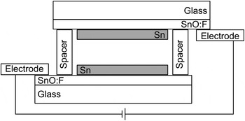
We have conducted the experiments of growing Sn whiskers in an electric field using true flat plate capacitor geometry shown in Fig. 1, where the surface area of the plates, and the area from which whiskers may grow, is of vastly greater dimension than the inter-plate spacing. While the field polarities are obviously opposite for the top and bottom Sn films, no differences in whisker developments were observed between these films.
Schematic cross-sectional view of the capacitive experimental setup.

We note that our first experiment conducted in the late summer showed dramatic differences in whisker growth between the samples exposed to an electric field and the no-field control. A subsequent experiment in the early winter did not show the same results; hence, it was hypothesized that humidity may have played a role. This lead to a 2 × 2 experiment, of high and low humidity, and high and no electric field. In summary, all samples in low humidity grew no detected whiskers; the sample in high humidity and no field grew a few short whiskers, comparable with the original control sample; and the sample in high humidity and high field grew many whiskers of various lengths.
More specifically, Sn films were grown on Pilkington TEC15-brand (SnO:F of nominal 15 Ω persheet resistance) glass chosen to allow for easy electrical connection to the film, and for improved film adhesion (due to surface roughness of the oxide layer). The films were grown through thermal evaporation, from pellets of 99.999% purity. The thickness of the deposited Sn films was 150 nm, determined by a quartz crystal microbalance thickness monitor. Such thickness is not considered as prone to significant whisker growth. We have also attempted several trials with films of similar thickness and chemical composition deposited through radio frequency (RF) magnetron sputtering.
The thermal evaporator used allows for simultaneous deposition of three samples providing a greater degree of confidence that subsequent differences are indeed due to intentional experimental factors and not variations in the depositions. In the original experiment, one deposition supplied the samples for both electrodes as well as the control sample. For the subsequent 2 × 2 (humidity and field), experiment, six samples from two depositions were needed. In this case, we used one sample for low field from each deposition. The electrodes for the high-field experiments, in high and low humidity, used one sample each from the two depositions, to indicate whether whisker growth in those situations were due to experimental conditions and not deposition batch.
Adhesive spacers were applied to the plates. Afterwards, the spacer thickness was confirmed with a stylus profilometer to have height 50 ± 5 µm as illustrated in Fig. 1. Bias was held at 14.5 V, using a BK Precision 1696 regulated power supply providing the average field between the plates 2900 V/cm. Electrical contact to the films was made with conductive tape applied to the SnO:F layer. Films were held at voltage for the time of 1 week. Whiskers were observed using a Hitachi S-4800 scanning electron microscope (SEM).
In the experimental rounds with humidity as an additional variable, a saturated sodium chloride salt bath was used to maintain the relative humidity at what we call here to be a “high” level, of 75%. This humidity was confirmed through a hygrometer within the humidity chamber. Simultaneously, a hygrometer was used to measure the ambient humidity of what we here call a “low” level, which, over the course of the experiment, was measured as 40 ± 6%. There were no ambient humidity readings made during the initial experimental phase, as a humidity effect was not considered at that time. However, the measured ambient indoor humidity readings were found to be correlated with outdoor temperature obtained from the national weather service, over the same time period. As the average temperature during the initial phase of the experiment was 298 K with a high of 305 K, and during the 2 × 2 experiment was 270 K with a high of 285 K, this correlation suggests that it may be reasonable that ambient laboratory humidity was higher during the initial phase of the experiment.
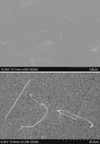
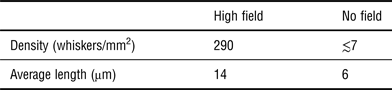
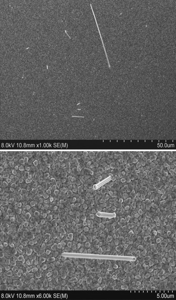
There were dramatic, order-of-magnitude differences in both whisker length and density between the experimental, high-field sample and the control, no field sample in our initial series of field induced whisker experiments. The differences in density and average length are summarized in Table I. Representative examples of the corresponding SEM observations are shown in Fig. 2.
Above—broad view of original high-field sample, demonstrating length and density of whiskers. Below—a closer view of a different region of the sample. Whiskers have high aspect ratios, but may have kinks or curves.

Summary of whisker length and density.

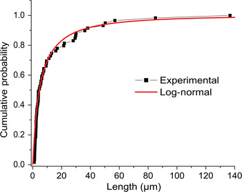
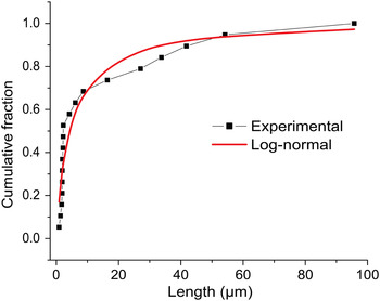
Other groups have found that metal whisker length follows a log-normal distribution.[ Reference Fang, Osterman and Pecht 15 – Reference Susan, Michael, Grant, McKenzie and Yelton 17 ] Our fit based on the data from random regions agrees with a log-normal curve too as shown in Fig. 3. The grain size appears to be normally distributed, with mean size 0.47 µm and standard deviation 0.1 µm. This also applied to the grain size in the subsequent experiment. The average whisker diameter was found to be 0.4 µm and standard deviation 0.1 µm. The similarity between the grain and the whisker diameter may suggest that whisker nucleation and growth are coupled to the grains themselves.
Cumulative fraction of sample of whisker length from our original experiment versus the log-normal fit with the parameters μ = 1.6 + ln (μm) and σ = 1.4 µm.

Whisker length and width were found to be uncorrelated.
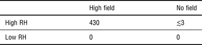
In our controlled humidity experiments, no whiskers grew in low-humidity samples, regardless of the field applied. In high humidity, the results were similar to those observed in the original experiment as summarized in Table II. Figure 4 illustrates the density and length of whiskers seen in the high-humidity, high-field sample. The statistical distribution of whisker lengths from the high-humidity, high-field experiment fits well the log-normal cumulative curve as shown in Fig. 5.
Above—from high-humidity, high-field sample. Below—close-up (from the same region) of short whiskers, illustrating whisker structure.

Cumulative fraction of the sample of whisker length from the high-field, high-humidity experiment versus the log-normal fit. The parameters of the fit are μ = 1.4 + ln (μm), σ = 1.6 µm.

Summary of the difference of whisker density (in mm−2) in the humidity–field experiment.
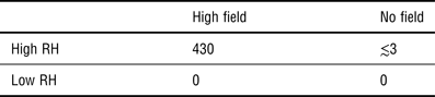
RH, relative humidity.
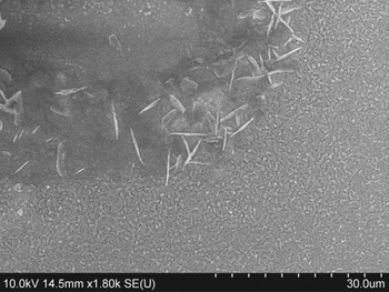
The RF sputtered Sn films in our experiments, even when exposed to high humidity and field, have not yet been observed to grow uniformly distributed whiskers in the manner of the evaporated films. However, we have observed holes of roughly 1 mm diameter in the films after 1 week of the high humidity and field conditions. These holes appear on both the electrodes. They are matched such that they appear on both electrodes in locations that would be overlapping when assembled as in Fig. 1. We speculate that these holes may have been created when a whisker caused a short circuit between the electrodes, and vaporized that region of the Sn film.[ Reference Mason and Eng 18 ] An SEM image of such a hole is seem in Fig. 6 with some short whiskers in the region of the edge of the hole.
Crater in a sputtered Sn film after exposure to high-humidity and electric field.

We conclude that in many cases the application of an electric field greatly accelerated the growth of Sn whiskers. In no case did a control, no-field samples grow whiskers at all without the high-field sample growing substantially more.
The result of the dramatic difference in whisker length and density in films grown in the presence of electric field is strong evidence that it may play an important role in whisker growth, which is consistent with the theoretical predictions.[ Reference Karpov 9 , Reference Karpov 10 ] However, inconsistencies between the various rounds of experiments point that these results remain preliminary, with need for further reproduction, and possibly inclusion of other factors. As an important practical application, further work in this direction has a potential of development of an accelerated life test in whisker-related reliability problems.
Acknowledgments
Microscopy for this study was done with equipment at the Center for Materials and Sensor Characterization center at the University of Toledo. We acknowledge A. D. Compaan for the deposition facilities and D. Strickler for the Pilkington TEC15-brand glass substrates. The financial support for this work was provided by NRC award No. NRC-HQ-12-G-38-0042 and NSF award No. 1066749.