Heart failure is a major concern for the developing as well as developed world and accounts for significant morbidity and mortality.Reference Levy, Garrison, Savage, Kannel and Castelli 1 Cardiac hypertrophy is an early response of the heart towards enhanced biomechanical stress. It is a part of progressive ventricular remodelling that involves the growth of cardiomyocytes, apoptosis, or necrosis, collagen deposition along with chamber enlargement, and pump dysfunction.Reference Levy, Garrison, Savage, Kannel and Castelli 1 , Reference Balakumar, Singh and Singh 2 Moreover, these alterations include enhanced expression of inflammatory cytokines, shift of myocardial substrate utilisation from fatty acids to glucose, constant myocardial extracellular matrix turnover, and activation of matrix metalloproteinase enzymes in cardiac tissues.Reference Finck and Kelly 3 , Reference Deschamps and Spinale 4 The intracellular oxidative stress plays a significant role in hypertrophic response.Reference Balakumar, Rose and Singh 5 Peroxisome proliferator-activated receptor-α is involved in the maintenance of fatty-acid metabolism in cardiomyocytes to generate energy. Their activation in the heart stimulates the upregulation of genes controlling mitochondrial fatty-acid uptake, thereby resulting in increased fatty-acid metabolism and generation of ATP.Reference Van der Lee, Vork and De Vries 6 , Reference Vosper, Khoudoli, Graham and Palmer 7 Fibrates such as fenofibrate, bezafibrate, and gemfibrozil are peroxisome proliferator-activated receptor-α agonists used clinically for the treatment of dyslipidemia and hypertriglyceridemia. The expression of peroxisome proliferator-activated receptor-α is reported to be downregulated in models of cardiac remodelling.Reference Balakumar, Rose and Singh 5 , Reference Erol 8 Therefore, fibrates may serve as potential therapeutic agents for the treatment of cardiac hypertrophy and failure. Major research regarding the role of fibrates in cardiac remodelling is restricted to fenofibrate that has proved its cardioprotective potential in various models of cardiac dysfunction; however, limited literature is available regarding its role in ameliorating cardiac hypertrophy. We have already reported gemfibrozil to protect against obesity-induced cardiac remodelling.Reference Singh, Singh, Singh and Krishan 9 The potential role of gemfibrozil against non-obese models, such as pressure overload-induced cardiac hypertrophy, still remains unexplored. The present study is an attempt to investigate the effect of gemfibrozil in partial abdominal aortic constriction-induced cardiac hypertrophy in rats.
Materials and methods
The experimental protocol used was duly approved by the institutional animal ethics committee. Young male wistar rats of 14–16 weeks of age weighing 200–225 g were selected for study. The rats were exposed to a 12-hour light and dark cycle in the institutional animal house and were maintained on group-specific rat feed and water ad libitum. The rats were divided into three groups, each comprising six animals.
Partial abdominal aortic constriction
The partial abdominal aortic constriction was performed as per the established procedure.Reference Singh, Singh, Singh and Krishan 9 , Reference Singh and Krishan 10
Morphological assessment of cardiac hypertrophy
The left ventricle including the interventricular septum was weighed, and values were expressed as milligrams per gram of body weight. The left ventricle was divided into three equal slices, and the wall thickness of each slice was noted at nine different points using an ocular micrometer. The mean values of all three slices were calculated.
Estimation of left-ventricular collagen and protein content
The left-ventricular collagen content was determined by measuring the hydroxyproline concentration,Reference Singh, Singh, Singh and Krishan 9 , Reference Singh and Krishan 10 whereas left-ventricular protein content was determined by the method of Lowry et alReference Lowry, Rosebrough, Farr and Randall 11
Estimation of oxidative stress
The oxidative stress in the cardiac tissues was measured in terms of thiobarbituric acid-reactive substances, superoxide anion generation, and reduced glutathione.Reference Singh, Singh, Singh and Krishan 9 , Reference Singh and Krishan 10
Serum analysis
Estimation of serum cholesterol and serum high-density lipoprotein was carried out using commercially available kit (Bayer Diagnostics Ltd, Bhopal, India). The serum triglycerides and serum glucose were measured using commercially available diagnostic kits (Transasia Biomedicals Ltd, Mumbai, India).
Histopathological studies
The heart was excised and immediately immersed in 10% buffered formalin, dehydrated in graded concentrations of ethanol, immersed in xylene, and then embedded in paraffin. The sections of 4 μm were cut and stained with haematoxylin–eosin and picrosirius red to observe cardiomyocyte diameter and collagen deposition, respectively.Reference Singh, Singh, Singh and Krishan 9
Experimental protocol
In group 1 (control group), no drug treatment was given to rats. In group 2 (partial abdominal aortic constriction control), the abdominal aorta was partially constricted and rats were allowed to survive for 4 weeks. In group 3 (partial abdominal aortic constriction+gemfibrozill 30 mg/kg, oral), the abdominal aorta was partially constricted and rats were allowed to survive for 4 weeks.
Drug and chemicals
Gemfibrozil was obtained from Nicolas-Piramal Research Labs (Mumbai, India). Folin–Ciocalteu reagent was procured from SRL (Mumbai, India) and bovine serum albumin was obtained from Sigma-Aldrich (Bangalore, India). All other reagents used in the study were of analytical grade.
Statistical analysis
The results were expressed as means±SE of the mean. The data obtained from various groups were statistically analysed using one-way analysis of variance followed by Tukey’s multiple range test. The p-value <0.05 was considered statistically significant.
Results
Effect of gemfibrozil treatment on the morphological parameters
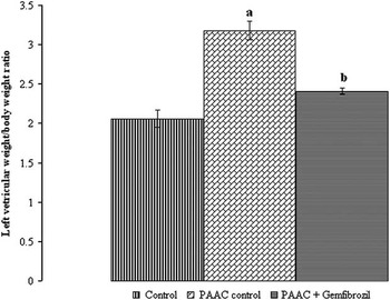
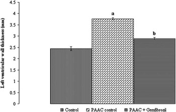
The partial abdominal aortic constriction-induced significant increase in left-ventricular weight to body weight ratio and left-ventricular wall thickness as compared with control rats. The treatment with gemfibrozil significantly attenuated partial abdominal aortic constriction-induced morphological changes in the heart (Figs 1 and 2).
Effect of gemfibrozil treatment on left-ventricular weight to body weight ratio. Values are expressed as mean±SEM ( a ) p<0.05 versus control; ( b ) p<0.05 versus partial abdominal aortic-banding control group.

Effect of gemfibrozil treatment on left-ventricular wall thickness. Values are expressed as mean±SEM ( a ) p<0.05 versus control; ( b ) p<0.05 versus partial abdominal aortic-banding control group.

Effect of gemfibrozil treatment on myocardial collagen and protein content
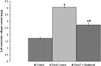
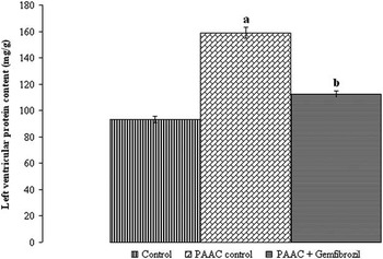
A significant increase in left-ventricular collagen and protein content was observed in rats with partial abdominal aortic constriction. The treatment with gemfibrozil significantly attenuated partial abdominal aortic constriction-induced increase in collagen and protein content (Figs 3 and 4).
Effect of gemfibrozil treatment on left-ventricular collagen content. Values are expressed as mean±SEM ( a ) p<0.05 versus control; ( b ) p<0.05 versus partial abdominal aortic-banding control group.

Effect of gemfibrozil treatment on left-ventricular protein content. Values are expressed as mean±SEM ( a ) p<0.05 versus control; ( b ) p<0.05 versus partial abdominal aortic-banding control group.

Effect of gemfibrozil treatment on the oxidative stress
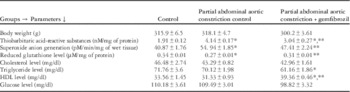
The partial abdominal aortic constriction-induced significant oxidative stress in the cardiac tissues is indicated by increase in thiobarbituric acid-reactive substances, superoxide anion generation, along with decrease in reduced glutathione level. The gemfibrozil treatment significantly attenuated partial abdominal aortic constriction-induced oxidative stress in the cardiac tissues (Table 1).
Effect of gemfibrozil on morphological, oxidative stress, and biochemical parameters.

Values are expressed as mean±SEM. p<0.05 is considered statistically significant
* p<0.05 versus control
** p<0.05 versus partial abdominal aortic constriction control
Effect of gemfibrozil treatment on serum parameters
No significant change was observed in serum cholesterol, triglycerides, and glucose level in various groups used in the present study. A significant increase in serum high-density lipoproteins was observed in gemfibrozil-treated group as compared with control and partial abdominal aortic constriction group (Table 1).
Effect of gemfibrozil treatment on histological parameters
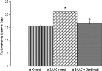
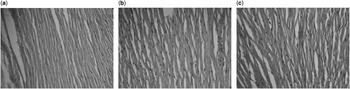
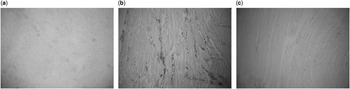
The haematoxylin–eosin staining revealed a significant increase in the cardiomyocyte diameter of rats with partial abdominal aortic constriction as compared with the control group. The treatment with gemfibrozil-attenuated partial abdominal aortic constriction-induced increase in the cardiomyocyte diameter (Figs 5 and 6). The picrosirius red staining of the rat heart observed marked increase in collagen deposition with partial abdominal aortic constriction as compared with normal rats that was attenuated with gemfibrozil treatment (Fig 7).
Effect of gemfibrozil treatment on cardiomyocyte diameter. Values are expressed as mean±SEM ( a ) p<0.05 versus control; ( b ) p<0.05 versus partial abdominal aortic-banding control group.

Haematoxylin–eosin staining of transverse sections of the left ventricle showing change in cardiomyocyte diameter at 400× magnification. ( a ) Normal control, ( b ) partial abdominal aortic constriction control, ( c ) partial abdominal aortic constriction+gemfibrozil group.

Picrosirius red staining of transverse sections of the left ventricle showing collagen deposition at 100× magnification. ( a ) Normal control, ( b ) partial abdominal aortic constriction control, ( c ) partial abdominal aortic constriction+gemfibrozil.

Discussion
During the progression of pathological cardiac hypertrophy in animal models and humans, the heart undergoes a shift from fatty-acid oxidation towards increased glucose utilization.Reference Bishop and Altschuld 12 The evidence implicates deactivation of the peroxisome proliferator-activated receptor-α signalling pathway as the mechanism driving the downregulation of fatty-acid oxidation genes in the hypertrophied heart. Reduced expression and activity of peroxisome proliferator-activated receptor-α closely correlates with decreased β-oxidation observed in cardiac hypertrophy.Reference Kanda, Nohara, Hasegawa, Kishimoto and Sasayama 13 The fibrate class of drugs, including fenofibrate and gemfibrozil, are synthetic peroxisome proliferator-activated receptor-α ligands and have been reported to ameliorate cardiovascular complications.Reference Erol 8 Major work involving exploration of the role of peroxisome proliferator-activated receptor-α has been conducted with fenofibrate, where its treatment has been found to attenuate the development of myocardial hypertrophy and fibrosis and to preserve in vivo contractile function in rats through the inhibition of NF-κB-mediated inflammation.Reference Ichihara, Obata and Yamada 14 Our research group has already reported the protective role of gemfibrozil treatment against high fat diet-induced cardiac hypertrophy in rats.Reference Singh, Singh, Singh and Krishan 9 To the best of our knowledge, the present study is the first one to explore the protective role of gemfibrozil against a non-obese model of cardiac hypertrophy in rats. The rats of age 3–4 months were selected for the present study, as they represent age of less than 20 years in humans.Reference Collier and Coleman 15 The partial abdominal aortic constriction model used in the present study is a well-established model to induce cardiac hypertrophy. The increase in the mean cardiomyocyte diameter, left-ventricular weight to body weight ratio, left-ventricular wall thickness, and protein content have been documented to be an index of experimental cardiac hypertrophy.Reference Balakumar, Rose and Singh 5 , Reference Singh, Singh, Singh and Krishan 9 The present study observed significant increase in all these parameters in rats with partial abdominal aortic constriction, whereas gemfibrozil treatment ameliorated partial abdominal aortic constriction-induced cardiac changes in rats, thus proving its cardioprotective effect.
Multiple stimulations, including static stretch of the cells, can promote fibroblast cell proliferation and collagen synthesis.Reference Porter and Turner 16 The partial abdominal aortic constriction-induced pressure overload stretches fibroblast cells that promote collagen synthesis in the rat heart. It was witnessed by a significant increase in the collagen content in rats with partial abdominal aortic constriction in the present study. The treatment with gemfibrozil significantly attenuated myocardial collagen deposition in rats.
Reactive oxygen species are involved in the induction of cardiac hypertrophy and fibrosis by activating tyrosine kinase, protein kinase C, mitogen-activated protein kinases, and transforming growth factor-β.Reference Zhao, Zhao, Chen, Ahokas and Sun 17 The free radicals generated within the cell react with lipids of the cell membrane, thereby forming lipid radicals and lipid peroxides that are measured in terms of thiobarbituric acid-reactive substances. A significant increase in left-ventricular thiobarbituric acid-reactive substances, superoxide anion generation along with significant decrease in reduced glutathione level indicated oxidative stress in partial abdominal aortic constriction control group. The treatment with gemfibrozil significantly attenuated oxidative stress as indicated by significant changes in various oxidative stress parameters used in the present study.
It is concluded that gemfibrozil treatment affords significant protection against partial abdominal aortic constriction-induced cardiac hypertrophy and oxidative stress in rats.
Acknowledgement
The authors highly acknowledge the Department of Pharmaceutical Sciences and Drug Research, Punjabi University, Patiala, for providing facility and support to carry out the research work.
Financial Support
This research received no specific grant from any funding agency, commercial, or not-for-profit sectors.
Conflicts of Interest
None.
Ethical Standards
The authors assert that all procedures contributing to this work comply with the ethical standards of the relevant national guidelines for the care and use of laboratory animals (male Wistar rats) and has been approved by the institutional committee (Department of Pharmaceutical Sciences and Drug Research, Punjabi University, Patiala).