Introduction
Trichodinids are common symbionts belonging to the family Trichodinidae (Phylum Ciliophora). These symbionts are epizoic on a broad range of invertebrates and vertebrates (mostly aquatic). The largest number of species are found on gills and skin of both freshwater and marine fishes (Van As and Basson, Reference Van As and Basson1986; Lom and Dyková, Reference Lom and Dyková1992; Basson and Van As, Reference Basson and Van As2002). Trichodinids have both ectozoic and endozoic forms (Lom and Dyková, Reference Lom and Dyková1992).
The ectozoic trichodinids, which often inhabit skin and gills of fishes, are more common than endozoic forms (Arthur and Lom, Reference Arthur and Lom1984; Lom and Dyková, Reference Lom and Dyková1992). They use the gills or skin of fish as a convenient substrate for gliding or temporary attachment. The symbionts feed on waterborne particles or detritus particles, disrupted cells and bacteria on the surface of the host. A severe infection (presence of a large number) of ectozoic trichodinids may result in irritation or damage to the epidermal cells under certain conditions (Lom and Dyková, Reference Lom and Dyková1992). In some cases, when young fish are seriously infested by certain ectozoic species, trichodinids may penetrate into the skin or gill tissue causing wounds (Lom and Dyková, Reference Lom and Dyková1992).
As a rule, endozoic trichodinids are likely to be host-specific and have a greater number of closely spaced denticles with narrow and long blades (Lom and Haldar, Reference Lom and Haldar1976; Lom and Dyková, Reference Lom and Dyková1992). Most endozoic fish trichodinids (Trichodina urinaria, T. algonquinensis, T. polycirra, T. urinicola, T. schizothoraci, Vauchomia nephritica, V. renicola, Paratrichodina phoxini, P. alburni and P degiustii) are found in the urinary tract including the urinary bladder and renal collecting duct in both marine and freshwater fish (Perca fluviatilis, P. flavescens, Notropis cornutus, N. heterodon, Notemigonus crysoleucas, Stizostedion lucioperca, Schizothorax intermedius, Rutilus rutilus, Abramis brama, Esox masquinongy, E. niger, Alburnus alburnus, Phoxinus phoxinus) (Lom, Reference Lom1958; Lom and Haldar, Reference Lom and Haldar1976; Li and Desser, Reference Li and Desser1983; Arthur and Lom, Reference Arthur and Lom1984; Lom and Dyková, Reference Lom and Dyková1992). Two endozoic trichodinid species have been reported from the intestine of marine fish, i.e. T. fariai from the checkered puffer (Sphoeroides testudineus) (Lom and Haldar, Reference Lom and Haldar1976; Lom and Dyková, Reference Lom and Dyková1992) and T. luba from the yellowfin surgeonfish Acanthurus xanthopterus (Basson et al., Reference Basson, Van As and Fishelson1990). Trichodina oviducti, T. rajae and T. rhinobatae were reported from the urogenital system including the oviducts, seminal grooves and copulatory sacs of skates (Raja radiate, R. scabina) (Khan, Reference Khan1972; Lom and Haldar, Reference Lom and Haldar1976) and guitarfish (Rhinobatos annulatus) (Van As and Basson, Reference Van As and Basson1996).
During a study designed to determine the health of shorthorn sculpins (Myoxocephalus scorpius) collected from the large industrial harbour in the vicinity of Nuuk, West Greenland, trichodinids were recorded from internal organs including spleen, kidney and liver of the shorthorn sculpin. Prevalence and intensity of the trichodinid infections were determined. The effect of contamination on the occurrence of trichodinid infections in sculpin was investigated.
Materials and methods
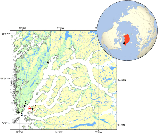
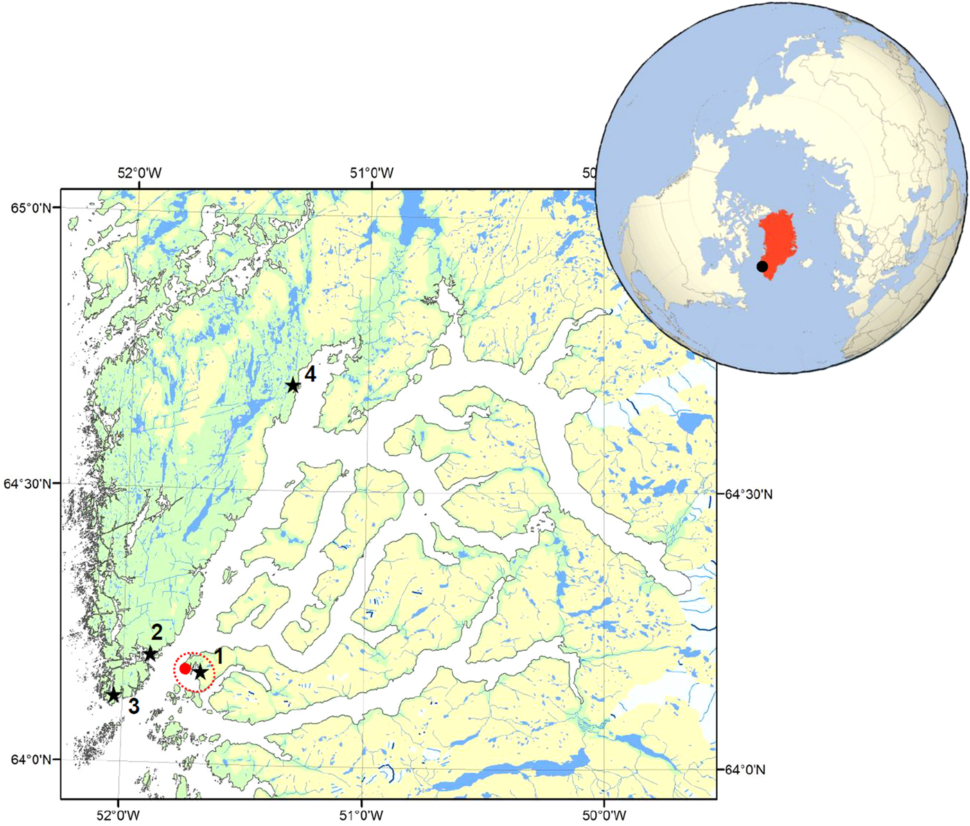
Shorthorn sculpins (n = 46) were caught using a fishing rod at four sampling locations (Fig. 1) in the vicinity of the industrial harbour in Nuuk, Greenland in 2016. Fish were kept alive in aerated tanks until dissection (maximum 3 h) and the gills and internal organs were then collected. Weight of fresh liver was recorded. Samples of the gills, spleen, kidney and liver were fixed using 4% neutral buffered formalin for histology. Each sample was sectioned using routine histology and one 4 µ m haematoxylin and eosin (HE) stained section was obtained for each sample. Histological examinations, including parasite detection, were undertaken using a light microscope (Nikon Eclipse Ni-U).
Map of sampling areas. Stars indicate sampling sites which were numbered following a reduced concentration of pollutants (Jacobsen and Asmund, Reference Jacobsen and Asmund2000; Strand and Asmund, Reference Strand and Asmund2003; Vorkamp et al., Reference Vorkamp, Strand, Christensen, Svendsen, Lassen, Hansen, Larsen and Andersen2010). Red dot and surrounding red dotted line indicate the town of Nuuk (~15.000 inhabitants).
Levels of trichodinid infections were assessed using prevalence (percentage of fish affected based on presence/absence) and intensity (estimated as number of the parasite per cm2 surface area of histological section). Image J software was employed for measuring surface areas using a calibrated scale. The intensity for each sampling location was averaged from individual intensities and values were presented as mean ± standard error (s.e.). Differences in the intensity of parasite among sampling locations were investigated using a non-parametric Kruskal–Wallis test and differences in prevalence were tested by the use of the χ 2 test of independence (IBM SPSS statistic 22). Difference in sex distribution of endozoic trichodinids (prevalence of endozoic trichodinid in male and female) were analysed with a χ 2 test of independence. Associations between intensity of endozoic trichodinids in all internal organs and body length, body weight, liver weight or intensity of ectozoic trichodinids were assessed using Pearson correlation analysis (IBM SPSS statistic 22). P < 0.05 was interpreted as statistically significant.
Results
General descriptions
Infected organs
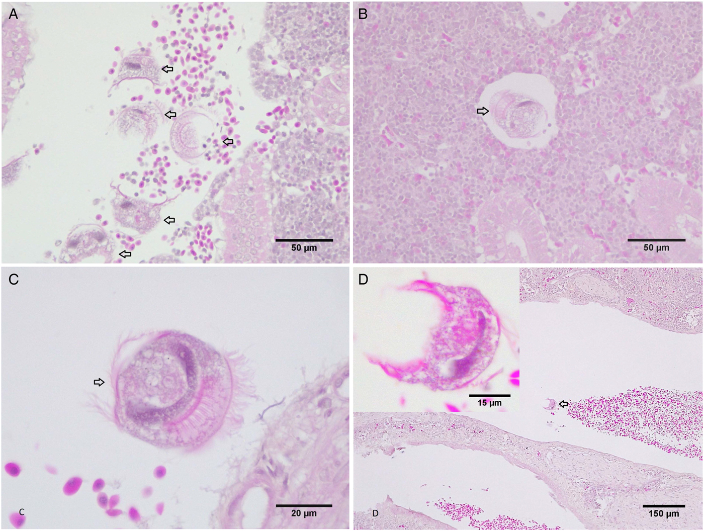
A high level of trichodinid infection was observed in internal organs of shorthorn sculpins. The endozoic trichodinids were found in blood vessels in the kidney, liver, spleen and connective tissue between spleen and pancreas (Fig. 2). Prevalence of trichodinid infections was highest in the kidney (15.2%), followed by the spleen (10.9%) and the liver (8.7%). The total prevalence in all examined internal organs (kidney, spleen and liver) reached 23.9%.
Histological sections of endozoic trichodinids in the internal organs of shorthorn sculpin (HE stains). (A and B) Trichodinids in the kidney. (C) Trichodinid in the liver. (D) Trichodinid in the spleen.

Morphology
The trichodinids in the internal organs had a typical horseshoe-shaped macronucleus, a relatively high body, probably cup-shaped, with a clearly deep concave adhesive disc. The dimensions of the parasite present in the internal organs (based on several well-oriented histological sections) were all morphologically similar [body diameter from 39 to 42 µ m; body height from 27 to 29 µ m (n = 3)]. The denticle blades were strongly developed and the rays appeared straight (not curved) (Fig. 2B and D). The trichodinid from the gills had a typical horseshoe-shaped macronucleus, the body diameter is roughly 32.5 µ m, denticle ring 20.2 µ m, with 25 denticles in the adhesive disc. Based on the body shapes, size ranges and denticle morphology, it is highly likely that both trichodinid species from the internal organs and gills belong to the genus Trichodina. It is likely that the trichodinids found in the spleen, kidney and liver belong to the same species and it is also likely that this species from the internal organs differ from the trichodinids observed on the gills of the same individual fish.
Clinical signs
Endozoic trichodinids did not cause any alterations to the colour of the liver. All infected fish had normal coloured livers (light brown). No endozoic trichodinids were detected from fish showing unusual colour of liver such as white, orange or dark brown. Endozoic trichodinids were not associated with any histological changes in spleen, kidney and liver.
Infection status
Effect of sampling location
There was no effect of sampling location on the prevalence and intensity of endozoic trichodinids in each organ (spleen, kidney and liver) and all internal organs (P > 0.05) (Table 1).
Prevalence (P, %) and intensity (I, no/cm2 surface area) (Mean ± s.e.) of trichodinids in the kidney, liver and spleen of shorthorn sculpins

Effect of sex
Infection level was not affected by sculpin sex (χ 2 = 0.996, df = 1, P = 0.318).
Correlations
There was a positive correlation between intensity of trichodinid infection in the internal organs and gills (R = 0.5, n = 46, P < 0.001). Fish with high numbers of ectozoic trichodinids on the gills tended to be infected by the endozoic trichodinids in either spleen, kidney or liver. Sculpins (n = 5), where the gills were not infected by ectozoic trichodinids, were free of the endozoic form in the internal organs.
There was no correlation between intensity of endozoic trichodinids and body length, body weight or liver weight of sampled fish (P > 0.05).
Discussion
To the best of our knowledge, this is the first report on the presence of endozoic trichodinids in organs such as liver and spleen and in the body cavity of a fish. Endozoic trichodinids were previously found in the renal collecting duct of the kidney (Lom and Dyková, Reference Lom and Dyková1992). Other endozoic trichodinid species were observed in the urinary tract, intestine, oviduct, urogenital and copulatory sacs of fish and amphibians (Lom and Haldar, Reference Lom and Haldar1976; Van As and Basson, Reference Van As and Basson1986; van der Bank et al., Reference van der Bank, Basson and Van As1989; Kruger et al., Reference Kruger, Basson and Van As1991; Lom and Dyková, Reference Lom and Dyková1992). Most of trichodinids are ectozoic and can be found in the gills, skin and nasal pits of fish (Lom and Dyková, Reference Lom and Dyková1992). While we report occurrence of trichodinids in internal organs where they were never seen before, the exact position or pathways of trichodinid transfer is an open question and will be addressed in a follow-up study.
In this study, a positive correlation was established between intensity of trichodinids on the gills and in internal organs. The correlation suggested that the environmental conditions, which were conducive for the proliferation of ectozoic trichodinids, seemed to support proliferation of endozoic trichodinids as well. Fish sex and age (size) had no apparent influence on trichodinid prevalence or intensity in the sculpins, so fish from different locations could be compared for endozoic trichodinid infections regardless of their sex or age.
Whereas most ectozoic trichodinids affect many host species, endozoic trichodinids tend to be more host specific (Lom and Dyková, Reference Lom and Dyková1992). Therefore, the endozoic trichodinids found in this study might possibly show clear host preference for sculpins.
Generally, the trichodinid species encountered in the liver, spleen and kidney appeared to display some of the typical characteristics of endozoic trichodinids (such as a high body and a relative deeply concave adhesive disc which strengthens the adhesive disc structure) (Lom and Haldar, Reference Lom and Haldar1977; Lom and Dyková, Reference Lom and Dyková1992). Sometimes endozoic trichodinids can show both ecto- and endo-characteristics suggesting a recent adaptation. For examples, the endozoic T. schizothoraci showing some of the typical surface-inhabiting characteristics (such as wide and strongly developed denticles), while urinary bladder trichodinids (Lom and Haldar, Reference Lom and Haldar1976; Gong et al., Reference Gong, Yu, Feng and Shen2005) or T. fugu reported from the gills shared some typical endozoic characteristic with T. urinaria found in urinary tract of freshwater fish (Xu, Reference Xu2007), i.e. typical delicate narrow denticles. Out of the more than 350 species in the family Trichodinidae, there has been no case of a single species occurring both ecto- and endozoically (Basson, unpublished). Unfortunately, in this study imprints of the kidney, spleen and liver were not collected; therefore, a complete morphological and taxonomical description was not possible.
Most studies on the effects of contaminants on trichodinids reported on ectozoic trichodinids from the gills and skin. Pollutant exposure induces pathological alteration in the gills and skin of fish and stress or impairs immune responses, which subsequently can provide beneficial conditions for the proliferation of some facultative ectosymbiotic ciliates including ectozoic trichodinids (Barker et al., Reference Barker, Khan, Lee, Hooper and Ryan1994; Khan et al., Reference Khan, Barker, Williams-Ryan and Hooper1994; Marcogliese et al., Reference Marcogliese, Nagler and Cyr1998; Muzzall and Bowen, Reference Muzzall and Bowen2002; Khan, Reference Khan2010, Reference Khan2011). Ectozoic trichodinids have been reported to increase on fish from sites polluted with pulp mill effluent or organic materials, municipal and domestic sewage (which associated with low dissolved oxygen concentration, high alkalinity and NH4 nitrogen concentration and toxins) and oil (Lehtinen et al., Reference Lehtinen, Notini and Landner1984; Khan and Thulin, Reference Khan and Thulin1991 ) but to decrease in areas contaminated by heavy metals (El-Seify et al., Reference El-Seify, Zaki, Razek, Desouky, Abbas, Hady and Zaid2011; El-Bouhy et al., Reference El-Bouhy, Reda and El-Azony2016). As far as we are aware, there is no published information on the effect of pollutants on endozoic trichodinids. In this study, there was no effect of location on the presence of trichodinids in sculpins; however, it does not necessarily mean that pollution has no effect on endozoic trichodinids. The quantification method for trichodinids in this study, which was only based on histological sections, might have underestimated the real intensity of trichodinid infections as some of the parasites could have be missed or lost during sampling. For future studies, we recommend to include more sampling methods specific for trichodinids such as mucous subsampling, formalin immersion technique (as suggested by Marcogliese et al., Reference Marcogliese, Pulkkinen and Valtonen2012), tissue imprints, sections, shaking and pooled shakes (Khan et al., Reference Khan, Barker, Williams-Ryan and Hooper1994) to achieve more accurate indications of infection levels of endozoic trichodinids found associated with shorthorn sculpin.
Financial support
This research received no specific grant from any funding agency, commercial or not-for-profit sectors.
Conflict of interest
None.