I. INTRODUCTION
Titanium dioxide (TiO2) is an inexpensive, chemically stable and nontoxic metal oxide that has been widely used as an advanced semiconductor and as a white pigment for many years.Reference Barnard and Zapol1–Reference Diebold3 TiO2 has generated much interest for photoelectrochemical applications, such as photocatalytic waterReference Du, Lai, Yang, Zhai, Kisailus, Su, Wang and Jiang4 and air purification and water splitting for hydrogen fuel generation, first reported by Fujishima and Honda.Reference Fujishima and Honda5, Reference Nowotny and Bahnemann6 Photoelectrolysis of water can be used as a renewable and environmentally benign route for hydrogen generation. Both applications require a photogeneration of electron-hole pairs that are involved in subsequent chemical reactions. Photoexcitation of a semiconductor-like TiO2 requires light with the energy equivalent to the band gap. The band gap energy of TiO2 in two of its phases (i.e., anatase and rutile) falls within the ultraviolet (UV) spectrum (i.e., 3.0 eV for rutile and 3.2 eV for anatase).Reference Gratzel7 Hence, TiO2 is an active photocatalytic material within the UV spectrum and absorbs only the UV part of solar spectrum.
Engineering TiO2 (size, phase, and band gap) for enhanced photocatalytic response and visible-light photogeneration has been investigated for decades. Modification of TiO2 through transition metal or anion doping has proven to be one of the most promising methods to modify the band structure.Reference Ohno, Akiyoshi, Umebayashi, Asai, Mitsui and Matsumura8–Reference Asahi, Morikawa, Ohwaki, Aoki and Taga12 Doped TiO2 has been studied from both experimental and theoretical aspects. Various transition metals have been used to substitute for Ti in TiO2 including Fe, V, W, Cr, Co, and Mn.Reference Zhou, Liu, Zhu and Zhang9–Reference Couselo, Einschlag, Candal and Jobbagy11, Reference Zhang, Zhao, Liu, Dong, Zhang and Chen13–Reference Goh, Chan and Liu15
For example, it was found that both VReference Zhou, Liu, Zhu and Zhang9 and MnReference Zhang, Zhao, Liu, Dong, Zhang and Chen13 atoms can substitute for Ti atoms within the TiO2 lattice, resulting in a shift of the photoluminescence (PL) emission peak toward longer wavelengths, which indicated reductions in the band gap. On the other hand, anion doping has also been utilized to reduce the band gap of TiO2.Reference Ohno, Akiyoshi, Umebayashi, Asai, Mitsui and Matsumura8, Reference Asahi, Morikawa, Ohwaki, Aoki and Taga12 Asahi reported the effects of N-doping on the band gap narrowing and photocatalytic improvements of TiO2.Reference Asahi, Morikawa, Ohwaki, Aoki and Taga12 Using a local-density approximation model, they demonstrated the density of states for N, C, P, F, and S substitutional dopants that also had a significant effect on the band gap. They confirmed their models by observing absorption of light in N-doped TiO2 at wavelengths less than 500 nm. Ohno et al.Reference Ohno, Akiyoshi, Umebayashi, Asai, Mitsui and Matsumura8 suggested that the photocatalytic activity of S-doped TiO2 improved with wavelengths above 440 nm. The selection of dopants for TiO2 is largely dependent on the desired application. For water/air purification applications, nontoxic transition metals can be used to reduce the band gap via reduction in the conduction energy band. However, for hydrogen generation applications, the band gap decrease should be the result of the valence energy band increase. The energy difference between the lower edge of the conduction band and the upper edge of the valence band of the band gap needs to overlap the range of standard potentials of water redox couples (H2O/O2 and H2/H2O).Reference Fujishima and Honda5, Reference Gratzel7
In this study, we incorporate Ta dopants uniformly within a TiO2 matrix via a solution route and investigate the dopant effects on the resulting optoelectronic properties. Ta doping of TiO2 has been investigated by different groups.Reference Wang, Geng, Guo, Jiang and Qu16–Reference Navale, Vadivel Murugan and Ravi18 For example, TiO2 was doped with Ta using a high temperature methodReference Navale, Vadivel Murugan and Ravi18 where TiO2 and Ta2O5 powders were mixed with acetone and ground for several hours, then annealed in air at 1573 K. This method requires high temperatures as a result of the large distances cations must diffuse within the TiO2 matrix. In this study, we use relatively low temperatures (∼453 K), via sol–gel chemistry processing utilized previously,Reference Cheng, Ma, Zhao and Qi2, Reference Wang and Ying19 to achieve molecular mixing that circumvents the need to anneal at high temperatures as previously suggested.Reference Navale, Vadivel Murugan and Ravi18 Wang et al. synthesized Ta-doped anatase TiO2 from sol–gel precursors templated with polystyrene.Reference Wang, Geng, Guo, Jiang and Qu16 The synthesized anatase Ta-doped TiO2 was prepared using different concentrations of dopants up to 7.5 at.%. The results indicated that Ta-doped TiO2 shows a decrease in the band gap, higher surface area, and improved photocatalytic properties in comparison to a commercially available TiO2 (i.e., Degussa P25). Others have investigated the effects of Ta-doped TiO2 using first-principle calculations.Reference Morgan, Scanlon and Watson17, Reference Long and English20
Here, we present an efficient and low-cost, hydrothermal approach for cation (i.e., Ta) doping (utilizing alkoxide precursors) of rutile phase TiO2 with a highly branched structure. We used a unique synthesis method to produce distinctive branched TiO2 structures, which we then doped with Ta using sol–gel chemistry. PL and ultraviolet-visible (UV–vis) spectroscopy results agree with previously reported results.Reference Wang, Geng, Guo, Jiang and Qu16, Reference Long and English20
II. METHODS
A. Preparation of materials
Ta-doped TiO2 materials were synthesized under solvothermal conditions with both precursors (native metal and dopant) present during the initial hydrolysis and condensation reactions that lead to nucleation clusters of Ta-doped TiO2. Ti–butoxide [Ti(OC4H9)4, Sigma-Aldrich, Morrisville, PA] and Ta-butoxide [Ta(OC4H9)5; Gelest, Inc., Morrisville, PA] were used as the metal oxide precursors. In a typical synthesis, thiourea [SC(NH2)2, Sigma-Aldrich] was mixed with 2 mL of HCl (37%). The mixture was added to 7 mL of toluene (C7H8, Sigma-Aldrich) and 180 μL of Ti–butoxide, cooled in an ice bath, immediately followed by the addition of the Ta precursor (the concentration of which was dependent on the amount of doping desired). The mixture was then stirred for 1 h in an ice bath to avoid rapid hydrolysis of the metal precursors. In addition, we utilized this mixture of toluene and water because its low miscibility at room temperature provides reduced rates of hydrolysis, facilitated only at interfaces and enabling molecular mixing of hydrolyzed metal species. The mixture was poured into 23-mL Teflon liners (Parr Instruments, Moline, IL) and sealed in stainless steel autoclaves, which were held at 180 °C for 12 h in a convection oven. The final product was centrifuged and washed several times with distilled water and finally ethanol, followed by drying at room temperature in air.
B. Material characterization
Crystal structure (phase and size) analysis of the product was carried out by x-ray diffraction (XRD, Philips X’Pert, Eindhoven, Netherlands) using Cu Kα radiation and x-ray photoelectron spectroscopy (XPS, Specs, Berlin, Germany). The XPS data were collected using a Leybold EA11-MCD (Leybold, Berlin, Germany) spectrometer equipped with an Al−Kα x-ray source (1486.6 eV) and a 100-mm concentric hemispherical electron energy analyzer. Scanning electron microscopy (SEM; FEI XL30, FEI, Eindhoven, Netherlands) at 20 kV and transmission electron microscopy (TEM; FEI T-20 and FEI Titan, FEI) at either 200 kV or 300 kV (FEI Titan, FEI) were used to identify size and morphological features of the product. In addition, TEM was used to look for any phase segregation that may have occurred during the nucleation process. The uniformity and distribution of dopant were studied using energy dispersive spectroscopy (EDS, XL30, FEI). Brunauer–Emmett–Teller (BET) surface area analysis via nitrogen adsorption (ASAP 2010; Micromeritics, Norcross, GA) was performed for both doped and nondoped TiO2 samples. The optical properties of the specimens were interrogated using PL [laser-325 nm line HeCd laser (KIMMON Inc., Tokyo, Japan), low-temperature cryostat (Cryogenic, Inc.), Oriel monochromator and detector (Newport, Inc., Fairfax, VA)]. SEM, EDS, and PL samples were deposited on a clean Si wafer. Dried powder specimens were used for BET, UV–vis, and PL analysis.
III. RESULTS AND DISCUSSION
A. Phase analysis
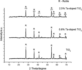
XRD analysis of nondoped, 0.6 at.%, and 2.5 at.% Ta-doped specimens, demonstrated in Fig. 1, reveals that all products are the rutile phase of TiO2 (JCPDS no. 01-073-1765). Modification of pH, temperature, and other hydrothermal conditions enables control of the phase and morphology of TiO2. Previously, it is shown that adjustment of pH will control the phase development of TiO2.Reference Diebold3, Reference Chen and Mao21, Reference Kinsinger, Wong, Li, Villalobos and Kisailus22 Our synthesis is performed at low pH (i.e., pH less than 0), and hence we expect the growth of rutile phase of TiO2 as previously observed.Reference Chen and Mao21, Reference Jiang, Yin, Jiang, Jiang, Feng, Chen, Zhou and Wada23 The fundamental crystal structure of anatase, brookite, and rutile consists of TiO6 octahedra with different spatial arrangements.Reference Cheng, Ma, Zhao and Qi2, Reference Fujishima and Honda5 Rutile is composed of primarily corner-sharing TiO6 octahedra (80%) and anatase is composed of 50% edge-and corner-sharing TiO6 octahedra.Reference Cheng, Ma, Zhao and Qi2, Reference Bokhimi, Morales, Aguilar, Toledo-Antonio and Pedraza24–Reference Burdett, Hughbanks, Miller, Richardson and Smith26 In order for an edge-sharing bond to form between two octahedra, two juxtaposed hydrolysis and condensation reactions must occur. Corner-sharing octahedra require just one hydrolysis and condensation reaction in the coordination sphere. The high HCl concentration in the reaction solution reduces OH− ions available to complex with the Ti center, thereby increasing Cl− ions in the feedstock solution. Increasing the number of Cl ligands suppresses the formation of edge-sharing bonds as a result of reduced OH ligands available for condensation while allowing corner-sharing bonds to be formed favoring the rutile phase.Reference Cheng, Ma, Zhao and Qi2
X-ray diffraction patterns of different TiO2 products: nondoped TiO2, 0.6 at.% and 2.5 at.% Ta-doped TiO2.

XRD confirms that no additional phases were detected, which suggests that Ta did not phase separate as a crystalline product and is likely incorporated within the TiO2 lattice. The initial mixing occurs in an ice bath to reduce the rate of hydrolysis of the Ti and Ta precursor to ensure homogeneous mixing and concurrent nucleation to incorporate Ta into the TiO2 network. The biphasic mixture of toluene and water was utilized to reduce hydrolysis during initial mixing. Once hydrolyzed, these polar species grow in the aqueous phase; although under hydrothermal conditions, it is likely that there was some miscibility of toluene in this phase.Reference Yang, Miller and Hawthorne27 Both Ti and Ta precursors are able to increase their coordination numbers from 4 to 6 and 5 to 8, respectively, which increases their chemical reactivity toward hydrolysis.Reference Cheng, Ma, Zhao and Qi2, Reference Livage and Sanchez28, 29 Ta also has a slightly higher electronegativity and lower partial charge than Ti, but the Ti precursor will hydrolyze marginally faster.Reference Zhou, Liu, Zhu and Zhang9, Reference Couselo, Einschlag, Candal and Jobbagy11, Reference Asahi, Morikawa, Ohwaki, Aoki and Taga12 Even a slight difference in the hydrolysis rates between the Ti and Ta precursors, if uncontrolled (i.e., through chelating ligands, reaction acidity, low temperature), may lead to phase separation due to incomplete mixing and premature formation of Ti–O–Ti networks and will inhibit uniform Ta doping within the TiO2 networks.Reference Cheng, Ma, Zhao and Qi2, Reference Zhou, Liu, Zhu and Zhang9, Reference Liu, Gao, Zhao and Tian10
The crystallite sizes of the samples were obtained from the XRD results using the Scherer formula:

where κ is the shape factor, λ is the wavelength of the radiation, β is full width at half maximum of the peak, and θ is the diffraction angle. The average crystallite size of the nondoped TiO2 sample is 36 nm, which subsequently decreases with increasing concentrations of the Ta dopant. The crystallite size of the 2.5 at.% Ta-doped TiO2 sample is reduced by half (∼14 nm) versus the nondoped TiO2. This is likely due to a slightly higher rate of nucleation due to the high concentration of precursor (Ta + Ti).Reference Cheng, Ma, Zhao and Qi2, Reference Zhang, Zhao, Liu, Dong, Zhang and Chen13 The decreased crystallite size indicates increased nuclei formation, which reduces nutrient available for subsequent growth of the crystals.
B. Chemical analysis
EDS was performed for each sample in four different regions to confirm the uniformity of dopant distribution. On the basis of our analyses, we found that the lightly doped sample contained 0.6 at.% (± 0.15 at.%) Ta, whereas the heavily doped sample contained 2.5 at.% (±0.5 at.%) Ta. The final concentration was averaged over five regions.
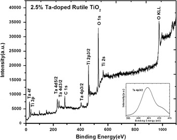
XPS (Fig. 2) was used to study the elemental surface composition of the heavily doped TiO2. C is the only impurity observed in the sample and it is used as a reference line. The spectrum revealed two peaks at 458 and 464 eV, which correspond to Ti 2p3/2 and Ti 2p1/2, respectively, bound to oxygen. The peak at 530 eV corresponds to oxygen 1s. Thus, the peaks at 458 and 464 eV correspond to TiO2.Reference Netterfield, Martin, Pacey, Sainty, Mckenzie and Auchterlonie30, Reference Fierro, Arrua, Nieto and Kremenic31 Evaluation of peaks at 25, 230, and 242 eV, revealed they were representative of Ta 4f5/2, Ta 4d5/2, and Ta 4d3/2, respectively. All peaks are assigned to Ta5+, indicating that Ta was oxidized within the lattice.Reference Siemens Meyer and Schultze32, Reference Ho, Contarini and Rabalais33
X-ray photoelectron spectrum of 2.5% Ta-doped TiO2.

C. Structural analysis
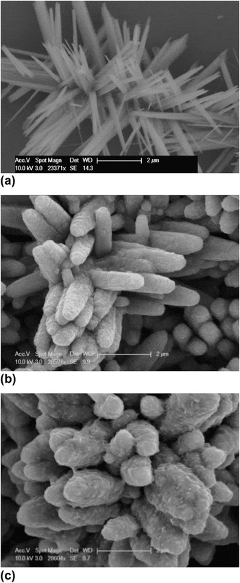
SEM images of the samples (Fig. 3) indicate the TiO2 samples with different concentrations of Ta show distinct morphologies. All samples demonstrate a spherulitic morphology, with a different frequency of secondary branches. The mechanism of the growth of such structures is elaborated in more detail elsewhereReference Granasy, Pusztai, Tegze, Warren and Douglas34, Reference Keith and Padden35, Reference Shen, Bao, Zheng, Gupta, An and Yanagisawa36 and is therefore not addressed in this article. We found that the addition of thiourea to our reaction solution had negligible effects on both microstructural and optoelectronic properties of resulting materials. However, we do observe morphological changes with increasing concentrations of Ta dopant. Both the surface roughness of the branches and their aspect ratio (i.e., L/d) change as the Ta concentration increases. For example, the aspect ratio (L/d) of the branches changes from 15 to 2.7 to 1.5 for nondoped, 0.6 at.% doped, and 2.5 at.% doped, respectively. This is likely due to the differences in the rates of hydrolysis and condensation between the Ti and Ta alkoxides. Although both metals have similar electronegativities, Ta, a large early transition metal, can coordinatively expand, accommodating up to eight ligands (versus six for Ti).Reference Cheng, Ma, Zhao and Qi2, Reference Livage and Sanchez28, 29 This expansion can enable the formation of less soluble speciesReference Sanchez, Livage, Henry and Babonneau37 and subsequently change the rate of nucleation of metal oxide, both of which would affect the crystallinity and mechanism of growth of the nanowires.
Scanning electron microscopy images of TiO2 synthesized at 180 °C (a) Nondoped, (b) 0.6 at.% Ta-doped TiO2, and (c) 2.5 at.% Ta-doped TiO2.

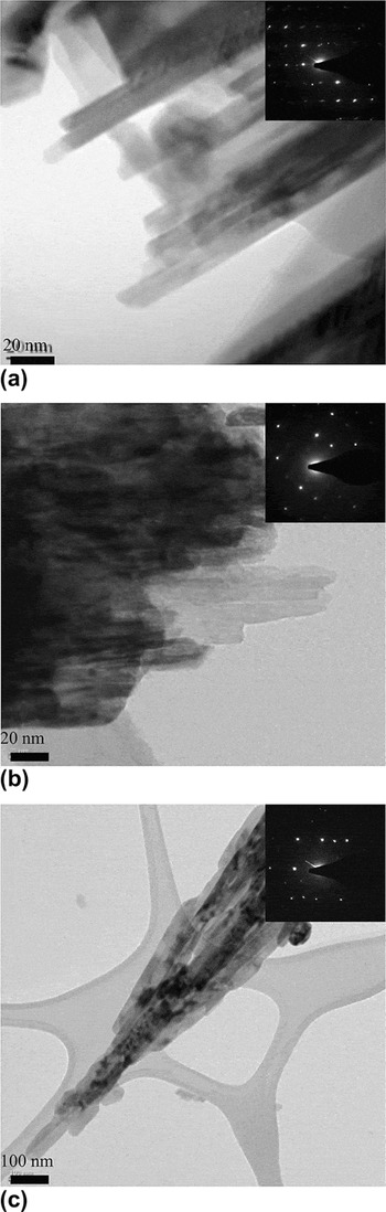
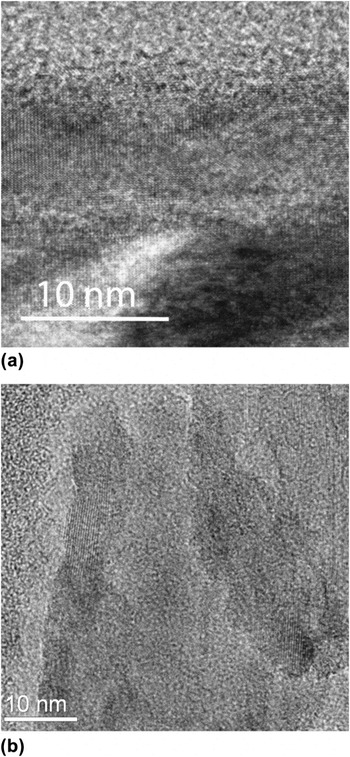
TEM bright-field images and selected area diffraction patterns (SADP) of nondoped, 0.6 at.%, and 2.5 at.% Ta-doped TiO2 nanostructures are shown in Fig. 4. The images reveal that the rods are constructed of smaller core nanowires.Reference Fierro, Arrua, Nieto and Kremenic31 These nanowires are oriented in the [001] direction and have likely formed via an oriented attachment mechanism.Reference Wang and Ying19, Reference Fierro, Arrua, Nieto and Kremenic31 On the basis of the observations from the bright-field images in Fig. 4, the aspect ratio of the rods (L/d) seems to decrease with increasing concentrations of Ta dopant and agrees with crystal sizes measured by XRD. The introduction of Ta likely lowers the solubility of the mixed metal oxide, which leads to an increase in the nucleation rate, and thus, a rougher surface.Reference Ma, Goh, Ma and White38 SADP confirmed the near single-crystalline nature and phase (i.e., rutile) of the rods. No additional structures/phases were detected from the bright-field and SADP, thus confirming phase uniformity. The high-resolution TEM images of nondoped and 2.5 at.% Ta-doped TiO2 are demonstrated in Fig. 5. The crystalline structure of both species is shown with no amorphous phases detected.
Bright-field transmission electron microscopy (TEM) image of TiO2 synthesized at 180 °C (a) nondoped TiO2 (b) 0.6 at.% Ta-doped, and (c) 2.5 at.% Ta-doped. Selected area diffraction patterns inserted for phase identification.

High-resolution TEM images (a) nondoped TiO2 and (b) 2.5at.% Ta-doped TiO2.

IV. PHOTOELECTRICAL CHARACTERIZATIONS
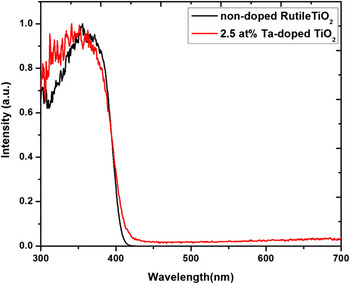
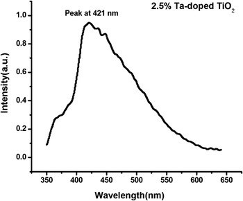
UV–vis spectroscopy (Fig. 6) results indicate a red shift for the Ta-doped TiO2. On the basis of this result, the estimated band gap for the nondoped rutile samples is ∼3.01 eV (413 nm) and 2.93 eV (424 nm) for the 2.5% Ta-doped rutile TiO2. PL measurements of the 2.5% Ta-doped TiO2 (illustrated in Fig. 7) indicate the sample has a band gap of 2.93 eV (423 nm), in excellent agreement with the UV–vis spectroscopy results. It should be mentioned that no observable red shift is recognized for 0.6% Ta-doped TiO2 and may be due to the limited resolution of the spectrometer. Band gap reduction due to Ta doping has been claimed in both experimentReference Wang, Geng, Guo, Jiang and Qu16 and theoryReference Long and English20 for TiO2 in anatase phase. Our results suggest the same trend for TiO2 in rutile phase. On the basis of our theoretical studies and other previous studies, the light absorption properties of TiO2 are attributed to 3d orbital of Ti (conduction band) and 2p orbital of O (valance band). Here, the conduction band is likely a combination of the 3d orbital of Ti and the 5d orbital of Ta, which are closer to the valance band. Thus, combinations of 5d and 3d orbitals of Ti and Ta, respectively, are mainly responsible for the reduction in the band gap.
Ultraviolet–visible spectroscopy results for nondoped and 2.5% Ta-doped rutile TiO2.

Photoluminescence of 2.5% Ta-doped rutile TiO2.

V. CONCLUSION
We have synthesized Ta-doped TiO2 with a highly branched morphology through a low-cost, hydrothermal method. Structural characterization of as-synthesized and Ta-doped TiO2 revealed that these nanostructures are rutile. Two different Ta-dopant levels were achieved, 0.6 at.% and 2.5 at.%. The dopants were incorporated in a uniform manner due to the low temperature of mixing of alkoxide precursors, which reduced the differences in the rates of hydrolysis and condensation of the Ti and Ta alkoxides. Increasing the concentration of Ta dopant had a significant effect on the crystallite sizes in the nanostructures, decreasing with increasing Ta dopant. UV–vis and PL spectroscopy confirmed the band gap reduction, which agrees with previous simulation results. The decrease in band gap is due to the formation of an energy level from a Ta 5d orbital, which reduces the conduction band. This reduction is likely a combination of the 3d orbital of Ti and the 5d orbital of Ta.