INTRODUCTION
During concurrent pregnancy and nematode infection, physiological demands for energy and protein required for foetal growth and maternal immunity may be met either by increased food intake (Bronson and Heideman, Reference Bronson, Heideman, Knobil and Neill1994), or by mobilizing stored triglycerides and adipose tissue (Spiegelman and Flier, Reference Spiegelman and Flier2001) or lean body mass (Wong et al. Reference Wong, Hildebrandt, Thrasher, Appleton, Ahima and Wu2007). If either food intake or energy reserves are insufficient, animals re-allocate resources to maintenance functions (Coop and Kyriazakis, Reference Coop and Kyriazakis1999), at the expense of functional immunity, current reproduction and lifetime reproductive success (Forbes, Reference Forbes1993). For example, pregnancy induces immunosuppression in livestock and rodents infected with nematodes (Houdijk et al. Reference Houdijk, Jessop, Knox and Kyriazakis2003; Ng'ang'a et al. Reference Ng'ang'a, Kanyari, Maingi and Munyua2006; Normanton et al. Reference Normanton, Jos, Houdijk, Jessop, Knox and Kyriazakis2007), and nematode infections reduce reproductive outcomes including litter size and pup mass (Kristan, Reference Kristan2002). Energy demands can also be accommodated through phenotypic plasticity in size and function of internal organ systems (Piersma and Lindstrom, Reference Piersma and Lindstrom1997), perhaps including bone.
Bone formation and resorption are regulated by hormones, soluble local growth factors as well as cytokines including interferon (IFN)-γ (Baron et al. Reference Baron, Vignery, Horowitz and Peck1983; Mann et al. Reference Mann, Jacobs, Buchinsky, Armstrong, Li, Ke, Ma, Jee and Epstein1994). Shifts in IFN-γ concentrations disrupt the tight cytokine balance involved in bone remodelling and lead to net bone loss as is observed during generalized inflammation (Lerner, Reference Lerner2006). IFN-γ is also an important regulatory cytokine in chronic nematode inflammation (Else et al. Reference Else, Finkelman, Maliszewski and Grencis1994; Tu et al. Reference Tu, Koski and Scott2008). Thus, up-regulation of the maternal immune system during nematode infection may induce bone remodelling in the mother as an adaptive response (Lochmiller and Deerenberg, Reference Lochmiller and Deerenberg2000) and reduce the maternal investment in foetal skeletal growth.
This study investigated energetic, immunological, physiological and skeletal responses to infection and pregnancy in the maternal and foetal compartments. We hypothesized that a stronger immune response during higher dose infection would increase resting metabolic rate (RMR). We further hypothesized that the shift in cytokine profiles in infected pregnant mice would promote nematode survival, induce maternal bone remodelling, and impair skeletal development in the foetus.
MATERIALS AND METHODS
Experimental design, mice and parasite
A 2×3 factorial design was employed that combined two conditions (pregnant and non-pregnant mice) with 3 doses of trickle infection (Sham: 3×0 L3; Low: 3×50±3 L3; High: 3×100±3 L3) with the murine gastrointestinal nematode (Heligmosomoides bakeri, previously named Heligmosomoides polygyrus; see Cable et al. (Reference Cable, Harris, Lewis and Behnke2006). The trickle infection protocol and the selected doses are typical of real world infection dynamics (Monroy and Enriquez, Reference Monroy and Enriquez1992) and induce a vigorous and persistent immune response (Behnke and Robinson, Reference Behnke and Robinson1985; Scott, Reference Scott1991). A total of 70 female, 8-week-old outbred CD1 mice (Charles River Breeding Laboratories, Quebec, Canada) were housed individually in Nalgene cages (Fisher Scientific, Montreal, Canada) with stainless steel covers in a temperature controlled mouse room (22–25°C), with a 14:10 h light:dark cycle. Normal pelleted rodent diet (8640 Teklad 22/5 mouse chow; 22·6% protein, 5·2% fat, 3·9% fibre, 3·1 kcal/g metabolizable energy) (Harlan Teklad, Madison, WI, USA) and water were provided to the mice ad libitum.
As it was impossible to process all mice at the same time, the experiment was staggered into 5 sets initiated over a period of 15 weeks. Previously uninfected, non-pregnant mice and primiparous pregnant mice (day 4 of pregnancy, defined as experimental day 0) were received from Charles River (Quebec, Canada). Mice were randomized into 3 infection doses, and infected on experimental days 2, 7 and 12 (days 6, 11 and 16 of pregnancy) to allow 3 infections prior to parturition. L3 were obtained by faecal culture of stock parasites (originally provided by Dr M. V. K. Sukhdeo, Rutgers University, New Brunswick, NJ, USA) maintained in outbred CD1 mice (Charles River) in our laboratory for over 15 years. Larvae were counted in 10 sham doses for accuracy before infecting the mice. The same larval culture was used for all infections within each set of mice.
Food intake and body temperature and weight were recorded between 09.00 and 10.00 h every second day, beginning on experimental day 1 (day 5 of pregnancy). At selected times during the experiment, resting metabolic rate (RMR) and maternal body composition were measured. At necropsy (experimental day 15, day 19 of pregnancy), mice were anaesthetized using Ketamine/Xylazine (100/10 mg/kg body weight) intraperitoneally (IP), then exsanguinated by cardiac puncture. Maternal serum samples were collected and frozen at −20°C until later analysis for cytokines and chemokines. Immediately after cardiac puncture, a cesarean section (C-section) was performed and intact uteri were removed. Amniotic fluid (AF), collected from each individual foetal sac using a 3 ml syringe fitted with a 22-gauge needle, was pooled for each dam, then immediately snap-frozen in liquid nitrogen. The pooled AF was stored at −80°C for later analysis of cytokines and chemokines. Foetuses were removed and then killed by exsanguination. Reproductive outcomes (number of foetuses, foetal mass and crown-rump length) were determined immediately thereafter. Foetal crown-rump length was measured from the top of the head to the base of the tail. Maternal organ masses for heart, spleen, liver, lungs, kidneys, pancreas, small intestine and small intestine length were also determined. All procedures were approved by the McGill Animal Care Committee according to guidelines of the Canadian Council on Animal Care (CCAC, 1993).
Energy expenditure
The RMR of each mouse was measured on 4 separate days chosen to correspond with key phases of infection. Baseline RMR was obtained on the day prior to first infection (experimental day 1). The RMR was also measured on experimental day 4 when the first dose of L4 would be within the serosal mucosa, on experimental day 9 when pre-adults would first be emerging into the intestinal lumen, and on experimental day 13 when mature adults would be present in the intestinal lumen. The RMR was assessed at room temperature (24±1°C) during the light phase (between 08.00 h and 17.00 h) when mice were in their inactive phase by an open-flow respirometry system (Multi-Channel Respirometry Gas Exchange System) connected to a paramagnetic oxygen analyser (Qubit Systems Research, Kingston, Ontario). Immediately prior to placing them in the respirometry chamber, non-fasted mice were weighed and their body temperature measured (to within 0·1°C) using a thermometer (Microtherma 2T, model TW2-193; Braintree Scientific, Braintree, MA), by inserting a pre-lubricated rectal probe for mice (RET-3, shaft ¾” long, 0·028” dia. Tip dia. 0·065”; Physitemp Instruments, Clifton, NJ) 2 cm into the rectum. The RMR was measured in groups of 4 mice at a time. Each mouse was placed in 1 of 4 cylindrical Perspex respirometry chambers with rubber stoppers (volume=625 ml). The flow of tank air (dried with self indicating, 4–8 mesh soda lime; Spectrum Chemical Mfg, Gardena, CA) was maintained by a pump and measured by a gas flow meter upstream of the chamber. Flow rate was 650–700 ml min−1. Gases leaving the mouse chambers were dried (magnesium perchlorate; Fisher Scientific, Montreal, Canada) and passed through the O2 and CO2 analysers (Qubit Systems Research) at approximately 150 ml min−1. We did not absorb CO2 before determination of O2 content of the excurrent gases, in order to maximize accuracy in the derived estimate of energy expenditure (Koteja, Reference Koteja1996). O2 consumption and CO2 production of each of the 4 mice were recorded sequentially for 10 min with a flush out with reference air for a period of 2·5 min between mice. This cycle was repeated 3 times over a period of 2·5 h for each group of 4 mice.
Data were automatically recorded by the customized software (Qubit Systems Research) as average O2 consumption and CO2 production for every 10 sec interval. For each mouse, values of O2 consumption (VO2, ml/min) and CO2 production (VCO2; ml/min) were calculated from the mean of the lowest stable 30 sec interval from each of the three 10 min runs, and these were used to determine the respiratory quotient (RQ). The RMR (Watts=Joules/sec) for each animal was then calculated from the derived RQ using the Weir equation (Weir, Reference Weir1949) and converted into KJ/day (Bartholomew, Reference Bartholomew and Gordon1977). We have used the term RMR, although we note that room temperature (22–25°C) was below the thermoneutral temperature for mice (30±1°C) (Hart, Reference Hart and Whittow1971).
Parasitological indicators
At necropsy the small intestine was excised, and the length and mass determined. The duodenum (10 cm immediately distal to the pyloric sphincter) was divided into 3 equal length portions (proximal, mid and distal) and the remaining small intestine was divided equally into the jejunum and ileum in order to assess worm distribution. The sex and number of adult H. bakeri in each section were determined by examining intestines that were slit open onto a glass plate and scanned using a dissection microscope. The number of 4th-stage larvae (L4) of H. bakeri remaining in the serosal musculature was also recorded. The percentage of worms and the percentage of L4 recovered were computed based on the cumulative infection dose (150 or 300).
Body composition and bone measurements
We used the dual-energy X-ray absorptiometry (DXA) technique (Model # QDR 4500A, version 12.5; Hologic Inc., Waltham, MA, USA) to measure maternal body composition (Nagy and Clair, Reference Nagy and Clair2000) and bone mineralization (Amman et al. Reference Amman, Rizzoli, Slosman and Bonjour1992). On each day, 2 manufacturer-specific aluminum/lucite bone phantoms were scanned for quality control prior to scanning animals. On experimental day 14 (day 18 of pregnancy), adult mice were weighed, immobilized by anaesthesia (Ketamine/Xylazine, 100/10 mg/kg body weight) IP, followed by whole body and high resolution regional scans. Mice were placed in the anterior-posterior position with limbs extended, spine straight, and tail placed around and not under the animal to permit regional analysis. Whole body composition was measured in triplicate without repositioning using the small animal software. Scans were conducted for approximately 8 min per mouse by one investigator (M.R.O.). The scan analysis recorded grams of body mass, lean and fat mass, percentage fat, bone mineral content (BMC), bone mineral density (BMD), and bone area (BA). The average intra-individual coefficients of variation for each mouse using the triplicate scans were 2·7% for whole body BMC, 2·2% for BMD, 1·4% for lean mass, and 9·2% for fat. The data were examined for the whole body and for specific skeletal regions (lumbar vertebrae 1–5, femur and tibia using high-resolution scanning) and values for triplicate scans were averaged.
Cytokine and chemokine measurements
Multiplex suspension bead array immunoassay was performed on serum and AF using a Luminex 200™ xMAP analyzer (Luminex Corp., Austin, TX, USA) to quantify cytokines and chemokines known to play an important role in pregnancy (Orsi et al. Reference Orsi, Gopichandran, Ekbote and Walker2006) and immunity to H. bakeri (Urban et al. Reference Urban, Noben-Trauth, Donaldson, Madden, Morris, Collins and Finkelman1998; Gause et al. Reference Gause, Urban and Stadecker2003). The Mouse Cytokine 10-Plex kit (LINCO Research, St Charles, MO, USA, # MCYTO-70K) was used to measure IL-1β, IL-4, IL-5, IL-6, IL-10, IL-12p70, IL-13, IFN-γ, tumor necrosis factor (TNF)-α, and monocyte chemotactic protein (MCP)-1, according to the manufacturer's specifications. Assays were performed in duplicate and assay sensitivity ranged between 0·3 and 10·3 pg/ml, depending on the analyte (manufacturer's details). Serial dilutions prepared from a lyophilized standard were transferred to appropriate microtitre wells (Microtiter 96-well filter plate, Millipore, Bedford, MA, USA, # MSBVN12), and 25 μl of assay buffer and 25 μl of serum matrix or serum or AF samples were added to the appropriate microtitre wells. After sonication (Branson Sonicator, Branson Co., Shelton, CT, USA, model # 3200), 25 μl of diluted antibody-coated bead complexes mixed with assay buffer were then added to each well. Samples were sealed, covered with aluminum foil and incubated with agitation on a plate shaker (Sanofi Diagnostics Pasteur, Chaska, MN, USA, model # 2202) overnight (16 h) at 4°C. Fluid was gently removed by vacuum filtration. After washing twice with wash buffer (200 μl/well), 25 μl of cocktail biotinylated detection antibody was added and sealed, and the covered plates were incubated for 1 h at room temperature with vigorous agitation, but without vacuuming after incubation. Thereafter, 25 μl of Streptavidin-Phycoerythrin was added and plates were incubated with agitation for 30 min at room temperature. All microtitre wells received a final wash (3 times with 200 μl/well), removing the wash buffer by vacuum filtration between each wash. Sheath fluid (100 μl) was added to all wells, and the beads were resuspended by shaking the sealed, covered plates on a plate shaker for 5 min. Samples were then analysed on the Luminex using MasterPlex® CT software version 1.2 (MiraiBio, Inc., Alameda, CA, USA). A minimum of 50 events (beads) was collected for each of the 10 cytokines and median fluorescence intensities (MFI) were obtained. Cytokine concentrations were automatically calculated based on standard curve data using MasterPlex® QT analysis software version 3.0 (MiraiBio, Inc., Alameda, CA, USA), and were expressed as pg/ml±s.e.m.
Statistical analysis
All analyses were performed using Proc Mixed model in SAS statistical software (v. 9.1; SAS Institute Inc., Cary, NC, USA) and P values <0·05 were considered statistically significant. Unless otherwise indicated, values are presented as least square means±s.e.m. All parameters were tested for normality prior to analysis. Post-hoc Bonferroni adjustment (where appropriate) was used to account for multiple comparisons.
For variables measured once, we used either a 2×2 or 2×3 design to study the main effects of pregnancy (non-pregnant vs pregnant) and either infection (uninfected vs infected) or dose of infection (sham, low and high), depending on the variable. When there were no significant interactions among the main effects, the data were pooled according to main effects, and re-analysed.
RMR, body temperature and food intake were analysed using repeated-measures analysis of variance (ANOVA) with the main effects of pregnancy, dose of infection, and time (experimental days 4, 9 and 13 for RMR, 7, 9, 11, 13, 15 and 17 for body temperature, and 3, 5, 7, 9, 11 and 13 for food intake) modelled as fixed effects and individual mice (dams) as random effects, after correcting for baseline values using a spatial power covariance structure for RMR or auto-regressive covariance structure for other variables. For foetal crown-rump lengths and foetal weight, infection status was modelled as a fixed effect and individual dams as random effects nested within infection status. Linear regression was used to determine whether the RMR was predicted by whole body mass, lean mass, food intake or litter size.
The cytokine data sets were not normally distributed. Therefore serum cytokines were compared between pregnant and non-pregnant mice using a one-way non-parametric Kruskal-Wallis ANOVA. The subset of data used to examine infection effects within pregnancy states was normally distributed; hence the effect of infection dose on serum and AF cytokine concentration was examined using one-way ANOVA.
RESULTS
Parasite outcomes
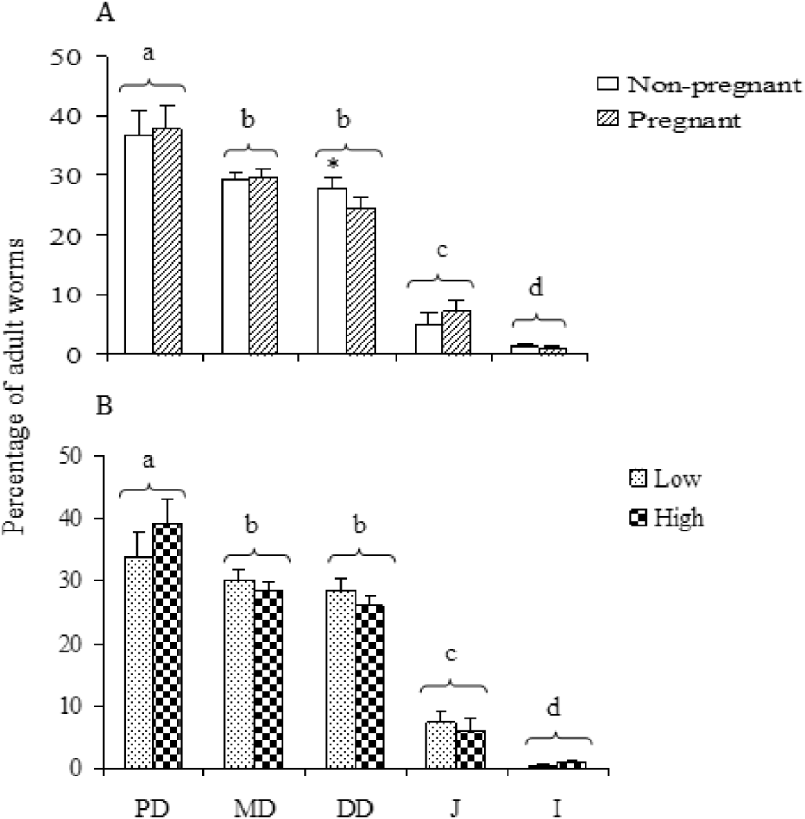
All mice infected with H. bakeri developed mature infections, and no sham mice became infected. Significant main effects of pregnancy and dose were found for percentage worm recovery and no significant interaction was detected. The percentage of worms recovered was higher in pregnant mice and in high dose infected mice compared to non-pregnant mice and low dose infected mice respectively (Fig. 1A). The percentage of L4 was also higher in pregnant compared to non-pregnant mice (Fig. 1B), but no main effect of dose or interaction between dose and pregnancy state were detected. In all infected mice, the highest percentage of worms was in the proximal duodenum (Fig. 2A, B) and few were found in the jejunum or the ileum. Within the duodenum, worms in pregnant mice were more anteriorad, as shown by the lower percentage of worms in the distal duodenum, compared with non-pregnant mice (Fig. 2A).
Effect of infection dose (low, high) and pregnancy on recovery of Heligmosomoides bakeri. (A) Combined adult worms and L4 recovery as a percentage of dose (main effect of dose: F 1,43·7=7·34, P=0·0096; main effect of pregnancy state: F 1,43·5=77·7, P<0·0001); interaction of pregnancy state * dose: F 1,43·1=0·56, P=0·4597). (B) L4 recovery as a percentage of dose (main effect of dose: F 1,43·6=0·34, P=0·5646; main effect of pregnancy state: F 1,43·4=98·48, P<0·0001); interaction of pregnancy state * dose: F 1,42·8=4·07, P=0·0598). Different lowercase letters represent significant differences between pregnant and non-pregnant mice, or among infection doses (P<0·05).

Percentage distribution of Heligmosomoides bakeri along the small intestine in non-pregnant vs pregnant mice (A) and between low and high dose infections (B). PD, MD, DD, J and I refer to proximal duodenum, mid duodenum, distal duodenum, jejunum and ileum respectively. Different lowercase letters represent significant differences among regions (pooled data for pregnant and non-pregnant mice); * indicates higher recovery in non-pregnant compared with pregnant mice within a region (P<0·05).

Food intake
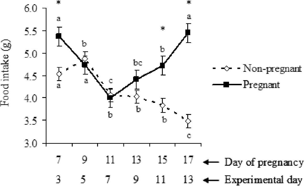
Parasite infection had no effect on food intake regardless of the dose, but the pattern of food intake over time differed between non-pregnant and pregnant mice (Fig. 3). Whereas average daily food intake gradually decreased in non-pregnant mice, food intake initially declined but then increased steadily during the final week of pregnancy (Fig. 3).
Effect of pregnancy on average food intake per day over time (data pooled across infection groups). Main effect of pregnancy state: F 1,66=14·38, P=0·0003; main effect of dose: F 2,64·2=0·87, P=0·4237; main effect of time: F 5,265=9·19, P<0·0001; interaction of pregnancy state * time: F 5,265=10·12, P<0·0001. Differences between pregnancy states at specific time-points are represented by *; different lowercase letters represent significant differences across time within each group (P<0·05).

Visceral organ responses
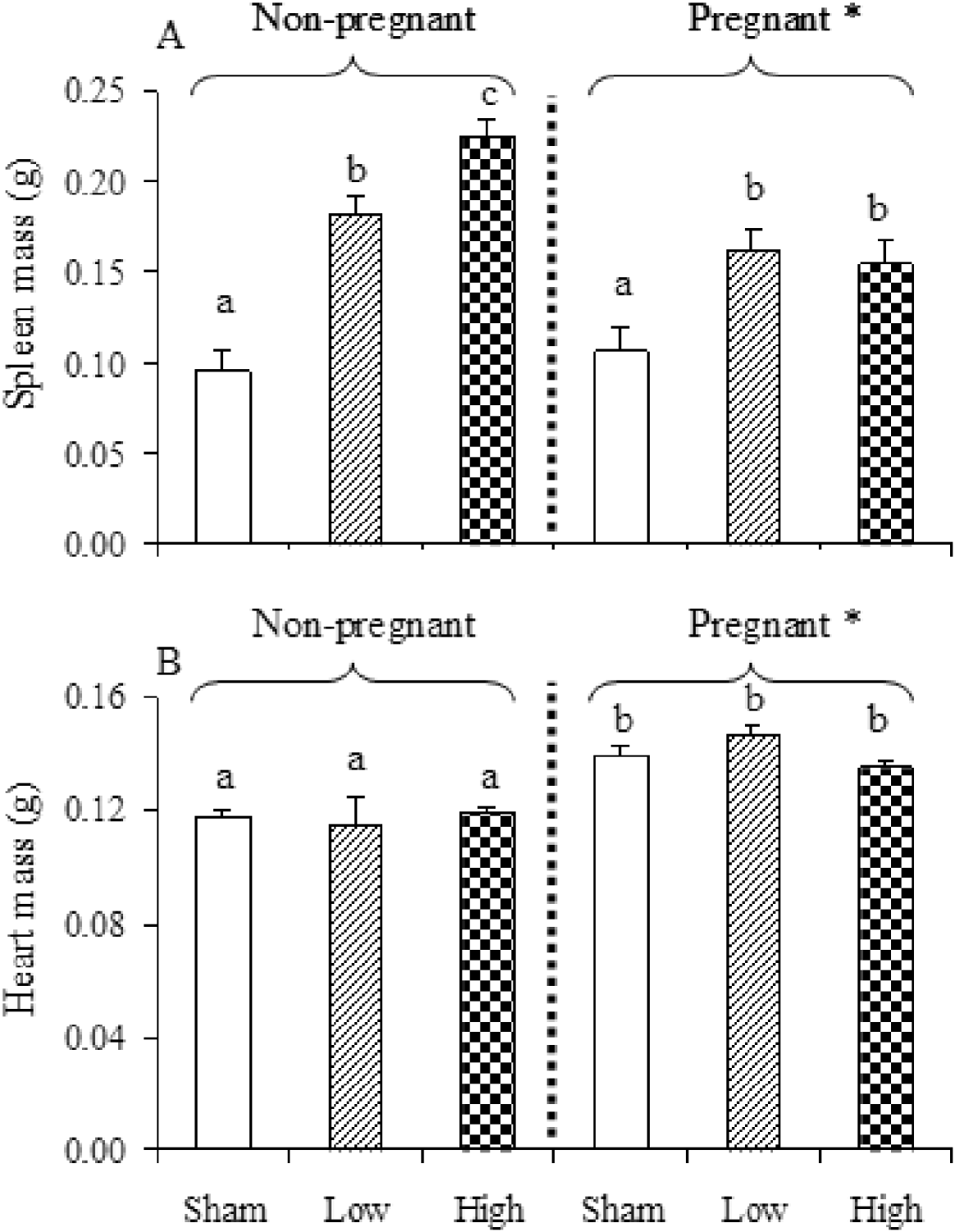
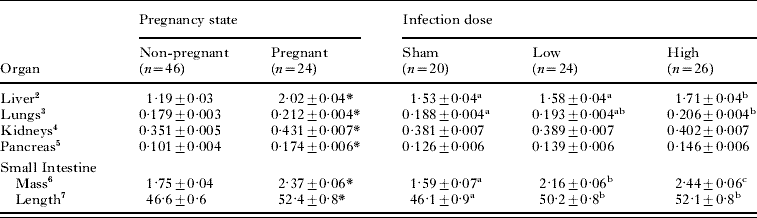
Both pregnancy and infection increased the length of the small intestine and the mass of most visceral organs (Table 1), with no statistical interaction. Whereas the effect of infection was only evident at the high dose for liver and lung mass, small intestine mass increased in a dose-dependent manner. In the case of spleen (Fig. 4A) and heart mass (Fig. 4B), there was a significant interaction between pregnancy and infection dose. Spleen mass increased in a dose-dependent manner in non-pregnant mice, and was elevated in infected pregnant mice, but not in a dose-dependent manner (Fig. 4A). Post-hoc multiple comparisons of heart mass showed that all pregnancy groups had higher heart mass than all non-pregnant mice (Fig. 4B).
Interaction effects of pregnancy and Heligmosomoides bakeri dose on spleen mass (A) (main effect of pregnancy state: F 1,60·6=8·09, P=0·006; main effect of dose: F 2,60·8=31·7, P<0·0001; interaction of pregnancy state * dose: F 2,61·8=5·99, P=0·0042); and heart mass (B) (main effect of pregnancy state: F 1,64=95·43, P<0·0001; main effect of dose: F 2,64=1·07, P=0·3495; interaction of pregnancy state * dose: F 2,64=3·84, P=0·0265). Different lowercase letters represent significant differences across all groups, based on multiple comparisons; * represents a significant difference between pregnant and non-pregnant mice.

Main effects of pregnancy (pooled across dose) and Heligmosomoides bakeri infection dose (pooled across pregnancy state) on organ masses (g) and small intestine length (cm)Footnote 1

1 A significant effect of pregnancy is represented by
*, whereas different superscripts represent differences among infection doses.
2 Pregnancy state: F 1,62·6=420·50, P<0·0001; Infection dose: F 2,62·7=7·81, P=0·0009; Pregnancy state×dose: NS.
3 Pregnancy state: F 1,63·3=47·30, P<0·0001; Infection dose: F 2,63·4=6·03, P=0·004; Pregnancy×dose: NS.
4 Pregnancy state: F 1,62·9=104·29, P<0·0001; Infection dose: F 2,63=2·80, P=0·0683; Pregnancy×dose: NS.
5 Pregnancy state: F 1,66=106·06, P<0·0001; Infection dose: F 2,66=3·08, P=0·0529; Pregnancy×dose: NS.
6 Pregnancy state: F 1,66=68·72, P<0·0001; Infection dose: F 2,66=45·72, P<0·0001; Pregnancy×dose: NS.
7 Pregnancy state: F 1,63·3=34·88, P<0·0001; Infection dose: F 2,63·4=13·37, P<0·0001; Pregnancy×dose: NS.
Body composition and bone measurements
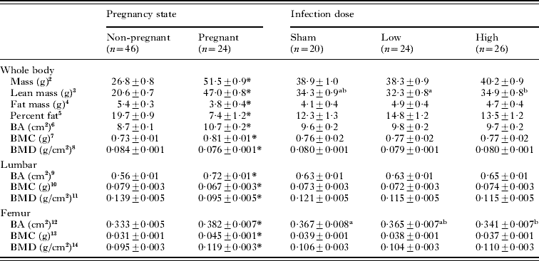
Pregnancy had a stronger impact on many dimensions of maternal body composition than infection (Table 2). On experimental day 14, pregnant mice had higher whole body mass and lean mass, whole body bone area (BA), bone mineral content (BMC), but lower fat mass, percentage fat and bone mineral density (BMD), compared with non-pregnant mice. Infection dose had no effect on whole body fat mass or percentage fat, BMC or BMD (Table 2) and no interaction was detected. However, lean mass was borderline greater (F 2,62·5=3·01, P=0·0566) in high dose than low dose infections.
Main effects of pregnancy (pooled across dose) and infection dose (pooled across pregnancy state) on maternal whole body mass measured on experimental day 14 (day 18 of pregnancy), and on lean and fat mass, and femur and lumbar bone area (BA), bone mineral content (BMC) and bone mineral density (BMD), as measured using a QDR 4500 Hologic densitometerFootnote 1

1 A significant effect of pregnancy is represented by *, whereas different lowercase letters represent significant differences among infection doses.
2 Pregnancy state: F 1,62·5=803·32, P<0·0001; Infection dose: NS; Pregnancy×dose: NS.
3 Pregnancy state: F 1,62·5=1089·69, P<0·0001; Infection dose: F 2,62·5=3·01, P=0·0566; Pregnancy×dose: NS.
4 Pregnancy state: F 1,63·2=10·58, P=0·0018; Infection dose: NS; Pregnancy×dose: NS.
5 Pregnancy state: F 1,63·2=70·17, P<0·0001; Infection dose: NS; Pregnancy×dose: NS.
6 Pregnancy state: F 1,62·8=125·52, P<0·0001; Infection dose: NS; Pregnancy×dose: NS.
7 Pregnancy state: F 1,62·7=22·06, P<0·0001; Infection dose: NS; Pregnancy×dose: NS.
8 Pregnancy state: F 1,62·3=83·85, P<0·0001; Infection dose: NS; Pregnancy×dose: NS.
9 Pregnancy state: F 1,66=95·22, P<0·0001; Infection dose: NS; Pregnancy×dose: NS.
10 Pregnancy state: F 1,62·2=24·18, P<0·0001; Infection dose: NS; Pregnancy×dose: NS.
11 Pregnancy state: F 1,62·1=236·72, P<0·0001; Infection dose: NS; Pregnancy×dose: NS.
12 Pregnancy state: F 1,66=30·20, P<0·0001; Infection dose: F 2,66=4·13, P=0·0204; Pregnancy×dose: NS.
13 Pregnancy state: F 1,62·6=127·87, P<0·0001; Infection dose: NS; Pregnancy×dose: NS.
14 Pregnancy state: F 1,62·6=57·13, P<0·0001; Infection dose: NS; Pregnancy×dose: NS.
As a further assessment of bone composition, regional analyses were performed for BA, BMC and BMD using lumbar, femur and tibia regions (Table 2). In no case was a significant interaction detected. Pregnant mice had higher femur BA, BMC and BMC but lower lumbar BMC and BMD, compared with non-pregnant mice. In contrast, femur BA was reduced in high dose compared with uninfected mice, but neither femur BMC or BMD were affected by infection dose. Post-hoc multiple comparisons demonstrated that pregnant mice had greater lumbar BA in all infection groups. Neither infection dose nor pregnancy had any effect on tibia BA, BMC and BMD (data not shown).
Reproductive outcomes
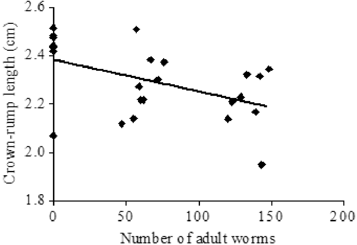
High dose infection decreased crown-rump length compared to sham infection (F 2,17=10·03, P=0·001). Furthermore, crown-rump length declined significantly with increasing adult worm burden (Fig. 5). However, infection dose had no impact on litter size (average litter size 12±1) or foetal weight (average weight of foetus 1·21±0·07 g).
Relationship between number of adult Heligmosomoides bakeri and foetal crown-rump length (mean of all pups in litter). y=2·3833−0·0013x, r 2=0·23, P=0·0172.

Energy expenditure and body temperature
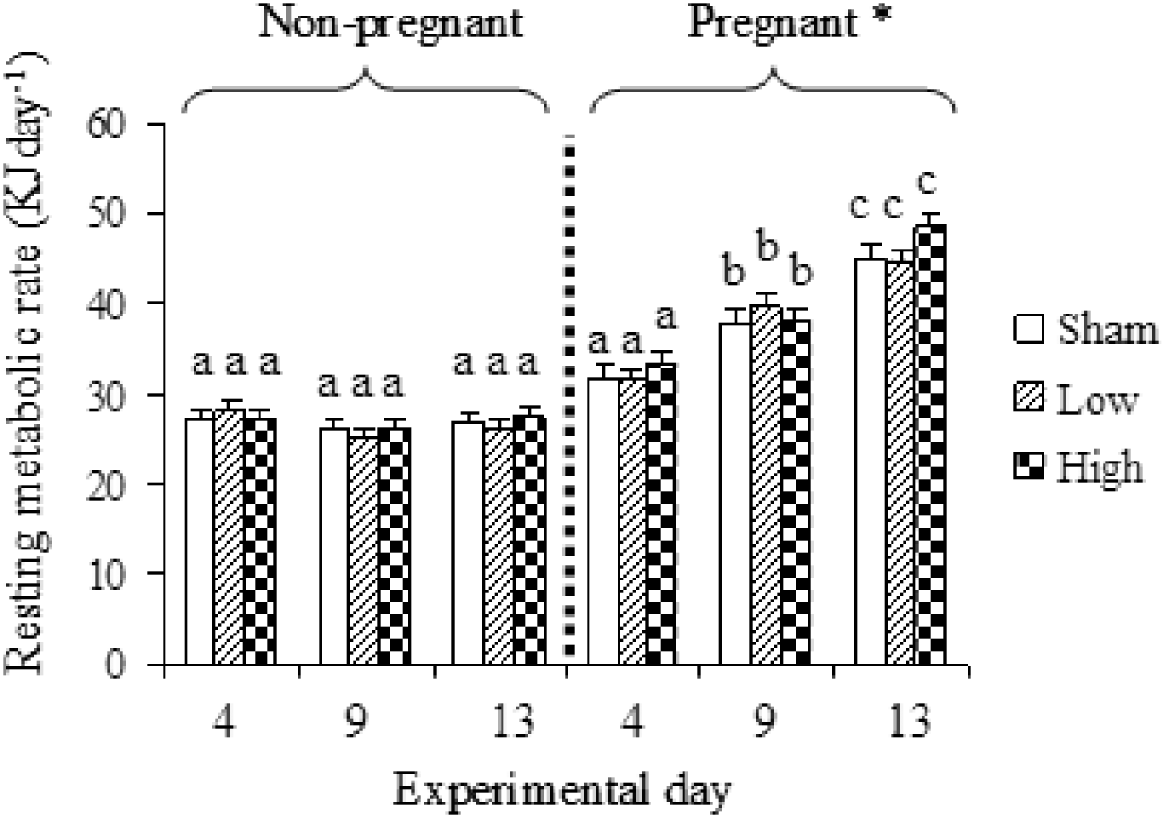
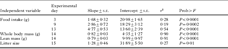
Pregnancy increased maternal RMR across all infection doses on experimental days 9 and 13 but infection had no detectable impact on RMR (Fig. 6). Linear regression showed a positive relationship between RMR and average food intake on experimental days 4, 9 and 13 (Table 3). The RMR also increased linearly with whole body mass, lean mass and litter size measured at the end of the experiment (Table 3).
Resting metabolic rate (RMR) (adjusted for baseline RMR) for non-pregnant and pregnant mice either uninfected (sham), or infected with low or high dose of Heligmosomoides bakeri. Main effect of pregnancy state: F 1,65=245·44, P<0·0001; main effect of dose: F 2,61·8=0·68, P=0·5123; main effect of time: F 2,107=73·24, P<0·0001; interaction of pregnancy state * time: F 2,107=83·1, P<0·0001; interaction of dose * time: F 4,107=1·65, P=0·166; interaction of pregnancy state * dose: F 2,61·1=0·33, P=0·7235; interaction of pregnancy state * dose * time: F 6,130=1·07, P=0·3817. Different lowercase letters represent significant differences across all groups, based on multiple comparisons; * represents a significant difference between pregnant and non-pregnant mice.

Relationship between maternal resting metabolic rate (RMR) and average daily food intake, whole body mass, lean mass and litter size

Body temperature was higher in pregnant (37·60±0·08°C) compared with non-pregnant (37·32±0·07°C) mice (F 1,62·9=9·75, P=0·0027). Body temperature was lower in mice with the high dose infection (37·36±0·08°C) compared with uninfected mice (37·63±0·09°C) (F 2,62·8=3·71, P=0·0301). There was no interaction between pregnancy state and infection. However, there was a significant interaction between pregnancy state (pregnant vs non-pregnant) and time (F 2,258=2·94, P=0·0135) whereby body temperature declined during pregnancy but remained constant in non-pregnant mice.
Cytokines in serum and amniotic fluid
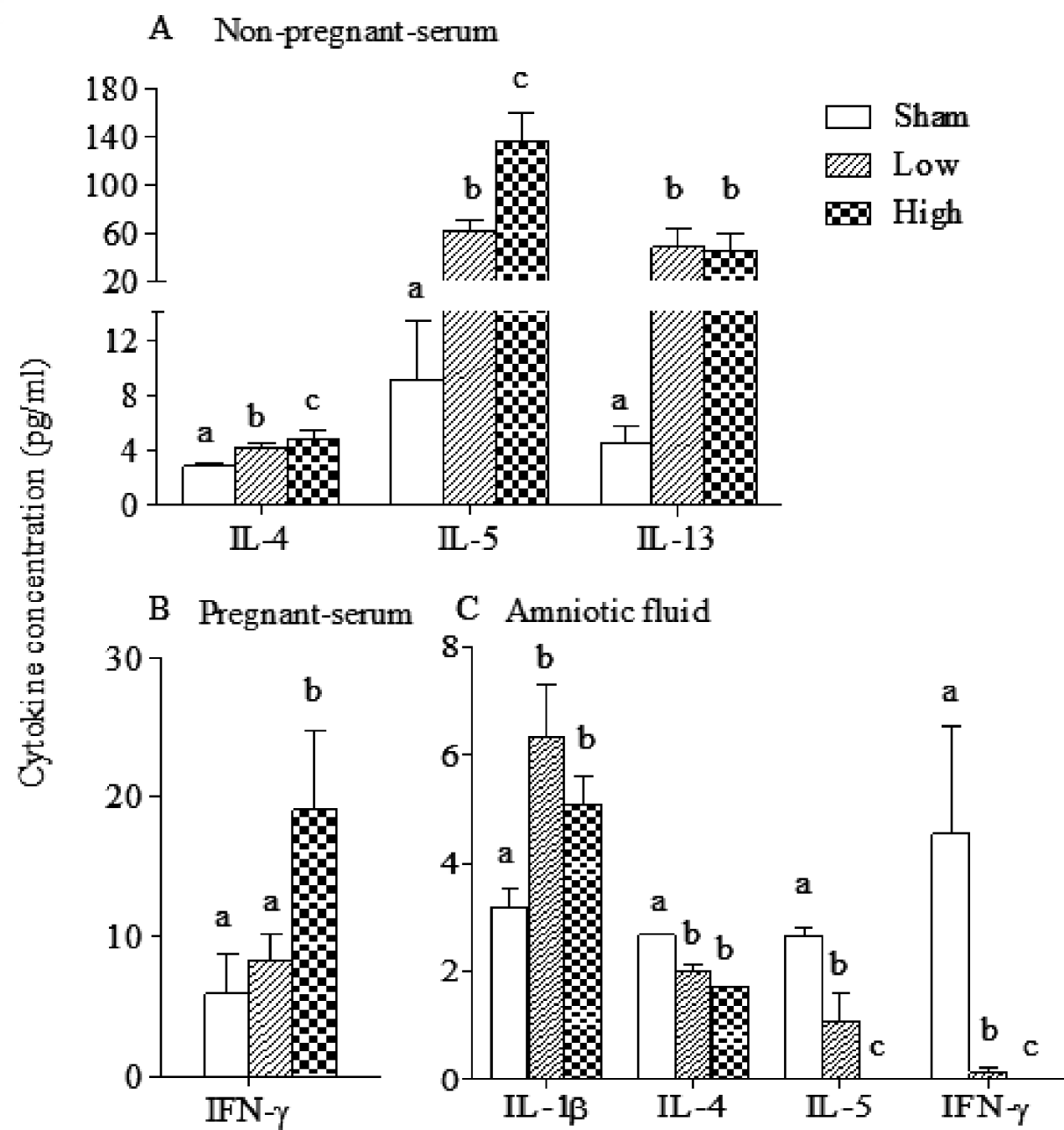
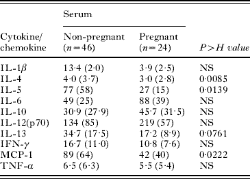
The effect of pregnancy on serum cytokines was evident through lower serum concentrations of IL-4, IL-5, IL-13 and MCP-1 (Table 4). Infection also influenced serum cytokines. Among the non-pregnant mice, serum IL-4 and IL-5 increased with dose (Fig. 7A) and serum IL-13 was higher in infected compared with uninfected mice (Fig. 7A). Among pregnant mice, IFN-γ was elevated in high dose compared with uninfected mice (Fig. 7B).
Effect of infection dose on cytokine profile in serum from non-pregnant (A) and pregnant mice (B), and in amniotic fluid (C). Different lowercase letters represent significant differences between infection doses (P<0·05).

Main effect of pregnancy (pooled across dose) on serum cytokine profiles (concentration in pg/ml) at day 19 of pregnancyFootnote 1

1 Values are arithmetic means obtained from Kruskal-Wallis analysis, and values in parentheses are medians.
In amniotic fluid, infected dams had elevated IL-1β (F 2,14=3·64, P=0·0535), but decreased IL-4 (F 2,15=36·59, P<0·0001), relative to uninfected dams. Both IL-5 (F 2,15=17·66, P=0·0001) and IFN-γ (F 2,14=3·41, P=0·05) were reduced in amniotic fluid in a dose-dependent manner (Fig. 7C).
DISCUSSION
This work highlights the largely independent ways in which the mouse responds to competing demands of infection and pregnancy. Whereas the energetic costs of infection were met through reduced body temperature, the costs of pregnancy and the associated increase in RMR were met through increased food intake and a reduction of fat reserves. Skeletal responses to infection and pregnancy also differed. Pregnancy increased the area of weight-supporting bones such as the femur at the expense of the lumbar region, whereas infection reduced maternal femur area and stunted foetal growth. These important shifts in bone both in the maternal and foetal compartments are consistent with known mechanisms of bone remodelling involving elevated IFN-γ (Nanes et al. Reference Nanes, Rubin, Titus, Hendy and Catherwood1990; Mann et al. Reference Mann, Jacobs, Buchinsky, Armstrong, Li, Ke, Ma, Jee and Epstein1994) and IL-1β (Nguyen et al. Reference Nguyen, Dewhirst, Hauschka and Stashenko1991) concentrations in maternal serum and amniotic fluid, respectively.
During pregnancy, the increased visceral organ lean mass, foetal growth, RMR and body temperature were accompanied by increased food intake and reduced fat mass. Given that increased food intake provided energy, but also led to an increase in the size of the digestive tract, as reported previously (Hammond and Diamond, Reference Hammond and Diamond1997; Piersma and Lindstrom, Reference Piersma and Lindstrom1997), its net energy benefit may have been minimal (Piersma and Drent, Reference Piersma and Drent2003). Instead, the RQ of 0·73 at experimental day 13 (data not shown) indicates that energy was generated through fat mobilization (McClave et al. Reference McClave, Lowen, Kleber, McConnell, Jung and Goldsmith2003).
Infection, in contrast, increased the mass of spleen, liver and small intestine, and also induced cytokine production, but was not accompanied by increased RMR or food intake, or reduced fat reserves. Only rarely has increased food intake been reported during H. bakeri infection (Wong et al. Reference Wong, Hildebrandt, Thrasher, Appleton, Ahima and Wu2007); in fact, Tu et al. (Reference Tu, Koski, Wykes and Scott2007) observed a transient decrease in food intake immediately following a challenge infection. Instead, our H. bakeri-infected mice reduced their body temperature by 0·27°C. A 0·5°C decrease in body temperature decreases the RMR by approximately 4% if conductance and evaporative heat loss remain the same (Kristan and Hammond, Reference Kristan and Hammond2006). This has been proposed as a mechanism to decrease energy expenditure during H. bakeri infection (Wong et al. Reference Wong, Hildebrandt, Thrasher, Appleton, Ahima and Wu2007). To date, literature has been inconsistent with regard to the effect of H. bakeri on RMR (Kristan and Hammond, Reference Kristan and Hammond2000, Reference Kristan and Hammond2001, Reference Kristan and Hammond2004, Reference Kristan and Hammond2006; Kristan, Reference Kristan2002). We suggest that the decreased body temperature without increased food intake served to reduce the energy costs of infection-induced immune responses and the increase in visceral tissue mass, resulting in no increase in RMR.
When infection was superimposed on pregnancy, the RMR and food intake remained elevated and fat mass remained low. In addition, the Th2 immune response that is necessary for intestinal epithelial cell turnover (Cliffe et al. Reference Cliffe, Humphreys, Lane, Potten, Booth and Grencis2005) leading to worm expulsion (Urban et al. Reference Urban, Madden, Cheever, Trotta, Katona and Finkelman1993) was suppressed and worm recovery was elevated compared with infected, non-pregnant mice. These results are consistent with other reports of Th2 suppression during reproduction of GI nematode-infected birds and mammals as evidenced by depressed antibody production (Sulila and Mattsson, Reference Sulila and Mattsson1990; Deerenberg et al. Reference Deerenberg, Apanius, Daan and Bos1997), down-regulated cell-mediated immunity (Medina et al. Reference Medina, Smithson and Kincade1993; Gustafsson et al. Reference Gustafsson, Nordling, Andersson, Sheldon and Qvarnstrom1994; Saino et al. Reference Saino, Calza and Moller1997), and elevated fecal egg output (Houdijk et al. Reference Houdijk, Jessop, Knox and Kyriazakis2003; Ng'ang'a et al. Reference Ng'ang'a, Kanyari, Maingi and Munyua2006; Normanton et al. Reference Normanton, Jos, Houdijk, Jessop, Knox and Kyriazakis2007). It is likely that suppression of the Th2 response may have resulted from up-regulation of IFN-γ, evident at the high dose infection in pregnant mice. Furthermore, pregnancy lowered MCP-1 concentrations, and this cytokine has been associated with infiltration of macrophages around the larval parasite (Sarafi et al. Reference Sarafi, Garcia-Zepeda, MacLean, Charo and Luster1997) and expulsion of adult H. bakeri (Tu et al. Reference Tu, Koski and Scott2008). Thus, both our elevated IFN-γ and reduced MCP-1 would have contributed to the higher worm recovery in pregnant mice.
Our results on maternal bone remodelling during pregnancy are consistent with previously reported increases in whole body and femur BA and BMC (Miller et al. Reference Miller, Shupe, Redd, Miller and Omura1986; Zeni et al. Reference Zeni, Di Gregorio and Mautalen1999) attributed to increased mechanical loading (De Boer and Wood, Reference De Boer and Wood1989; Glauber et al. Reference Glauber, Vollmer, Nevitt, Ensrud and Orwoll1995). Furthermore, foetal demands for calcium are first met from those maternal bone regions with a high metabolic turnover such as trabecular-rich areas (Zeni et al. Reference Zeni, Weisstaub, Di Gregorio, Ronanre De Ferrer and Portela2003), including the lumbar region. This would explain the lower lumbar BMC and BMD in our pregnant mice. In contrast to pregnancy, high dose infection reduced maternal femur BA. Studies in sheep have shown that GI nematode infection leads to thinning of the trabecular structure and reduced bone formation (Thamsborg and Hauge, Reference Thamsborg and Hauge2001) and is associated with reduced deposition of phosphorus and calcium in bone (Sykes et al. Reference Sykes, Coop and Angus1979; Coop and Field, Reference Coop and Field1983). The reduced maternal femur BA in our infected mice may be an indirect consequence of elevated maternal serum IFN-γ concentrations, either through its promotion of bone resorption as occurs in long bones such as the femur under pathological conditions (Nanes et al. Reference Nanes, Rubin, Titus, Hendy and Catherwood1990; Mann et al. Reference Mann, Jacobs, Buchinsky, Armstrong, Li, Ke, Ma, Jee and Epstein1994), or through its inhibition of bone formation (Mann et al. Reference Mann, Jacobs, Buchinsky, Armstrong, Li, Ke, Ma, Jee and Epstein1994) in mice that have not yet reached their peak femur bone mass (Ward et al. Reference Ward, Piekarz and Fonseca2007).
In the foetal compartment, linear growth of the foetus was reduced in response to H. bakeri infection of the dams, an effect that increased in severity with increasing numbers of intestinal worms. To our knowledge this has not been observed with other GI nematode infections, although growth stunting in nematode-infected children is common (Casapía et al. Reference Casapía, Joseph, Núñez, Rahme and Gyorkos2007; Payne et al. Reference Payne, Koski, Ortega-Barria and Scott2007; Jardim-Botelho et al. Reference Jardim-Botelho, Brooker, Geiger, Fleming, Souza Lopes, Diemert, Correa-Oliveira and Bethony2008). We also observed a high concentration of IL-1β in amniotic fluid, over and above that required as a preparatory step in the initiation of labour (Romero et al. Reference Romero, Brody, Oyarzun, Mazor, Wu, Hobbins and Durum1989). IL-1 β has been reported to inhibit bone formation in vivo (Nguyen et al. Reference Nguyen, Dewhirst, Hauschka and Stashenko1991) which could explain our observed decrease in foetal crown-rump length in response to infection.
In summary, GI nematode infection induced region-specific shifts in bone area and mineralization in the maternal compartment and reduced foetal crown-rump length. Evidence is provided that pro-inflammatory cytokines in maternal serum and amniotic fluid may be involved. We propose that the H. bakeri – pregnancy model may provide a useful animal system for studying bone remodelling during GI nematode infection.
ACKNOWLEDGEMENTS
We thank Mr Alastair Dorreen for assistance with the cytokine assays. This study was supported by Natural Sciences and Engineering Research Council of Canada (NSERC) Discovery Grants to M. E. S. and K. G. K., and Fonds Québécois de la recherche sur la nature et les technologies (FQRNT). Research at the Institute of Parasitology was supported by a Regroupement stratégique from FQRNT. The DXA equipment was purchased through a Canadian Research Chair – Canada Foundation for Innovation Award (HAW).