The pathogenesis of viral myocarditis involves both the direct viral injury and the host immune response. The cytotoxicity mediated by cytotoxic T lymphocytes is an important mechanism of cardiomyocytic damage.Reference Podack1, Reference Hassin, Fixler and Shimoni2 Cytotoxic T lymphocytes kill their target cells predominantly by inducing programmed cell death, or apoptosis,Reference Jerome, Sloan and Aubert3 and eliminate pathogenic cells in large part through the perforin/granzyme pathway.Reference Zhang, Chu and Wang4 Multiple characteristics of porforin passageways have been shown in the myocardium using immuno-electron microscopy, confirming that the killing cells damage the myocardium by releasing the porforin.Reference Seko, Shinkai and Kawasaki5 The clearance of Ebola virus has also been shown to be perforin-dependent.Reference Gupta, Greer and Mahanty6 The perforin-mediated cytotoxin effect has been shown to have a double action in viral myocarditis, removing the virus, but also harming the myocardium.Reference Xiong, Yang, Hu and Chen7, Reference Xiong, Yang, Hu and Chen8 Perforin is one kind of glycoprotein similar to complement C in its structure and function, and is expressed mainly in the cytoplasm of the natural killer cells, cytotoxic T lymphocytes. Granzymes are a group of serine proteases which are stored in the granules of cytotoxic lymphocytes. They are released into the intercellular space together with perforin via exocytosis. Perforin forms cylindrical pores in the target cell membrane. These pores can serve as passive conductors of granzymes through the target cell membrane. Perforin can lyse the target cells, but granzymes have no function of cytolysis. They cause fragmentation of deoxyribonucleic acid and lead to apoptosis of target cells. In apoptosis itself, however, it is Caspase-3 that plays the roles of executioner.Reference White, Amini, Khalili, Kogan, Donaldson and Darbinian9, Reference Gobe10
Based on these theories and discoveries, we hypothesized that blocking the perforin/granzyme pathway might reduce the myocardial damage mediated by immunity. To the best of our knowledge, as yet, there are no published studies on this topic. Hence, our study was designed to explore the role of perforin/granzyme-mediated cytotoxicity of cytotoxic T lymphocytes in mice with myocarditis, and to observe the effect of anti-perforin neutralizing antibody on the pathological changes seen in the myocardium, myocardial viral titers, cardiomyocyte apoptosis and expression of caspase-3 in the setting of viral myocarditis.
Materials and methods
We purchased 45 male Balb/c mice, aged 4 weeks, from the animal experiment center of Shandong University. Their weight ranged from 15 to 20 grams. The animals were housed 5 mice per cage in a laminar air flow room maintained at 22 plus or minus 2°C with relative humidity of 55 plus or minus 5%.
The mice were divided randomly into 3 groups of 15 animals, namely a normal control group, a group inoculated with coxsackie virus B3, and a further group inoculated with virus but also inoculated with anti-perforin neutralizing antibody. Mice in the second and third groups were inoculated with 0.15 milliliters coxsackie virus B3 obtained from Shandong Academy of Medicine, Jinan, China, the tissue culture infective dose being 109 per milliliter, while the animals in the normal control group received 0.15 milliliters Eagle. The mice in the third group were inoculated with anti-perforin neutralizing antibody, obtained from the MBL company, Japan, at 0.1milligrams/kilogram, at 6 hours and 3 days, respectively, after infection. The extra mice in each group were sacrificed on the tenth day after infection. Half of the hearts was fixed in 10% neutral buffered formalin for staining with haematoxylin and eosin, immunohistochemistry, and Tunel assay. The other half was stored at −80°C for reverse-transcription polymerase chain reaction and measuring viral titers.
The specimens were dehydrated, embedded in paraffin, sectioned at a thickness of 5 microns, mounted on adhesive microscope slides, and stained with haematoxylin and eosin. After staining, we observed the myocardial pathological change and calculated a pathologic score by light microscope. Each slice was observed randomly using 5 high power fields, scoring inflammation from zero to 4 based on the ratio of lesions area presented in a section. No pathological change was scored as zero, with a ratio of less than 25% score at 1, around 50% at 2, 75% at 3, and more than 75% at 4.
The stored myocardium was homogenated in a copper screen after washing by buffered phosphate. After undergoing three thaw-freeze cycles, they were centrifuged for 10 minutes at 1000 revolutions per minute. We obtained the clear supernatant liquid, inoculated Hela cells, and observed the state of cell growth. Viral isolation was positive if the cells had pathological change. Otherwise the result was negative. The positive was titrated on HeLa cells, which were cultured in 96 shadow mask.
Tunel (Terminal transferase-mediated 2′-deoxyuridine 5′-triphosphate-biotin nick end labelling) staining on paraffin-embedded sections was used to demonstrate cells dying of apoptosis, using the protocol provided by the manufacturer. Briefly, 5 microns thick sections were dewaxed, rehydrated, and incubated with terminal deoxynucleotidyl transferase enzyme for 1 hour in a humidified chamber at 37°C. The reaction was demonstrated by incubation with anti-digoxigenin-peroxidase for 30 minutes in a humidified chamber at room temperature and visualized in a buffer containing diaminobenzidine. The specimens were counterstained by immersion in haematoxylin. The cells with clear nuclear labeling were deemed positive cells. The results of positive cells and their distribution were observed under 400× microscope. Five random high power fields were required for counting, and then the apoptotic ratios were calculated and analyzed.
Immunohistochemistry were performed for detecting the expressions of caspase-3 according to the instructions of the manufacturer. Briefly, myocardial sections were deparaffinized in xylene and rehydrated in graded ethanol. Protein antigenicity was enhanced by boiling sections with microwaves in citrate buffer for 15 minutes. Sections were treated with 3% hydrogen peroxide in phosphate buffer solution for 20 minutes at 37°C to inactive endogenous peroxidase activity. Sections were incubated with the primary antibody (Santa Cruz company, United States of America) at 4°C overnight, followed by staining with a streptavidin–biotin–peroxidase kit (Beijing Zhongshan biological company, China). The sections were reacted in a 3,3′ diaminobenzidine solution (Beijing Zhongshan biological company, China), counterstained with haematoxylin and mounted. For negative controls, an equivalent volume of phosphate buffer solution was used in place of the specific primary antibody. We observed the result by light microscope, and calculated the mean positive area by five high power fields (×400) randomly. It reflected the level of the expression of caspase-3 protein.
Total ribonucleic acid was extracted using Trizol, obtained from the Shanghai Biology Company, China. The first-strand complementary deoxyribonucleic acid was reverse transcripted via avian myeloblastosis virus reverse transcription reagent. The complementary deoxyribonucleic acid for Caspase-3 or β-actin was amplified by polymerase chain reaction using Taq deoxyribonucleic acid polymerase. The following set of primers was used (forward and reverse, respectively) to estimate gene expression: for Caspase-3 antigen: 5′-GGA GCT GGA CTG TGG CAT TGA- 3′ and 5′-CAG TTC TTT CGT GAG CAT GGA-3′; for β-actin: 5′-AAG ATC CTG ACC GAG CGT GG-3′ and 5′-CAG CAC TGT GTT GGC ATA GA G G-3′. The product were 322 base pairs Caspase-3 complementary deoxyribonucleic acid and 327 base pairs β-actin complementary deoxyribonucleic acid. Gene expression levels were represented as ratios of target gene to co-amplified internal standard.
The data were described as mean plus or minus standard deviation and analyzed by SPSS version 10.0 to evaluate the statistical difference. One way ANOVA was used to test statistic significance among the groups. A probability value of less than 0.05 was considered statistically significant.
Results
Pathological change of myocardium
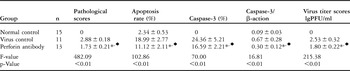
Extensive inflammatory cells had infiltrated the myocardium in the mice inoculated with coxsackie virus B3, with multiple disrupted myocytes seen, along with partial myocardial necrosis, changes typical for viral myocarditis (Fig. 1). The myocardial histological structure was markedly improved in the mice receiving anti-perforin neutralizing antibody therapy (Fig. 1). The myocardial pathological score was significantly lower in the treated mice than in those inoculated only with coxsackie virus B3 (p < 0.01) (Table 1). In the Table 1, we also show the results for viral titers, which were lower in the treated mice (p < 0.01)
Haematoxylin and eosin staining for the pathological change of myocardium. The images are shown at original magnification 100×. (a) In the myocardium of mice in coxsackie virus B3 control group, there were massive infiltrating inflammatory cells, lots of broken myocytes, and partial necrotic myocardium tissue. (b) There were only focal infiltrating inflammatory cells in the myocardium, with reduced myocardial tissue necrosis in mice receiving anti-perforin neutralizing antibody.

Myocardial pathological scores, rate of apoptosis, Caspase-3 positive percentage, Caspase-3 messenger ribonucleic acid expression and virus titer level ( ).

*Compared with normal control group, p < 0.01.
⧫Compared with coxsackie virus B3 control group, p < 0.01.
Apoptosis of cardiomyocyte
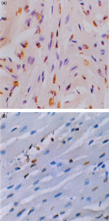
The apoptotic nucleuses were coloured brown, most being detected around the endocardium, epicardiumm, and areas of inflammation. In addition, some vascular endothelial cells and infiltration lymphocytes were stained positively. Only a few apoptotic cardiomyocytes were seen in the mice receiving anti-perforin neutralizing antibody, with the rate of apoptosis rate being lower in the treated mice (p < 0.01) (Fig. 2, Table 1).
Tunel staining for the apoptosis of cardiocytes, the images shown at original magnification 400×. (a) There were extensive apopotic cells in the animals infected with coxsackie virus B3, the apoptotic cell nucleuses distributed around the endocardium, epicardium, and areas of inflammation. In addition, some infiltration lymphocytes were stained positively. (b) Only a few apopotic cells were seen in the animals treated with anti-perforin neutralizing antibody, the rate of apoptosis being lower in the treated animals.

Expression Caspase-3 protein in myocardium
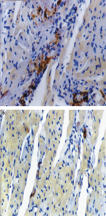
There was no positive staining cell in the normal mice. In those infected with coxsackie virus B3, and those treated with anti-perforin neutralizing antibody, the Caspase-3 protein positive staining cells were mostly distributed in the myocardium around the areas of inflammation, findings consistent with the distribution of apoptotic cardiomyocytes. The expression of Caspase-3 protein was lower in the mice receiving anti-perforin neutralizing antibody than in those infected only with coxsackie virus B3 (p < 0.01) (Fig. 3, Table 1).
Immunohistohemical staining for Caspase-3 in myocardium. Shown at original magnification 200×. (a) In the infected animals, the Caspase-3 protein positive staining cells are mostly located in the myocardium around the sites of inflammation, with a few infiltrating lymphocytes and blood vessel endothelial cells. (b) The expression of Caspase-3 protein is significantly decreased in the treated animals.

Expression of Caspase-3 messenger ribonucleic acid in myocardium
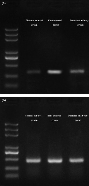
All products of Caspase-3 and β-actin amplification showed the banding 322 base pairs and 327 base pairs respectively after the agarose gel electrophoresis. They conformed to the design fragment size. The banding density scanning and the semi-quantitative analysis demonstrated that the expression of Caspase-3 messenger ribonucleic acid gene was lower in the treated mice (Fig. 4, Table 1).
Reverse-transcription polymerase chain reaction for the expression of Caspase-3 messenger ribonucleic acid gene in myocardium of three groups. Myocardium was lysed, and purified ribonucleic acid was reverse-transcribed and then amplified with the specific primers. (a) Reverse-transcription polymerase chain reaction showed decreased Caspase-3 messenger ribonucleic acid expression in the animals receiving anti-perforin neutralizing antibody therapy compared to the infected mice. (b) β-actin-specific nucleic acid fragments are shown, their level serving as a control for the reverse-transcription polymerase chain reaction.

Relevant analysis of the cardiomyocyte apoptosis rate, the Caspase-3 protein expression level and the myocardial pathological scores
The straight line correlation analysis demonstrated that Caspase-3 positive area expression rate and myocardium apoptosis rate were positively correlated with myocardial pathological scores (r = 0.73 p < 0.01 and r = 0.81, p < 0.01, respectively).
Discussion
The pathogenesis of viral myocarditis involves the direct viral injury and the host immune response. The cytotoxicity mediated by cytotoxic T lymphocytes plays a very important role in the myocardial damage of viral myocarditis. Cytotoxic T lymphocytes kill their target cells predominantly by inducing apoptosis, damaging the myocardium mainly through the perforin/granzyme pathway, but also via the Fas/FasL pathway.Reference Lee, Bar-Haim and Machlenkin11–Reference Kim, Kang and Noh13 After the cytotoxic T lymphocytes join the target cells, they release perforin into the intercellular space by exocytosis, the perforin adhering rapidly to the membranes of the target cells. Once anchored, perforin begins the polymerization in the presence of calcium to form cylindrical pores with an internal diameter of 5 to 20 nanometers.Reference Voskoboinik, Thia and Fletcher14–Reference Sauer, Pratsch, Tschopp, Bhakdi and Peters16 The pores produced can serve as passive conductors of granzymes and granulysin through the membranes, resulting in depolarization of the target cells. This leads to changes in osmotic pressure, with extracellular water flowing into the cell, while some electrolytes and the macro-molecule material flow out. The end result is lysis of the target cells. Granzymes that enter the target cells can decompose specifically the proteins of the nucleuses, causing fragmentation of deoxyribonucleic acid, and producing apoptosis.
Evidence of apoptosis of cardiomyocytes in active myocarditis has been demonstrated,Reference Zorc-Pleskovic, Alibegović, Zorc, Milutinović, Radovanović and Petrović17 and apoptosis has been shown to play a critical role in mediating cardiac injury in the setting of viral myocarditis.Reference DeBiasi, Robinson and Sherry18–Reference Saraste, Arola and Vuorinen20 We found rates of apoptosis to be higher in our infected mice than in our normal control animals, with the rates of apoptosis correlating positively with our myocardial pathological scores, showing that apoptosis of cardiomyocytes is part of the pathophysiology of myocarditis.
The activation of caspases, a conserved family of cysteine proteases, is a main event in the initiation and execution of programmed cell death or apoptosis.Reference Cooper, Granville and Lowenberger21 Caspase-8 and Caspase-9 are considered the initiators,Reference Fujikawa, Shinmei, Zhao and Aviles22–Reference Ghosh, Khazaei and Moien Afshari24 with Caspase-3 acting as the executioner of apoptotic death. We showed that cardiomyocytic apoptosis, Caspase-3 protein and gene expression all significantly increased in the infected but untreated mice, with Caspase-3 distributed mainly around the areas of inflammation, findings consistent with the distribution of the apoptotic cardiomyocytes. Others have already made similar observations,Reference DeBiasi, Robinson and Sherry18, Reference Purevjav, Nelson and Varela25 showing that Caspase-3 indeed plays a very important role in cellular apoptosis. Moreover, we found Caspase-3 expression to be strong positively correlated with the myocardial pathological score, indicating that Caspase-3 might also contribute to the myocardial damage in the setting of viral myocarditis.
We found the myocardial pathological score, the rate of cardiomyocytic apoptosis, and expression of Caspase-3 protein and messenger ribonucleic acid all to be lower in the animals treated with anti-perforin neutralizing antibody. We infer that the binding between anti-perforin neutralizing antibody and perforin molecules located at the membrane surface have blocked the the effect of perforin perforation, preventing the formation of channels across the membranes. Caspase-3 is mainly activated by granzymes.Reference Zapata, Takahashi, Salvesen and Reed26, Reference Galvin, Spaeny-Dekking, Wang, Seth, Hack and Froelich27 Once the perforin perforation effect is blocked by anti-perforin neutralizing antibody, granzymes can not enter the target cells, and Caspase-3 activation is also reduced. Therefore, the target cell apoptosis is suppressed, and the myocardial pathological scores is decreased. We also found the virus titer to be significantly lower in the treated animals, showing that anti-perforin neutralizing antibody may relieve viral replication.
Our results, therefore, provide further confirmation that the apoptosis mediated by the perforin/granzyme pathway is another important mechanism of cytotoxic T lymphocytes causing the myocardial damage in viral myocarditis. They verify that anti-perforin neutralizing antibody can relieve myocardial pathological change, cardiomyocytic apoptosis, and viral replication in the setting of viral myocarditis. For the first time, our study demonstrates that the anti-perforin neutralizing antibody reduces the myocardial damage by blocking the perforin/granzyme pathway and downregulating the expression of messenger ribonucleic acid and protein of Caspase-3. This approach may provide promising novel therapeutic strategies for the clinical treatment of viral myocarditis.
Acknowledgements
This study was supported by Shandong Medical Board in China (CA1DBA11).