INTRODUCTION
While parasites and their effects have been well documented for shallow-water communities, especially those with economic importance, there are few studies of micro-parasites in the deep sea. This may partly be due to limited sample availability, but, more importantly, it is simply due to their inconspicuous nature (reviewed by De Buron and Morand, 2004). Hydrothermal vent ecosystems are characterized by exceptionally high density that may render the vulnerability of these organisms against parasites by means of their facilitated transmission and elevated prevalence values (Ward et al. 2004). Even more, hydrothermal vents were classified as oases for parasitic protists that may have caused some of the mysterious sudden mortality episodes shaping vent fauna (Moreira and Lopez-Garcia, 2003).
Bathymodiolid bivalves are biomass-dominant species located at deep-sea hydrothermal vent ecosystems of the Mid-Atlantic Ridge (MAR) where they can reach densities as high as 700 individuals per m2 (Kádár et al. 2005a). They are sessile organisms harbouring endosymbionts belonging to sulphide-oxidizing chemoautotrophs and methanotroph bacteria (Fiala-Medioni and Lepennec, 1987; Kádár et al. 2005a). Parasitism in species of Bathymodiolus species from both cold seeps and deep sea vents were studied by Ward and co-workers (2004). They identified 10 parasitic agents in various organs including viral-like gut inclusions, rickettsia-like inclusions in gill and mantle, bacterial gill rosette, chlamidia-like gut inclusions, gill-dwelling ciliates and an unidentified inclusion in the gut tissue. Another health-assessment study of mussels from petroleum seeps carried out by Powell and co-workers (1999) also described body inclusions in digestive glands that resemble Chlamydial/Rickettsial inclusions. This fragmentary literature suggests the acute need for further indepth parasitological investigations for the accurate assessment of the health status of mussel populations located at vents.
In spite of being anchored by byssal threads, bathymodiolids are capable of migrating towards the nutrient flow (Desbruyeres et al. 2000) that can be very variable given the dynamic nature of the vent ecosystem (Kádár et al. 2005b). Undisturbed byssogenesis therefore, is a means of survival and thus merits the attention of the scientific community. Additionally, being aware of the extreme environmental conditions typical to deep sea hydrothermal vents, in terms of high pressure, temperature, low pH and presence of elevated concentrations of toxicants (Kádár et al. 2005b) under which byssus threads of the vent mussel are formed, its ultrastructure per se deserves closer scrutiny. Byssus threads are extracellular collagenous structures secreted by the byssus gland of the foot, and are composed of 3 distinct parts, originally described in Mytilus by Brown (1952), and its composition in the hydrothermal mussel has not been addressed to date.
This study is part of a wider-ranging assessment of additional physiological roles played by this attachment organ, i.e. its potential role in metal detoxification (Kádár et al. 2005c, 2006). During our ultrastructural observations on byssus micro- morphology, an unexpected presence of a series of prokaryotic organisms with potential parasitic influence was found, and is described here in order to draw attention of the pertinent specialists. Employing classical histology and electron microscopy we provide evidence of the presence of a novel membrane-bound inclusion within the byssus threads of the vent mussel Bathmodiolus azoricus (Von Cosel et al. 1999) that may cause byssus failure and thus impairment in the normal functioning of this organ. The biological implications of a potential parasitic influence are also discussed.
MATERIALS AND METHODS
Sample collection
Samples were collected in July 2005 with the R/V ‘Atalante’, using the equipment secured to the Remotely Operated Vehicle (ROV) Victor 6000. Mussels from Eiffel Tower at Lucky Strike (−1700 m, 37 °18′N, 32 °16′W) (Langmuir et al. 1997) were collected by the telemanipulated ROV arm and brought to surface in an insulated box. Individuals were measured, dissected and byssus fragments were fixed for light and electron microscopy following the protocol described below. Byssus threads from 5 mussels were studied.
Tissue preparation for light and electron microscopy
Byssal fragments from the 3 distinct regions of the mussel B. azoricus: adhesive plaque, distal portion and proximal portion, were fixed in modified Trump's fixative (3% glutaraldehyde and 3% paraformaldehyde made up with a fixation buffer containing: 0·15 M Na cacodylate, 0·3 M sucrose, 0·2 M NaCl and 0·008 M CaCl2) according to the method described by Distel and Felbeck (1987). Following primary fixation, samples were washed in 0·1 M cacodylate buffer (pH 7·8), post-fixed in 1% OsO4, dehydrated in an ascending ethanol series and embedded in Epon. Semi-thin (2 μm) sections were stained with methylene blue, while ultra-thin sections were double stained with uranyl acetate and lead citrate before examination with a JEOL 100CXII transmission electron microscope operated at 60 KV.
RESULTS AND DISCUSSION
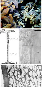
The edifice walls of Eiffel Tower in Lucky Strike vent field, the sampling location for this study, were covered by B. azoricus that thickly coated virtually all available rock surfaces. A typical picture of the organization of the biota (Fig. 1A) reveals that mussels are biomass-dominant over other co-inhabitants such as hydrothermal shrimps (Mirocaris fortunata and Rimicaris exoculata) and crabs (Segonzacia mesatlantica). Mussels attach to the substratum and/or to each other by means of byssal threads. The general macro-anatomy of the byssus thread of B. azoricus, composed by the stem emerging from the byssal gland and continuing by the proximal and distal parts of the thread and ending in the adhesive plaque (Fig. 1B), was similar to those described in other mytilid bivalves (Lucas et al. 2002). The adhesive plaque consists of a coat of collagen-like fibres that is attached to a protective coating of about 3 μm thickness (Fig. 1C) and which is layered over an inner spongy matrix (Fig. 1D). However, its biochemical composition might vary given its functionality under the extreme hydrothermal conditions. Thus, adhesion of B. azoricus to underwater surfaces in itself is of great scientific and biotechnological interest because it is (i) strong and durable, (ii) opportunistic and (iii) not undermined by the presence of water and the corrosive environment of the hydrothermal vent.

Fig. 1. Byssus structure in the vent mussel Bathymodiolus azoricus. (A) Typical organization of the mussel clumps attached via byssus, virtually covering all rocky surfaces at Eiffel Tower in Lucky Strike vent field of the Mid-Atlantic Ridge. (B) Schematic presentation of the byssus thread consisting of the stem emerging from the gland, the proximal- and distal-regions of the thread and the adhesive plaque that comes in contact to the attachment surface (adapted with permission from Herbert Waite). (C) Electron micrograph showing the outer fibrous coat (oc) of the adhesive plaque covered by a protective coating (asterisks). Scale bar: 1 μm. (D) Ultrathin transversal section through the adhesive plaque showing the outer coat (oc) layered on the spongeous collagen matrix (scm). Scale bar=2 μm.
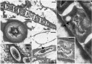
It was interesting to note the occurrence of an unusual filamentous organism embedded within the spongy matrix of the adhesive plaques in all byssus samples analysed. Observed under TEM, they appeared as a string of maximum length approximately 7 μm, containing up to 10 anucleated cells bound within a single membrane (Fig. 2A). The cells within the string had a double membrane, were regularly rod shaped, measured approximately 1 μm in diameter (Fig. 2B), and exhibited DNA strands in the centre of an electron-translucent area (Fig. 2Bi). Some transverse sections showed a very interesting concentric membranous organization (Fig. 2C) seemingly distinct from the filamentous form, and thus its connection to the former was not clear. The filamentous organism seemed to cause coagulation-like damage to the collagen fibres (Fig. 2D). In some favourable sections possible division could be seen (Fig. 2E).

Fig. 2. Byssus parasite in the vent mussel Bathymodiolus azoricus. (A) Full length of the string formed by the filamentous prokaryote. Scale bar=1 μm. (B) Higher magnification of 1 cell within the string of the filamentous organism showing the double membrane (arrows). Scale bar=0·5 μm. (Bi) Higher magnification of central part of the cell from (B) showing DNA strands in the centre of an electron-translucent area (asterisks). Scale bar=0·2 μm. (C) Transverse sections showing unusual concentric membranous organization of a cell. Scale bar=0·2 μm. (D) Transversal section through the filamentous organism showing the damage caused by the organism in the organized collagen structure (asterisks). Scale bar=0·5 μm. (E) Section showing a putative fission of the cell by invagination of the membrane (arrow). Scale bar=0·6 μm.
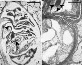
Another intra-byssus organism was a small (15 μ length) cystic organism with a barrel-shaped body containing a series of lobes filled with lipid-like droplets (Fig. 3A). It did not seem to cause any damage to the hosting byssus ultrastructure as suggested by the organized network of the surrounding collagen fibres (Fig. 3A). In a more peripheral section it was possible to observe that the body was encircled by at least 8 hoops of membranes (Fig. 3B). The invagination of these membranes separated the anterior part from the posterior lobe (Fig. 3B). Also, in the anterior lobe an electron-dense vesicle with possible ‘visual’ role could be observed. There was no indication of active movement of the organism within the byssus string, i.e. the cilia were absent.

Fig. 3. Intra-byssal organism in Bathymodiolus azoricus. (A) Cystic organism with a series of lobes filled with lipid-like droplets (arrowheads). Note the lack of disturbance and no damage to the spongy byssus matrix. Scale bar=4 μm; (B) A more peripheral section of the same organism whose body is separated into 2 lobes by an invagination (asterisk) of the 8-fold membrane (arrows). Scale bar=1 μm.
Symbiotic relationships between marine invertebrates and bacteria are a prominent feature in hydrothermal vent systems as an adaptation to utilize the chemical energy of the system (Hoffmeister and Martin, 2003). Arguably, the best example for such ecological success is the co-habitation of the deep-sea hydrothermal bivalve B. azoricus with its endosymbiotic chemoautotrophic bacteria. In addition, prokaryotes were also described in vent and seep mussels as non-gill symbioses and/or parasitoses (Johnson and Lepennec, 1995; Johnson and Fernandez, 2001; Ward et al. 2004; Powell et al. 1999). However, the nature of their symbiotic association in the case of non-gill symbiosis, whether it is nutritional, shelter or illness-related, is not completely elucidated to date. Our unusual finding of the ubiquitous presence of these intra-byssal organisms, one with putative parasitic influence, raises interesting questions related to the host-prokaryote reciprocal functions in the vent mussel. Byssus is a structure constructed with the greatest energy dissipation or hysteresis (Papov et al. 1995), and has a very high energetic value that could provide nutritional support and a suitable settlement surface for these organisms. They are clustered within the inner spongy net of the byssus string, and are exclusively found in the adhesive plaque, i.e. the distal end of the byssus that is in contact with the rock surfaces. There were no indications on how these organisms enter the thread. It is unlikely that they have accessed the byssus after its injection into the water since its protective protein, designated Mefp-1 (Mytilus edulis foot protein 1) protects the threads from degradation by bacteria (Qin and Waite, 1995). Moreover strong antibacterial activity in modiolids was demonstrated in byssus by Haug and co-workers (2004), and also in our lab in B. azoricus (Bettencourt et al., manuscript in preparation). Furthermore, the integrity of these organisms may indicate that they have not undergone digestion and/or decomposition. On the contrary, they seemed to divide in some cases. At least one of these microorganims may cause failure of the string by disruption of the collagen fibres that make up the spongy inner part of this organ. Their exact physiological role, however, remains to be elucidated by further, more in-depth biochemical, immunological and microbiological investigations. Because byssogenesis is a significant factor in the survival of mussels, a proper parasitological screening is essential both in mussel populations at vents and in those selected for post-capture investigations. Therefore, identification and physiological description of these novel symbiont and/or parasitic organisms inside the byssus threads of B. azoricus is not only of great theoretical, but also of practical importance.
This study was undertaken under the framework of FISIOVENT-Physiological adaptations to extreme conditions at deep-sea hydrothermal vents, project (POCTI/MAR/55547/2004) funded by FCT, Portugal. A post-doctoral fellowship was awarded to Eniko Kádár by FCT (SFRH-BPD-19625/2004). We thank the crew of R/V Atalante and the ROV team for technical assistance during sample collection and Ana Colaço for providing mussel samples.