Implications
Research aimed at altering cattle growth to optimize carcass composition often focuses on manipulating postnatal nutrition and management. However, there is emerging evidence that the prenatal period may provide opportunities to influence muscle and fat development and, in response, carcass composition. Results of this study indicate that although some genes responsible for fetal myogenesis and intramuscular adipogenesis were differentially expressed, fetal growth and muscle histology were not affected by the alterations in maternal energy applied during mid-gestation. Data suggest that minor changes in dietary energy provided to gestating cows during mid-gestation will not impact fetal growth.
Introduction
During gestation there is a tremendous draw of nutrients to meet the demands of the developing fetus, and if these demands are not satisfied, alterations in body composition of the adult animal could result. Muscle and adipose tissue grow by hyperplasia, primarily during fetal life and while the number of muscle fibers is set late in gestation, adipocytes can continue to develop into adult life in cattle (Bonnet et al., Reference Bonnet, Cassar-Malek, Chilliard and Picard2010). Adipogenesis is the creation of new adipocytes, and refers to the proliferation, differentiation and conversion of undifferentiated cells into fat tissue. Adipoblasts originate from fibroblast or mesenchymal stem cells, and are the early precursors to adipocytes. The stages of adipose development are strictly regulated by a series of transcription factors as reviewed by Bonnet et al. (Reference Bonnet, Cassar-Malek, Chilliard and Picard2010). During development perirenal adipose tissue appears at 80 days after conception, followed by subcutaneous and intermuscular adipose tissue at 180 days after conception (Bonnet et al., Reference Bonnet, Cassar-Malek, Chilliard and Picard2010). Du et al. (Reference Du, Wang, Fu, Yang and Zhu2010) proposed that the fetal stage offers the greatest chance to improve the ability of livestock to deposit intramuscular fat due to a greater abundance of non-committed pluripotent cells. Studies utilizing sheep have reported adipose tissue in the fetus can be manipulated by maternal nutrition (Bispham et al., Reference Bispham, Gopalakrishnan, Dandrea, Wilson, Budge, Keisler, Broughton, Stephenson and Symonds2003; Ford et al., Reference Ford, Hess, Schwope, Nijland, Gilbert, Vonnahme, Means, Han and Nathanielsz2007). Research in sheep also indicates that maternal nutrition during gestation influences gene expression necessary for intramuscular adipocyte development (Tong et al., Reference Tong, Zhu, Underwood, Hess, Ford and Du2008). Therefore, it may be possible to influence the distribution of fat within beef cattle through manipulation of gestational nutrition.
Skeletal muscle is the dominant tissue in beef carcasses and muscle fiber number corresponds with increased leanness and faster growth rates. Muscle development requires myogenic precursor cells to migrate from the dermomyotome to the myotome beginning with embryonic myoblast, followed by fetal myoblast, and later satellite cells (Biressi et al., Reference Biressi, Molinaro and Cossu2007). These myoblast then align and fuse with surrounding myoblast to form multinucleated embryonic myotubes. As myotubes mature, myofibrillar proteins actin and myosin are added and accumulation of myofibrillar proteins push nuclei to the periphery (Biressi et al., Reference Biressi, Molinaro and Cossu2007). Once differentiation has occurred the collection of fused myotubes is referred to as a muscle fiber or myofiber. This embryonic generation of myofibers is observed from 30 days after conception (Bonnet et al., Reference Bonnet, Cassar-Malek, Chilliard and Picard2010). Successive groups of myoblast will migrate, align and fuse to form secondary myofibers later in the fetal stage of myogenesis. It is reported that the fetal stage is the only time in development when fiber number can be altered, more specifically between 2 and 8 months of gestation (Russell and Oteruelo, Reference Russell and Oteruelo1981; Du et al., Reference Du, Wang, Fu, Yang and Zhu2010). This period of gestation coincides with secondary myogenesis, and research has determined that secondary fibers are susceptible to maternal nutrient restriction (Du et al., Reference Du, Wang, Fu, Yang and Zhu2010). Evidence in sheep indicates that restriction of total digestible nutrients during gestation can negatively influence muscle fiber number in lamb fetuses and subsequent postnatal growth (Zhu et al., Reference Zhu, Ford, Natanielsz and Du2004). Further, sows on low-protein diets produced piglets with less primary and secondary myofibers after birth and persistently restricted muscle growth potential as reported by Rehfeldt et al. (Reference Rehfeldt, Lefaucheur, Block, Stabenow, Pfuhl, Otten, Metges and Kalbe2012). Therefore, the maternal environment in which the fetus develops may be important in determining postnatal muscle mass.
Studying the response of genes to maternal over-feeding and nutrient restriction during mid-gestation could demonstrate how management decisions influence fetal adipose and muscle development. Therefore, it was hypothesized that the expression of genes involved in muscle fiber and adipocyte development will differ as a result of altered maternal nutrition during mid-gestation. The objective of this experiment was to determine whether maternal energy supply that is inadequate, adequate or supersedes the nutrient requirements of beef cattle (NRC) for growing heifers in mid-gestation results in differences in gene expression regulating myogenesis and adipogenesis in the bovine fetus.
Material and methods
Animals
Before initiation of this experiment animal care, handling and sampling were approved by the South Dakota State University Institutional Animal Care and Use Committee. In total, 23 Angus cross-bred heifers approximately 14 months of age and of similar breed composition and body condition were artificially inseminated using sexed (female selected) semen from a single Angus bull to reduce potential effects of genetics and sex. Heifers were synchronized using a common synchronization protocol and inseminated within the same week. Heifers received a common diet until breeding, and throughout early gestation (day 85). At day 85 of gestation heifers were stratified by weight and assigned randomly to a dietary treatment.
Dietary treatments
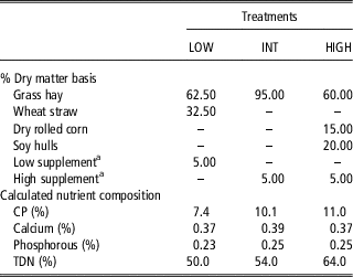
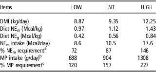
During treatment, heifers were housed at the South Dakota State University Beef Nutrition unit. Dietary treatment groups were low (n=8; LOW; 523±14.5 kg of initial BW), intermediate (n=7; INT; 535±15.5 kg of initial BW) and high (n=8; HIGH; 522±14.5 kg of initial BW). All dietary treatments were formulated in accordance to the Beef Cattle NRC (2000). Heifers were offered feed twice daily; diets are reported in Table 1. The Beef Cattle NRC (2000) Table Generator was used to predict the average calories of net energy and metabolizable protein required during months 3, 4 and 5 of gestation for the normal growth and pregnancy of an Angus heifer with a mature weight of 725 kg. Based on model-predicted intake and treatment diet formulation, the LOW, INT and HIGH treatment were predicted to supply 80%, 100% and 150% of the calories needed for normal growth and pregnancy. Based on actual measured dry matter intake (DMI), LOW, INT and HIGH diets provided 72%, 87% and 146% of the predicted net energy for maintenance (NEm). Metabolizable protein requirements for normal growth and pregnancy were met or exceeded by all dietary treatments. Treatment diets were initiated on day 85 of gestation and continued through day 180 of gestation when cesarean sections were performed. The treatment period was selected to coincide with a period of time pregnant cows may encounter a nutrient restriction (mid-gestation), which coincides with secondary myogenesis and early stages of adipogenesis (Du et al., Reference Du, Wang, Fu, Yang and Zhu2010). BW was recorded at the beginning and end of treatment period and used to determine dam growth during treatment. Ultrasound measurements for dam fat thickness (FT) and longissimus muscle area (LMA) were measured between the 12th and 13th ribs at the three-quarter position of the longissimus muscle (LM) using an Aloka 500V real-time ultrasound machine (Aloka, Wallingford, CT, USA) 12 days before treatment initiation and at day 170 of gestation to determine dam body condition changes during treatment.
Ingredient and nutrient content of dietary treatments offered to dams from 85 to 180 days gestation

LOW=72% net energy for maintenance (NEm); INT=87% NEm; HIGH=146% NEm; TDN=total digestible nutrient.
a Provided vitamins and minerals to meet nutrient requirements (Beef Cattle NRC, 2000).
Sample collections
In total, 23 heifers were used at trial initiation; however, one heifer in the INT treatment aborted at 164 days of gestation. Therefore, 22 heifers received a standing left side cesarean section, on days 179, 180 or 181 of gestation. A university veterinarian performed anesthesia and cesarean sections. Immediately, following the cesarean section fetal BW and crown rump length were recorded, and each fetus was exsanguinated via the umbilical vein. Samples from the LM were collected from the left and right sides of the fetus by removing a 7.62 cm section of muscle on each side of the 13th rib; muscle from the right side and any visible subcutaneous fat located over the LM were quickly diced, snap frozen in liquid nitrogen and stored at −80°C until analysis. The section of the LM that was removed from the left side of each carcass and the semitendinosus (ST) from the left side was prepared for histological analysis. These muscles were chosen due to their importance to the meat industry, ease of collection and common use in research allowing for comparison with other studies. Samples for histology were fixed in paraformaldehyde and stored in 70% ethanol. The right hind limb (RHL) of each fetus was removed at the hip joint, stored on dry ice and frozen at −20°C until subsequent dissection for compositional analysis.
Histology
Fixed LM and ST samples were embedded in paraffin and 10 μm transverse sections were cut and stained with Harris modified hematoxylin and counterstained with eosin Y for visualization of fiber diameter under microscopy. A minimum of 300 muscle fibers were analyzed for each fetus to determine fiber area, diameter and number using NIS Element BR 3.0 technology under 40× magnification (Nikon Instruments Inc., Melville, NY, USA). The field of view measurement was 285×213 μm. Muscle fiber number was determined by manually counting mature distinguishable muscle fibers within the field of view. Average muscle fiber was determined using four slides per fetus.
Hind limb dissection and composition analysis
Each RHL was allowed to thaw at 4°C for 48 h before dissection. The ST was dissected and individual muscle weight was recorded. Approximately 5 g of muscle from the medial portion of the ST were immersed in liquid nitrogen and powdered using a mortar and pestle. Samples (2 g) were extracted with petroleum ether in a side arm soxhlet for 60 h (Association of Official Analytical Chemists, 1990) followed by drying at 60°C for 24 h. Percent crude fat was calculated as the difference between the extracted weight and the lyophilized sample weight multiplied by 100. Percent moisture was calculated as the difference between the sample weight and the dry weight, divided by the sample weight multiplied by 100. Femur bones were denuded of muscle tissue using a scalpel and boiled for 90 min in 2 l of distilled water. Joints were removed at the epiphyseal line and the femur bone was allowed to air dry and then oven dried for 48 h at 70°C and dry weight and length were recorded.
RNA extraction
Samples were immersed in liquid nitrogen and powdered using a mortar and pestle, and liquid nitrogen. TRI Reagent® RT (Molecular Research Center Inc., Cincinnati, OH, USA) was used to extract RNA from fetal LM and subcutaneous adipose tissue. Approximately 100 mg of powdered sample was placed into 1 ml of TRI Reagent RT. To achieve phase separation, 50 μl of bromoanisol (Molecular Research Center Inc.) was added to each sample, vortexed for 10 s, and centrifugated at 12 000×g for 15 min at 4°C. The sample separated into three phases: the aqueous RNA phase, DNA interface and protein. An aliquot of 500 μl was removed from the aqueous layer; RNA was precipitated by adding 500 μl of isopropanol. The sample was incubated at room temperature for 5 min followed by centrifugation at 12 000×g for 5 min at 4°C. With the RNA pellet firmly attached to the tube a mixture of 75% ethanol and 25% RNA-free water was added, followed by centrifugation at 5000×g for 5 min at 4°C to wash the sample. The ethanol wash solution was decanted and tubes were inverted for 3 min to air dry. The RNA pellet was then rehydrated with 100 μl of RNA-free water. RNA concentration was determined using a Nanodrop spectophotometer (Thermo Scientific, Wilmington, DE, USA). To improve RNA quality a DNase I Amplification Grade kit (Invitrogen, Roche Molecular Systems Inc., Foster City, CA, USA) was used to remove genomic DNA contamination, and RNA concentration was diluted to 200 ng/μl. A high-capacity cDNA reverse transcription kit (Applied Biosystems, Carlsbad, CA, USA) converted RNA to complementary DNA (cDNA) using a Bio-Rad MyCycler Thermocycler (Bio-Rad Laboratories, Hercules, CA, USA).
Real-time PCR
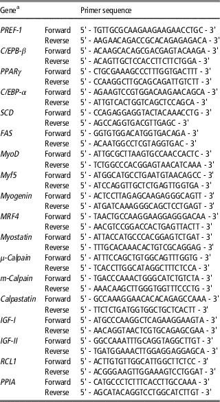
Real-time (RT) quantitative PCR analysis using reverse transcribed cDNA was performed in triplicate with a SYBR Green RT-PCR kit from Bio-Rad (Bio-Rad Laboratories). Specific primer sets for genes of interest and housekeeping genes are listed in Table 2. Genes of interest were selected based on their relevance during myogenesis and adipogenesis. Briefly, specific primers were designed by utilizing the National Center for Biotechnology Information (United States Library of Medicine, Bethesda, MD, USA) database; primers were designed with PrimerQuest software from Integrated DNA Technologies (Coralville, IA, USA). RT-PCR was performed with an ABI 7900 HT High-Throughput Real-Time Thermocycler (Applied Biosystems). Each cDNA sample was amplified in triplicate and triplicates were averaged to determine cycle threshold values. The reaction program for amplification of cDNA using SYBR Green was as follows: Stage 1 – 10 min at 95°C; Stage 2 – 15 s at 95°C followed by 1 min at 60°C (40 cycles). Reaction specificity was determined by melting curve analysis and agarose gel electrophoresis of amplicons.
Primer sequence for genes of interest and housekeeping genes in fetal adipose and longissimus muscle tissue

PREF-1=preadipocyte factor-1; C/EBP-β=CCAAT/enhancer-binding protein β; PPAR γ=peroxisome proliferator-activated receptor γ; C/EBP-α=CCAAT/enhancer-binding protein α; SCD=stearoyl-CoA desaturase; FAS=fatty acid synthase; MyoD=myoblast determination protein 1; Myf5=myogenic factor 5; RCL1=RNA terminal phosphate cyclase-like 1; PPIA=peptidylprolyl isomerase A.
a The reaction program for amplification of complementary DNA using SYBR Green is as follows: Stage 1 (hold) – 10 min at 95°C; Stage 2 (denature and annealing) – 15 s at 95°C followed by 1 min at 60°C for 40 cycles; Stage 3 (melting curve analysis) – 15 s at 95°C, 15 s at 60°C, and 15 s at 95°C.
Gene expression analysis
Fold change was calculated using REST 2008 (Relative Expression Software Tool V2.0.07, Corbett Research Pty Ltd, Sydney, Australia). The REST 2008 program incorporates RT-PCR reaction efficiencies, reference gene normalization, and cycle threshold values to determine statistical differences between a treatment sample and the control. Reaction efficiencies were calculated from cycle threshold values of serially diluted pooled cDNA in triplicate. RNA terminal phosphate cyclase-like 1 (RCL1) and peptidylprolyl isomerase A (PPIA) were used as housekeeping genes. Both PPIA and RCL1 have been identified as suitable housekeeping genes in bovine LM and adipose tissue (Ghinis-Hozumi et al., Reference Ghinis-Hozumi, Gonzalez-Gallardo, Gonzalez-Davalos, Antaramian, Villarroya, Shimada, Varela-Echavarria and Mora2011) and were validated for our samples using BestKeeper (Pfaffl et al., Reference Pfaffl, Tichopad, Prgomet and Neuvians2004).
Statistical analysis
Dam performance and fetal growth characteristics were analyzed using the GLM procedure of SAS (SAS Institute Inc., Cary, NC, USA) with dietary treatment group as the main effect. DMI was calculated using pen as the experimental unit. Least square means were used to separate differences in treatment means that resulted from dietary treatment. RT-PCR gene expression data were analyzed using REST 2008 to calculate fold change difference between LOW, or HIGH, and INT which served as the control. Statistical significance was determined by permutation testing by REST 2008 software; significance was determined at P<0.05 and a statistical tendency was determined at P<0.10.
Results
Dam performance during mid-gestation
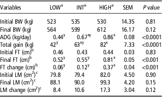
Actual DMI, net energy intake and metabolizable protein intake for the dams is reported in Table 3. Based on actual DMI and analyzed nutrient profiles, LOW, INT and HIGH heifers were estimated to have consumed 72%, 87% and 146%, respectively, of the net energy needed for normal growth as predicted by the Beef Cattle NRC (2000). Actual metabolizable protein intake exceeded the requirement for growing heifers in each treatment. Initial dam BW was similar between treatment groups (P>0.10). Dams in the HIGH treatment had increased (P<0.05) total gain during the treatment period compared with the LOW dams, however, total gain of HIGH and LOW treatments were not different from INT (Table 4). Average daily gains for LOW, INT and HIGH were 0.44, 0.67 and 0.86 kg/day, respectively, and HIGH and LOW differed (P<0.05) as a result of treatment. According to the NRC Table Generator, the predicted growth of a pregnant Angus heifer with a mature weight of 725 kg and fed to meet NRC nutrient requirements during months 3, 4 and 5 of gestation is 0.66 kg/day (Beef Cattle NRC, 2000). Relative to NRC-predicted growth, the LOW, INT and HIGH dams in this study achieved 67%, 102% and 130% of the predicted ADG, respectively.
Dry matter and nutrient intake of dietary treatments offered to dams from 85 to 180 days gestation

LOW=72% net energy for maintenance (NEm); INT=87% NEm; HIGH=146% NEm; DMI=dry matter intake.
a % NEm requirement calculated using NEm intake for each treatment divided by the predicted calories required during months 3, 4 and 5 of gestation for the normal growth and pregnancy of an Angus heifer with a mature weight of 725 kg, provided by the Beef Cattle NRC (2000) Table Generator.
b Predicted based on degradability of protein sources included in the diet.
c % Metabolizable protein (MP) requirement calculated using MP intake for each treatment divided by the predicted MP required during months 3, 4 and 5 of gestation for the normal growth and pregnancy of an Angus heifer with a mature weight of 725 kg, provided by the Beef Cattle NRC (2000) Table Generator.
Growth and ultrasound body composition of dams during days 85 to 180 of gestation

ADG=average daily gain.
x,yMeans within rows that do not have common superscripts differ, P<0.05 as a result of dietary treatment.
a Dietary treatment offered to beef dams from 85 to 180 days of gestation; LOW 72% net energy for maintenance (NEm); INT 87% NEm; HIGH 146% net energy requirement for normal growth and pregnancy of a bred Angus heifer (Beef Cattle NRC, 2000).
b Ultrasound-measured fat thickness (FT); initial measure 12 days before initiation of dietary treatment and final measurement following 84 days of dietary treatment.
c Ultrasound-measured longissimus muscle (LM); initial measure 12 days before initiation of dietary treatment and final measurement following 84 days of dietary treatment.
The treatment diets also contributed to differences in dam body condition as evaluated by ultrasound (Table 4). FT increased (P<0.05) during treatment for LOW, INT and HIGH heifers (0.06, 0.12, 0.37 cm, respectively). Final ultrasound FT was measured at day 84 of the treatment period and HIGH heifers had accumulated greater subcutaneous fat (P<0.05) than INT and LOW heifers; but FT was similar for INT and LOW dams. Initial and final ultrasound LMA measurements were similar across treatments.
Fetal growth, histology and composition
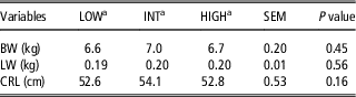
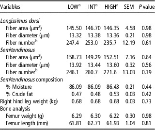
Although changes in dam weight and composition were observed, dietary treatment did not influence fetal BW, crown rump length or liver weight (Table 5). Fiber diameter, area and number were similar across treatments in the LM and ST samples (Table 6). Analysis of the RHL revealed no differences in percent moisture of the ST, percent crude fat in the ST, femur weight or femur length between treatments (P>0.10).
Effect of maternal nutrition from 85 to 180 days gestation on measures of fetal growth

LW=fetal liver weight; CRL=crown rump length.
a Dietary treatment offered to beef dams from 85 to 180 days of gestation; LOW 72% net energy for maintenance (NEm), INT 87% NEm, HIGH 146% net energy requirement for normal growth and pregnancy of a bred Angus heifer (Beef Cattle NRC, 2000).
Effect of maternal nutrition from 85 to 180 days gestation on fetal muscle, fat and bone characteristics

a Dietary treatment offered to beef dams from 85 to 180 days of gestation; LOW 72% net energy for maintenance (NEm), INT 87% NEm, HIGH 146% NEm requirement.
b Per field of view (285×213 μm). Four transverse sections were evaluated per fetus.
Expression of genes in fetal longissimus dorsi and subcutaneous fat
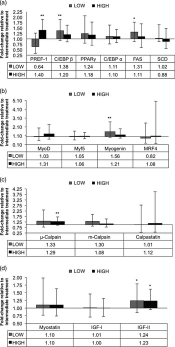
RT-PCR analysis of fetal subcutaneous (SUBQ) fat samples revealed no differences between treatments. However, differences were detected between treatments in fetal LM samples. For analysis, the LOW and HIGH treatments were compared with the INT, which served as the control. The HIGH diets resulted in an up-regulation (P<0.05) of preadipocyte fator-1 (PREF-1) when compared with fetuses from INT dams (Figure 1a), however, expression of PREF-1 was similar between LOW and INT fetuses. Expression of CCAAT/enhancer-binding protein-β (C/EBP-β) was up-regulated in the LOW fetuses (P<0.05) as compared with INT (Figure 1a). Dietary treatments did not induce gene expression differences for peroxisome proliferator-activated receptor γ (PPARγ), C/EBP-α, or stearoyl-CoA desaturase (SCD), however, there was a tendency (P<0.10) for up-regulation of fatty acid synthase (FAS) in LOW compared with INT fetuses (Figure 1a).
Real-time PCR analysis of the influence of high- and low maternal nutrition on expression in fetal bovine longissimus of (a) transcription factors involved in adipogenesis: preadipocyte factor-1 (PREF-1), CCAAT/enhancer-binding protein α and β (C/EBP-α, C/EBP-β), peroxisome proliferator-activated receptor γ (PPARγ), fatty acid synthase (FAS), stearoyl-CoA desaturase (SCD); (b) myoregulatory factors: myoblast determination protein 1 (MyoD), myogenic factor 5 (Myf5), myogenin, myogenic regulatory factor-4 (MRF4); (c) calpain system: μ-calpain, m-calpain, calpastatin; (d) growth factors: myostatin, insulin-like growth factor-I (IGF-I), insulin-like growth factor-II (IGF-II) at 180 days of gestation. Statistical comparisons were made using REST 2008, a program designed to compare expression difference between the control and a single treatment. INT was used as the control with LOW or HIGH being compared with INT; bars indicate standard error. *P<0.10, **P<0.05.

Analysis of fetal LM samples revealed no differences in expression of myogenic factor 5 (Myf5), myoblast determination protein 1 (MyoD) or myogenic regulatory factor-4 (MRF4) (Figure 1b). However, myogenin was up-regulated in the LOW fetuses (P<0.05) as compared with INT. Relative gene expression for μ-calpain, m-calpain and calpastatin was also determined by RT-PCR in the fetal LM samples (Figure 1c). Expression of m-calpain and calpastatin were similar between treatments; however, there was an up-regulation of μ-calpain in the HIGH fetuses as compared with INT (P<0.05). Numerically, μ-calpain was greater in the LOW fetuses, however, the variation between samples within the LOW treatment fetuses for expression of this enzyme resulted in no significant difference as compared with INT (P>0.10). Analysis of fetal samples revealed no differences between treatment groups for relative expression of myostatin or IGF-I (Figure 1d). However, there was a tendency for IGF-II expression to be up-regulated (P<0.10) in both the HIGH and LOW fetuses as compared with INT.
Discussion
Influence of maternal diet on fetal growth and muscle histology
In this study, the level of maternal dietary manipulation did not influence measures of fetal growth. This is in agreement with Meyer et al. (Reference Meyer, Reed, Vonnahme, Soto-Navarro, Reynolds, Ford, Hess and Canton2010) who reported nutrient restriction during mid-gestation did not reduce fetal BW or liver weight evaluated at 125 days of gestation. Rasby et al. (Reference Rasby, Wetterman, Geisert, Rice and Wallace1990) reported no differences in fetuses at 260 days gestation from cows fed to a body condition score (BCS) 4 at 195 days gestation as compared with cows fed to a BCS 6. The evidence that maternal nutrition did not influence fetal weight indicates that the dam was able to provide the necessary nutrients to the fetus, regardless of nutrient status of the diet. However, the majority of prenatal growth occurs in the last 2 months of gestation, which may make restrictions during early to mid-gestation less important for fetal growth. Previous research in sheep indicates that maternal nutrient restriction can result in reduced muscle weights, decreased fiber number and increased percentage of type II glycolytic fibers (Zhu et al., Reference Zhu, Ford, Means, Hess, Nathanielsz and Du2006; Daniel et al., Reference Daniel, Brameld, Craigon, Scollan and Buttery2007). It appears that the extent of energy restriction in the current trial, where protein supply was adequate, did not negatively impact muscle fiber diameter, number or area at this stage of development.
Response of fetal adipogenic gene expression to maternal nutrition
Adipogenesis is initiated at approximately day 120 of gestation in the bovine fetus and continues throughout the remainder of gestation and postnatal growth (Du et al., Reference Du, Wang, Fu, Yang and Zhu2010). In this experiment, the pregnancy was terminated at day 180 of gestation. Therefore, lack of differences in gene expression from SUBQ samples indicates that tissues may have been collected before appreciable SUBQ adipose tissue development. During tissue collection only extremely small amounts of SUBQ adipose tissue were recovered from the rib section of the fetus.
The most important stage for intramuscular adipogenesis in the growth and development of an animal is during the fetal stage. This is primarily due to increased amounts of pluripotent mesenchymal cells located within the muscle and their potential commitment to an adipogenic lineage (Du et al., Reference Du, Wang, Fu, Yang and Zhu2010). Although both skeletal muscle cells and adipocytes are derived from the mesoderm during embryonic development, Du et al. (Reference Du, Zhu, Means, Hess and Ford2015) describes early myogenesis and adipogenesis as competitive processes. Increasing adipogenesis in the developing muscle increases intramuscular fat and thereby has the potential to increase beef flavor in the mature animal (Du et al., Reference Du, Zhu, Means, Hess and Ford2015). Although new adipocytes are readily generated during postnatal growth, they are primarily located in fat depots other than intramuscular fat, as evident in rodent models (Faust et al., Reference Faust, Johnson, Stern and Hirsch1978; Valet et al., Reference Valet, Tavernier, Castan-Laurell, Saulnier-Blache and Langin2002), and therefore do not contribute to beef flavor. Similar findings have been reported based upon serial ultrasound measurements and serial slaughters in beef cattle. Intermuscular and subcutaneous adipose tissue deposition accumulates at a much greater rate than does intramuscular adipose tissue in beef cattle (Brethour, Reference Brethour2000). Wertz et al. (Reference Wertz, Berger, Walker, Faulkner, McKeith and Rodriguez-Zas2002) demonstrated that early weaning and early placement on concentrate allowed for increased marbling before over-finishing occurred. This supports the proposed hypothesis that improvements in the distribution of adipose tissue can be achieved during a more efficient stage of growth, such as the fetal stage (Du et al., Reference Du, Wang, Fu, Yang and Zhu2010; Underwood et al., Reference Underwood, Tong, Price, Roberts, Grings, Hess, Means and Du2010). Two prominent transcription factor families appear to be essential for differentiation of preadipocytes: the C/EBPs and PPARs (Chawla and Lazar, Reference Chawla and Lazar1994; Darlington et al., Reference Darlington, Ross and MacDougald1998). In addition, proteins such as PREF-1 have been reported to maintain a proliferative state and delay differentiation (Sul et al., Reference Sul, Latasa, Moon and Kim2000b).
Preadipocytes produce and secrete PREF-1, which can be useful in identifying cells that are preadipocytes (Sul et al., Reference Sul, Latasa, Moon and Kim2000b). Greater expression of PREF-1 in LM of HIGH fetuses suggests that a greater number of preadipocytes are present in the LM of HIGH fetuses. Conversely, this result may suggest that differentiation of preadipocytes in the LM adipose tissue is slowed in HIGH fetuses and proliferation of additional preadipocytes is occurring because PREF-1 inhibits preadipocyte differentiation. In cultured 3T3-L1 cells, PREF-1 inhibited the expression of transcription factors C/EBP-α and PPARγ (Sul et al., Reference Sul, Latasa, Moon and Kim2000b). Regardless, the increased expression of PREF-1 points to a potential for increased adipocyte development within the LM. This finding in beef fetuses is similar to work by Tong et al. (Reference Tong, Zhu, Underwood, Hess, Ford and Du2008) who reported fetuses from over-fed ewes had an up-regulation of PREF-1 as compared with ewes fed a diet that met NRC requirements for maintenance.
An up-regulation of C/EBP-β corresponds with the early stages of adipogenesis and appears necessary to initiate PPARγ expression, which is essential for adipocyte differentiation (Darlington et al., Reference Darlington, Ross and MacDougald1998). Increased C/EBP-β expression in the LOW fetuses could indicate that differentiation of preadipocytes to lipid-filling mature adipocytes has been initiated. The initial activation of C/EBP-α stops the proliferation of preadipocytes, and initiates the up-regulation of PPARγ in forming adipocytes (Darlington et al., Reference Darlington, Ross and MacDougald1998). However, the greatest up-regulation of C/EBP-α occurs later in adipogenesis, and is required for the continued expression of PPARγ (Lazar, Reference Lazar1999). The expression of PPARγ and C/EBP-α are up-regulated in the cell downstream of C/EBP-β expression (Lazar, Reference Lazar1999), and may be influenced by nutrition in latter stages of gestation.
FAS is a key regulating enzyme necessary for de novo fatty acid synthesis, and FAS expression is susceptible to nutrient status (Sul et al., Reference Sul, Smas, Mei and Zhou2000a). The tendency for increased expression of FAS in fetuses from the LOW treatment group may suggest a shift from preadipocytes to mature, lipid-filling cells. The lack of differences between treatments for PPARγ, C/EBP-α or SCD expression is possibly due to the fact that samples were collected early in adipogenesis, a phase with limited lipid accumulation.
Response of fetal myoregulatory gene expression to maternal nutrition
The primary MRFs are Myf5 and MyoD, and these transcription factors are essential for determination of myogenic lineage and myoblast proliferation. Myf5 and MyoD are more highly expressed in undifferentiated myoblasts (Braun et al., Reference Braun, Buschhausen-Denker, Bober, Tannich and Arnold1989), and an up-regulation of Myf5 and MyoD could indicate greater proliferation of myoblasts ultimately resulting in increased muscle development. However, in this study LOW and HIGH fetuses did not differ from INT fetuses for expression of Myf5 or MyoD. Myogenin and MRF4 are considered secondary MRFs, and are thought to be responsible for the terminal differentiation and maintenance of differentiated myofibrils (Perry and Rudnicki, Reference Perry and Rudnicki2000). The up-regulation of myogenin in the LOW fetuses could indicate enhanced terminal differentiation. This could result in a reduction in the proliferation of myoblasts, earlier fusion and therefore potentially reduce the number of myofibers formed. However, the lack of differences in muscle fiber number observed between treatments indicates that this change in myogenin expression was not sufficient to cause changes in fiber number. The expression of MRF4 was similar between treatments and as expected was not highly expressed. The expression of MRF4 occurs later during myogenesis and is the dominant MRF in adult skeletal muscle (Hannon et al., Reference Hannon, Smith, Bales and Santerre1992).
The calpain system is generally recognized as being essential in protein turnover; however, m- and μ-calpain activity is also necessary for myoblast organization and fusion (Barnoy et al., Reference Barnoy, Glaser and Kosower1997). The up-regulation of μ-calpain in LM of HIGH fetuses could indicate greater myoblast migration and fusion. It is suggested that increased myoblast proliferation and fusion as a result of increased calpain activity could lead to increased myonuclei resulting in potentially larger muscle fibers as well as affecting the number of fibers in the adult muscle tissue (Goll et al., Reference Goll, Thompson, Taylor and Ouali1998). However, no differences in fiber size or number were detected in this study. In addition, the calpain system regulates muscle accretion via its inhibitor calpastatin. Up-regulation of calpastatin preserves protein by reducing degradation caused by μ-calpain activity (Goll et al., Reference Goll, Thompson, Taylor and Ouali1998). Du et al. (Reference Du, Tong, Zhao, Underwood, Zhu, Ford and Nathanielsz2004) identified a down-regulation of calpastatin expression in beef cows subjected to a diet that supplied 68% NEm required, and concluded that this occurred in order to provide nutrients to the fetus. Interestingly, Du et al. (Reference Du, Tong, Zhao, Underwood, Zhu, Ford and Nathanielsz2004) found an up-regulation of calpastatin expression in the fetuses from restricted dams. This response was most likely a method of preserving the developed myofibrils by slowing protein turnover, evident by similar fetal weights between treatment groups. These findings differ from our study; while fetal weights were similar, relative calpastatin expression did not differ across treatments (Figure 1c). This discrepancy could be the result of differences between treatment diets between the two studies. Dietary energy was similar between these two studies; Du et al. (Reference Du, Tong, Zhao, Underwood, Zhu, Ford and Nathanielsz2004) offered restricted dams a diet that supplied 68% of the NEm required, whereas the current study provided LOW dams 72% of the NEm required. However, a greater amount of metabolizable protein was made available to dams in the current study. The LOW dams received 120% of their metabolizable protein requirement, whereas Du et al. (Reference Du, Tong, Zhao, Underwood, Zhu, Ford and Nathanielsz2004) provided 86.7% of the metabolizable protein requirement to dams during the first 120 days of gestation.
Myostatin is a powerful negative regulator of skeletal muscle growth, and when absent muscle growth increases. Expression of myostatin is present throughout myogenesis and myoblast proliferation and differentiation is mediated by the expression of myostatin (McPherron et al., Reference McPherron, Lawler and Lee1997). No differences in myostatin expression were detected between treatments in this study. These results were expected, given that fetuses were of similar genetics and metabolizable protein was in abundance for both the LOW and HIGH treatment dams.
In contrast to myostatin, expression of IGF-I and IGF-II promote muscle growth (Glass, Reference Glass2003). IGF-I is a contributor to embryonic development but primarily is responsible for postnatal growth, whereas IGF-II is essential for embryonic growth and development. Fetal expression of IGF-I was not influenced by maternal nutrition during gestation in this study. Similar IGF-I results were reported in nutrient-restricted sheep fetuses (Brameld et al., Reference Brameld, Mostyn, Dandrea, Stephenson, Dawson, Buttery and Symonds2000). In the current study, IGF-II expression tended to be increased in the LM of both HIGH and LOW fetuses. However, Brameld et al. (Reference Brameld, Mostyn, Dandrea, Stephenson, Dawson, Buttery and Symonds2000) reported the abundance of IGF-II protein was greater in skeletal muscle from nutrient-restricted sheep fetuses. Results may differ as Brameld et al. (Reference Brameld, Mostyn, Dandrea, Stephenson, Dawson, Buttery and Symonds2000) subjected dams to nutrient restriction early in gestation, whereas the present study investigated maternal nutrient during mid-gestation. Whether the tendency for up-regulation of IGF-II in both the LOW and HIGH fetuses would manifest into skeletal muscle mass differences in mature market animals remains to be determined.
Conclusions
Data collected from the LM samples indicate that although fetal growth characteristics were similar between LOW, INT and HIGH treatment groups, maternal nutrition can influence expression of some genes responsible for fetal myogenesis and intramuscular adipogenesis. Given that small fold changes in gene expression can be biologically significant, the effect of maternal nutrition on the expression of fetal genes regulating adipose and muscle growth warrants further investigation. The subsequent effects of altered fetal gene expression on carcass quality could generate greater potential revenue for beef producers and warrants further investigation.
Acknowledgments
This research was funded in part by grants from the South Dakota Beef Industry Council. Salaries and Research support was provided by State and Federal Funds appropriated to South Dakota State University.