Introduction
Colorectal cancer is the most common cancer of the gastrointestinal tract. It is the third most common cause of death for men and women in the Western World.Reference Siegel, Miller and Jemal1 The majority of colorectal cancer cases are adenocarcinomas and polyps. Worldwide, about 1·57 million patients are diagnosed and over 771,000 are expected to die from colon cancer every year.Reference Salem, Hartley, Unger and Marshall2
One of the major problems in the treatment of rectal cancer is local recurrence.Reference Kapiteijn, Marijnen and Nagtegaal3 Conventional surgery causes high rates of local recurrence, and in many cases, it fails to remove cancerous tumour.Reference Wiggers, de Vries and Veeze-Kuypers4 Studies have proven the superiority of preoperative radiotherapy compared to postoperative in terms of local recurrence rates and overall survival.Reference Holm, Cedermark and Rutqvist5,Reference Påhlman6 Lower local recurrence rates have been found with radiation therapy in most of these studies, especially those who use preoperative radiation.Reference Frykholm, Glimelius and Påhlman7 From a study by Camma et al., we can conclude that preoperative radiotherapy is more effective.Reference Camma, Giunta, Fiorica, Pagliaro, Craxi and Cottone8 All grade toxic effects such as diarrhoea, haematology and dermatology were 27% with preoperative versus 40% with postoperative treatment.
Preoperative radiotherapy is usually administered with three-dimensional conventional radiotherapy (3DCRT).Reference Jabbour, Patel and Herman9 The aim of this technique is to provide a conformality high dose rate to the target tumour, while reducing the dose to adjacent healthy tissue. This should reduce both acute and chronic morbidity.Reference Simson, Mitra, Ahlawat, Sharma, Yadav and Mishra10 Technological developments in radiation oncology, such as the use of intensity-modulated radiation therapy (IMRT) and volumetric-modulated arc therapy (VMAT) show greater accuracy and increase in dose.Reference Zhao, Hu and Cai11 With the advent of these improved techniques, the use of conventional radiotherapy is gradually decreasing.Reference Simson, Mitra, Ahlawat, Sharma, Yadav and Mishra10,Reference Zhao, Hu and Cai11
Several studies have been performed around the world to prove the most effective radiotherapy technique for rectal cancer when compared to treatments such as 3DCRT, IMRT and VMAT.Reference Zhao, Hu and Cai11 Dosimetric studies demonstrate the superiority of the IMRT technique in the overall reduction of bowel bag and tumour dose in high dose areas associated with reduced gastrointestinal toxicity.Reference Reyngold, Niland and ter Veer12–Reference Duthoy, De Gersem and Vergote15 Other studies have shown that target tumour coverage improves as well as dose homogeneity and conformity while simultaneously, there is decreasing doses in organ at risks (OARs) for preoperatively designed rectal cancer cases.Reference Richetti, Fogliata and Clivio16,Reference Arbea, Ramos, Martínez-Monge, Moreno and Aristu17 However, dosimetric studies show that the evaluation of techniques like 3DCRT, IMRT and VMAT is rare and includes a small sample size, usually less than 10 patients.
While preoperative treatment results in long-term disease-free survival, it often displayed high rates of acute toxicity particularly in gastrointestinal, genitourinary and hematologic toxicity.Reference Meier, Mascia, Wolf and Kharofa18 The incidence of clinically significant complications associated with the radiation is approximately 5–20%.Reference Rupam, Balaji, Sereen and Patil19 Due to irradiation of the pelvis and the rectal region in particular, the gastrointestinal tract becomes vulnerable, resulting in adverse effects most common among them being small bowel obstruction.Reference Martínez Hernández Magro20 The main problem is it causes interruption of treatment in haematological toxicity after pelvic radiotherapy, which increases the total time of treatment.Reference Li, Liu and Zhai21 In pelvic irradiation, due to its anatomical position, the bladder is exposed to radiation to cause undesirable side effects such as nocturia, frequency and urinary urgency.Reference Elliott and Malaeb22
The purpose of this study is to compare both dosimetrically and radiobiology of the OARs sparing and the target coverage among VMAT, IMRT and 3DCRT plans in 24 patients. Target coverage and target dose distribution, conformality, normal tissue avoidance and irradiated body volume were evaluated and compared for the different plans.
Methods and Materials
Study population
This study including 24 patients’ staged T1–3 without metastasis M0 and with positive lymph nodes N+ local advanced rectal cancer underwent neoadjuvant chemoradiation therapy (CRT) in the ‘Theageneio’ Anticancer Hospital of Thessaloniki, Greece from September to June 2019. All the patients received preoperative radiation therapy with VMAT technique with a prescribed dose of 50·4 Gy.
Radiation therapy planning
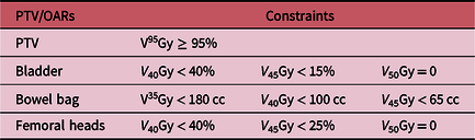
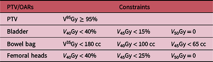
Computed tomography (CT)-based simulation (Siemens, Illinois, USA) was performed. Patients were simulated in supine position with full bladder. The CT-Sim files were transferred to Monaco TPS (Monaco™ Version 11.02; Elekta, Stockholm, Sweden), where all relevant volumes were contoured according to the International Commission on Radiation Units 62 protocol guidelines. The prescription dose of target volume (PTV) is 50·4 Gy in 28 fractions of 1·8 Gy. All plans have been performed to try to meet the constrains (both for PTV and OARs) presented in Table 1.
Constrain of OARs in rectal cancer
Dosimetric and radiobiological comparison
Dosimetric and radiobiological evaluation of all the plans was performed using dose–volume histogram (DVH). The homogeneity of the plans was measured in terms of the homogeneity index (HI), which was expressed as
 $HI = {{{D_{ \ge 95\% }}} \over {D \ge 5\% }}$
. The conformality of the plans was also evaluated with a conformality index, which is defined as the ratio of the target volume receiving 95% of the prescribed dose divided by the total volume receiving that dose level. D
max, D
mean and volumes of V
40, V
45 and V
50 were evaluated in accordance with international standards in order to assess the irradiation of OARs.
With regard to the radiobiological comparison of the radiotherapy plans, the Lyman–Kutcher–Burman model has been used. The formula for the tumour control probability (TCP) calculation is
 $$TCP = {({1 \over 2})^{\mathop \sum \nolimits_i {V_i}\exp \left[ {{{2{\it \gamma _{50}}} \over \it ln { \left( 2 \right)}}\left( {1 - {{{D_i}} \over {{D_{50}}}}} \right)} \right],}}$$
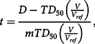
where γ 50 is a slope of dose–response, D 50 is a position of dose–response and (D i , V i ) are the bins of the DVH and the formula for normal tissue complication probability (NTCP) for the different OARs is
 $$NTCP = {1 \over {\sqrt {2\pi } }}\int_{ - \infty }^t {{e^{ - {t^2}/2}}} \;dx, {\rm and}$$
 $$t = {{D - T{D_{50}}\left( {{V \over {{V_{ref}}}}} \right)} \over {mT{D_{50}}\left( {{V \over {{V_{ref}}}}} \right)}},$$
where TD 50 is the uniform dose given to the entire organ volume that results in 50% complication risk, m is a measure of the slope of the sigmoid curve represented by the integral of the normal distribution, and V ref is the entire volume of relevant organ.
Based on the literatureReference Fiorino, Valdagni, Rancati and Sanguineti23–Reference Emami25 it was chosen to study specific endpoints for the OARs listed in Table 2.
NTCP parameters for specific endpoints for OARs

Statistical analysis
Twenty-four patients were treated with VMAT therapy. Four radiotherapy plans (3DCRT, 3 and 4 fields IMRT, VMAT) were performed on each patient. The dataset contains two sets of variables: the dosimetric (D max, D mean, HI, CI, V 40, V 45) and the radiological (TCP, NTCP) variables. Comparison of the mean value of the aforementioned variables is presented. The statistical comparisons both for dosimetric and radiobiological variables resulted from Shapiro test for normality checking. When the variables are normally distributed, we used t-test, while Mann–Whitney test was used for non-parametric data. p-values < 0 · 05 were considered as statistically significant. All the statistical analysis was performed using R programme.
Results
Dosimetric comparison
Dose comparison and coverage of the tumour
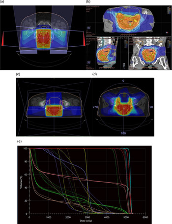
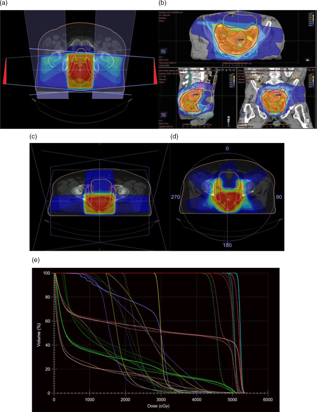
The prescribed dose, for all patients in this study with locally advanced rectal cancer, was 50·4 Gy. An example of beam configuration, dose distribution and DVH comparison for the four different treatment plans is shown in Figure 1. The PTV was outlined as a green region in all images.
Beam configuration and dose distribution for (a) 3DCRT, (b) 3-Field IMRT, (c) 4-Field IMRT, (d) VMAT techniques and (e) Comparative DVH of PTV and OARs for the techniques used.
In Table 3, all the measured dosimetric parameters for the PTV and for all techniques are presented.
Dosimetric parameters of four treatment planning techniques for PTV

Τ-test for the difference between two means could not be performed since our dependent variable (3DCRT, 3- and 4-field IMRT and VMAT) were not approximately normally distributed – a Shapiro test for normality has been performed – for each group of the independent variable (YES, NO). So, we performed a non-parametric test, the Mann–Whitney–Wilcoxon Test, where the parameters were normal distribution and a t-test was performed. Tables 4 and 5 present the results of this statistical analysis for PTV.
Dosimetric comparison of four treatment planning techniques for D max

Dosimetric comparison of four treatment planning techniques for D mean, CI and HI

All the radiotherapy plans met the clinical requirement of
 $PT{V_{95}} \ge 47\cdot88{\rm{\;Gy}}$
for each patient. 3DCRT exhibited better conformity index (p = 1·938e-05) than the other techniques, and VMAT exhibited better HI (p = 1·938e-05) than the other three radiotherapy techniques. The mean value of D
max was significantly lower in 3DCRT than the 3- and 4-field IMRT (p = 0·0002 and p = 0·0002, respectively) and VMAT (p = 0·0002).
Dose comparison of OARs
Bladder
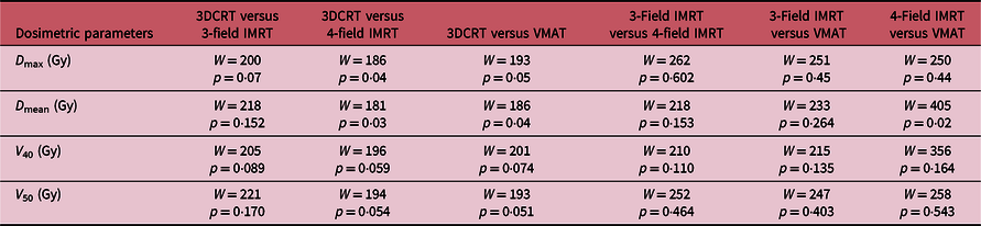
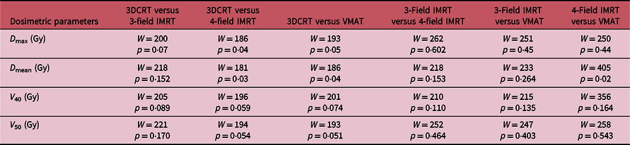
In the case of bladder, all the four radiotherapy techniques met the constraints (Table 6).
Dosimetric parameters of four treatment planning techniques for bladder

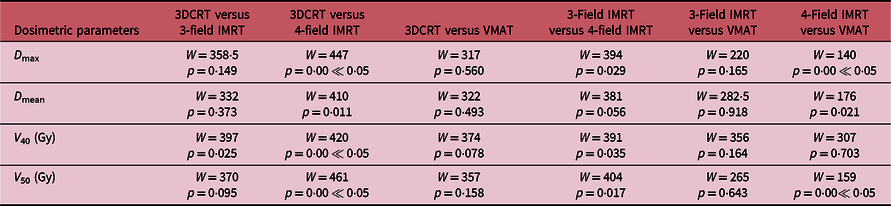
There were no significant differences between radiotherapy techniques on V 40 and V 50 (Table 7). Table 7 shows a statistically significant difference between 3DCRT and 4-field IMRT concerning the maximum dose to the bladder.
Dosimetric comparison of four treatment planning techniques for bladder
Bowel bag
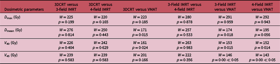
As far as the bowel bag is concerned, all the four radiotherapy techniques met the constraints (Table 8).
Dosimetric parameters of four treatment planning techniques for bowel bag

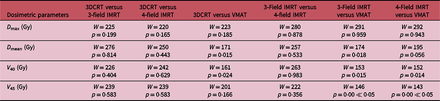
The bowel bag was checked for the Dmax, Dmean, V 40 and V 45 (Gy) variables. Regarding V 40, Table 9 shows a statistically significant difference between the 3DCRT and VMAT techniques. In addition, when comparing the VMAT and 3-Field IMRT techniques with respect to that same variable (V 40), we observe that in this case also, namely in VMAT radiotherapy plans, the bowel bag receives a larger dose of radiation. Finally, concerning the D max variable, we see that there is no statistically significant difference between separate radiotherapy plans.
Dosimetric comparison of four treatment planning techniques for bowel bag
Femoral heads
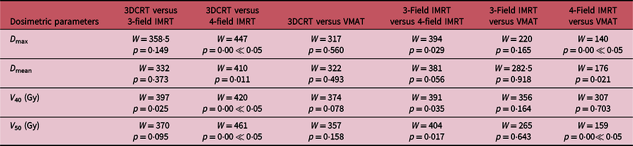
The dosimetric parameters measured for the femoral heads are presented in Table 10. In 3DCRT, it is statistically significant with lower dose on D max than 4-field IMRT.
Dosimetric parameters of four treatment planning techniques for femoral heads

Comparing the IMRT plans in a paired test, it proves that the 3-field IMRT has lower D max than 4-field (Table 11). However, 4-field IMRT has lower D max than VMAT. For V 40 and V 50 in 3DCRT, there was statistically significant lower dose than 4-field IMRT.
Dosimetric comparison of four treatment planning techniques for femoral heads
Radiobiological comparison
Tumour control probability
With regard to TCP, there was no statistically significant difference between the four radiotherapy plans (Tables 12 and 13). With regard to radiobiological comparison of plans, Table 12 shows that VMAT radiotherapy plans exhibit the highest Tumor Control Probability (80.76%) while 3DCRT plans have the lowest (75.87%); however, these results do not present a statistically significant difference in the calculated TCP, which was 80·76% and 75·87%, respectively.
Radiobiological parameter of four treatment planning techniques for PTV

Radiobiological comparison of four treatment planning techniques for TCP

Normal tissue complexity probability
Bladder
The NTCP for the bladder has been calculated for two endpoints: α/β = 6 for asymptomatic bladder (Table 14) and α/β = 3·4–4·5 for bladder contracture and volume loss (Table 15).
Radiobiological endpoints of four treatment planning techniques for symptomatic bladder

Radiobiological endpoint of four treatment planning techniques for bladder contracture and volume loss

As you can notice from Tables 14 and 15, both for symptomatic bladder and volume loss (α/β = 6 and α/β = 3·4–4·5), the complication probabilities of bladder for four radiotherapy techniques are in low rates. The highest probability was the nocturia, and it was identified in VMAT plans, 1·567% and 3·747, respectively.
Bowel bag and femoral heads
Comparison for four radiotherapy techniques (3DCRT, 3-field and 4-field IMRT and VMAT) showed that both the probability of obstruction of the bowel bag and the probability of grade 3 haematologic toxicity were in the order of 10–14 and 10–41, respectively, practically zero.
Discussion
This study compared dosimetrically and radiobiologically the target dose and OARs avoidance among the VMAT, two IMRT plans (three and four fields) and 3DCRT for the rectal cancer radiotherapy. Few studies have been performed to prove the most effective radiotherapy technique for rectal cancer by comparing treatments such as 3DCRT, IMRT and VMAT.Reference Zhao, Hu and Cai11–Reference Duthoy, De Gersem and Vergote15 This is one of the first attempts to compare simultaneously dosimetric and radiobiological in the case of rectal cancer.
All the radiotherapy techniques achieved comparable results for most of the dosimetric and radiological parameters. 3DCRT achieved lower D max in the tumour and better conformity index than the other three radiotherapy techniques. However, VMAT and 4-field IMRT were more homogeneous than 3DCRT and 3-field IMRT. Regarding OAR dose sparing, 3DCRT techniques proved to be significantly superior to both IMRT and VMAT techniques since they could easily meet the dosimetric constraints for the bladder (D max, V 40, V 50) and the bowel bag (D max, V 40). But for femoral heads, 4-field IMRT had lower D max, V 40 and V 50.
Jun Zhao et al. show that the IMRT and VMAT techniques have a lower dose sparing in OARs compared to the 3DCRT technique, with regard to most of the dosimetric variables evaluated (target response, OARs and normal tissue sparing). In addition to our study, 3DCRT achieved lower D max in the tumour and better conformity, and in case of dose sparing in bladder and bowel bag, it was significantly superior to both IMRT and VMAT plans. Lu Liu et al. prove that VMAT was the best choice in terms of conformal index and in addition to our study, 3DCRT has better CI.
Regarding the possibility of tumour control, VMAT plans have higher rates (80·76%) than the other three radiotherapy techniques, without this result identified as statistically significant. Also, in this study, the complication probabilities of the bladder, bowel bag and femoral heads were very low in the order of 2, 10–14 and 10–41, respectively, so that patients with rectal cancer are not at risk. This is mainly due to comparative low prescription dose of 50·4 Gy, which is not enough to overpass any significant dose–volume constrain for the relevant OARs related to clinical endpoints for severe side effects.
The number of patients in this study was limited. Based on the international literature, for the treatment of rectal cancer, preoperative radiotherapy is performed in stage patients T1–3 N+ M0. Patients included in the study were given radiotherapy with VMAT technique. The contouring of the PTV’s plan was not performed by the same radiotherapist, as a result, an operator error was introduced. For this study, the radiotherapy plans were developed for the same medical physicist.
Conclusion
Twenty four patients participated in this study, and the small sample size could raise questions concerning the validity of its results. However, comparing these findings to those of separate dosimetric and radiobiological studies shows that the 3DCRT technique is suitable for preoperative radiotherapy of stage T1–3 N+M0 rectal cancer. VMAT and 3- and 4-field IMRT achieved comparable dose sparing in OARs, and VMAT was more homogeneous and has the highest rate in TCP (80·76%).