Kawasaki disease is an acute systemic vasculitis and is the commonest cause of acquired heart disease in children. The exact aetiology of Kawasaki disease is still unknown; it is considered to have an infectious trigger followed by the activation of inflammatory process. Here, we report two cases of extremely rare Kawasaki disease with tsutsugamushi disease.
Case 1
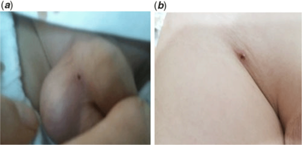
A 4-year-old boy presented with fever of 7 days duration and skin rash and bilateral conjunctival injection. He had a history of visiting a rural area with his grandmother. On admission, he had fever of 39.4 °C. His heart rate was 90/minute and his blood pressure was 90/60 mmHg. His pharynx was slightly injected and there was red lip. His neck was swollen with cervical lymphadenitis. He had erythematous macular rash on his trunk. Examination of his skin revealed an eschar on penile base of right scrotum (Fig 1a). His laboratory results showed a white blood cell count 4,720/mm3, 42% polymorphonuclear leukocytes, 39% lymphocytes, haemoglobin 10.3 gm/dl, platelet count 148,000/mm3, C-reactive protein 3.23 mg/dl, and pro-brain natriuretic peptide 316.5 pg/ml. Mycoplasma pneumonia IgM was negative. Orientia tsutsugamushi Ab test was positive. Echocardiographic findings 1 day after admission revealed mild dilatation of left coronary artery (right coronary artery = 1.8 mm, left coronary artery = 3 mm). He was treated on oral roxithromycin for diagnosis of tsutsugamushi disease along with clinical features of Kawasaki disease which resolved after therapy with intravenous immunoglobulin and aspirin. Over the next 48 hours, he became afebrile and his rash improved. He was placed on low-dose aspirin for 8 weeks. His echocardiogram 6 months after the onset of his illness was within normal limits (right coronary artery = 1.9 mm, left coronary artery = 2.7 mm).
Eschars on scrotum (a) and inguinal area (b).

Case 2
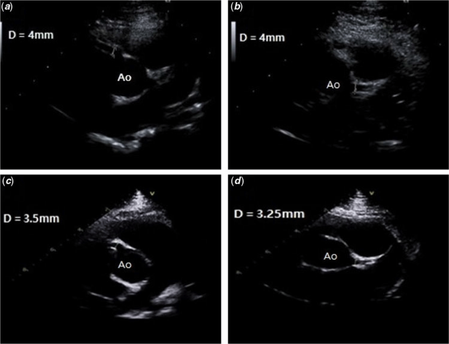
An 8-year-old boy presented with fever of 9 days duration, skin rash, bilateral conjunctival injection, and desquamation of bilateral finger tips. He had a history of climbing a mountain 10 days before symptoms occurred. On admission, he had fever of 38.3 °C. His heart rate was 80/minute and his blood pressure was 110/70 mmHg. His pharynx was injected and there was dry and red lip. He had erythematous macular rash on his trunk and lower extremities. Examination of his skin revealed an eschar on right inguinal area (Fig 1b). His laboratory results showed a white blood cell count 9,880/mm3, 53% polymorphonuclear leukocytes, 38% lymphocytes, haemoglobin 12.1 gm/dl, platelet count 267,000/mm3, C-reactive protein 1.23 mg/dl, and pro-brain natriuretic peptide 92.1 pg/ml. Mycoplasma pneumonia IgM was positive (24, normal < 10). Orientia tsutsugamushi antibody test was positive with the antibody titer 1:1280. Echocardiographic findings 1 day after admission revealed dilatation of both coronary arteries (right coronary artery = 4 mm, left coronary artery = 4 mm) (Fig 2a and b). He was treated on IV azithromycin and then oral roxithromycin for both mycoplasma infection and tsutsugamushi disease along with clinical features of Kawasaki disease which resolved after therapy with intravenous immunoglobulin and aspirin. Over the next 24 hours, he became afebrile and his rash improved. He was placed on low-dose aspirin for 8 weeks. His echocardiogram 2 months after the onset of his illness was within normal limits (right coronary artery = 3.5 mm, left coronary artery = 3.25 mm; Fig 2c and d).
Transthoracic echocardiography, parasternal short-axis view showing dilatation, 4 mm in diameter, involving (a) the right main coronary artery; (b) left main coronary artery soon after its origin from the aorta. Follow-up echocardiography showing normalised (c) right main coronary artery, 3.5 mm in diameter; (d) left main coronary artery, 3.25 mm in diameter.

Discussion
Kawasaki disease causes vasculitis syndrome in children which is characterised by clinical manifestations including fever persisting for 5 days or more, conjunctival infection, changes in oral mucosa and the tongue, skin rash, swelling of the cervical lymph nodes, and redness of the palms and soles of the feet.Reference Kawasaki1 Vasculitis in various tissues and organs, including the heart and blood vessels, is associated with several cardiovascular complications, such as myocarditis, pericarditis, ventricular dysfunction, valvular regurgitation, coronary ectasia or aneurysm, coronary thrombosis, coronary stenosis or occlusion, and ischaemic heart disease. Therefore, Kawasaki disease became one of the most common causes of acquired heart disease in infants and children.
Clinical and epidemiologic features suggest infectious agents as a possible cause of Kawasaki disease; however, the aetiology of Kawasaki disease still remains unknown. A number of microorganisms were hypothesised as an aetiology of the illness. Unfortunately, no specific agent that provides reproducible evidence has yet been reported.
Owing to the similarities in the clinical presentation, such as fever and skin rash, of Kawasaki disease and other infectious conditions, it is hypothesised that certain microbial infections may either cause Kawasaki disease or be associated with its pathogenesis. Although there is no definitive evidence that Kawasaki disease is an infectious disease, recent studies support the view that a dysregulated immune response to a variety of infectious stimuli is likely to contribute to Kawasaki disease pathogenesis.Reference Nakamura, Ikeda and Hamaoka2
Scrub typhus caused by Orientia tsutsugamushi can be transmitted by many species of chiggers encountered in high grass and brush. And it is endemic to Asia, especially Korea, Pakistan, and Australia. It infects vascular endothelial cells, causing systemic vasculitis, and macrophage and cardiomyocytes.
In one study in south India, which investigated clinical profile of 108 children diagnosed with scrub typhus, there was one case with complication of coronary artery dilatation, which was not been described in children.Reference Krishnan, Pillai, Elizabeth, Shanavas and Bindusha3
There was a study that electron microscopic findings of intracellular Rickettsia-like bodies have been found in biopsy specimens from the skin and lymph nodes, peripheral blood of patients with Kawasaki disease.Reference Hamashima, Kishi and Tasaka4 But ours is the first reported case of Kawasaki disease with tsutsugamushi disease with positive Orientia tsutsugamushi Ab. These support the hypothesis that rickettsial infection can be aetiologically related to Kawasaki disease.
These case reports suggest that Kawasaki disease can rarely occur concurrently or immediately after a rickettsial illness such as tsutsugamushi disease.
In addition, as there is possibility of Kawasaki disease preceded by tsutsugamushi disease, thorough physical examinations are needed to patients who are suspected for associated tsutsugamushi disease. And if there are features of Kawasaki disease, for example, conjunctival injection, red lip, cervical lymphadenitis, skin rash, etc., further evaluation including echocardiography should be considered for early diagnosis and treatment to prevent severe complication of Kawasaki disease and tsutsugamushi disease.
Acknowledgments
None.
Financial support
This research received no specific grant from any funding agency, commercial or not-for-profit sectors.
Conflict of interest
None.
Ethical standards
This article does not involve experimentation on humans or animals.