Key policy highlights
• Seasonal morphological changes in A. japonicus intestine were observed.
• Intestinal muscle and mucosal layers thinned in high water temperatures.
• Lipofuscin-like granules were observed in the mucosal and submucosal layers at high water temperatures, suggesting structural degradation of the tissue.
• Intestinal retraction and degeneration in A. japonicus under high temperatures may result from energy conservation, fasting, or reduced food intake during aestivation and winter dormancy.
Introduction
The Japanese sea cucumber (Apostichopus japonicus) is an important fishery species widely distributed along the coasts of Japan, China, Korea, and Russia (Takahashi, Reference Takahashi, Ueda, Maeda, Shimada and Takami2003). It is primarily exported to China (Shibuya, Reference Shibuya, Takahashi and Okumura2012), raising concerns about stock depletion due to increasing fishing pressure (Sakurai et al., Reference Sakurai, Yamada, Asabatake and Ohno2017).
After metamorphosis, A. japonicus initially resides and grows in intertidal zones before migrating to deeper offshore habitats (Hamano et al., Reference Hamano, Amio and Hayashi1989; Yamana et al., Reference Yamana, Hamano and Goshima2009a, Reference Yamana, Hamano and Goshima2010). Hamano et al. (Reference Hamano, Amio and Hayashi1989) reported that individuals in the post-migratory phase aestivate in sheltered algal reef areas during summer, while Yamana et al. (Reference Yamana, Hamano and Goshima2009a) observed aestivation among the narrow crevices of oyster-clusters and behind overhanging structures in subtidal zones. Aestivation is associated with weight loss (Nakahara et al., Reference Nakahara, Tokaji, Nakayama and Goshima2018; Nakajima and Gouda Reference Nakajima and Gouda2015; Tazono et al., Reference Tazono, Goda, Nakajima, Sato, Nagata and Motomae2016; Yang et al., Reference Yang, Yuan, Zhou, Mao, Zhang and Liu2005), including a reduction in the digestive tract weight-to-body wall weight ratio (Liu et al., Reference Liu, Zhou, Ru, Zhang, Cao and Yang2016; Masaki et al., Reference Masaki, Yamaura, Aoto and Okuma2007; Tachiyama et al., Reference Tachiyama, Shinohara and Fukagawa1997). In advanced stages of aestivation, the intestine may regress or disappear entirely (Nakajima and Gouda, Reference Nakajima and Gouda2015).
A. japonicus typically enters aestivation at water temperatures between 20.0°C and 30.5°C (Gao et al., Reference Gao, Yang, Xu, Wang and Liu2009; Ji et al., Reference Ji, Dong and Dong2008; Tachiyama et al., Reference Tachiyama, Shinohara and Fukagawa1997; Yang et al., Reference Yang, Yuan, Zhou, Mao, Zhang and Liu2005, Reference Yang, Zhou, Zhang, Yuan, Li, Liu and Zhang2006), and this state is likely triggered by sustained high temperatures rather than short-term changes. Photoperiod may also influence aestivation, as its onset and duration vary with latitude (Gao et al., Reference Gao, Yang, Xu, Wang, Liu and German2008). However, despite the critical importance of understanding environmental influences on this behaviour, few studies have explored how light conditions affect the aestivation of A. japonicus.
When studying aestivation in A. japonicus, it is essential to establish day lengths and water temperatures that closely mimic natural conditions. Such an approach enhances the accuracy of findings and facilitates a comprehensive understanding of the species’ physiological and behavioural responses during aestivation. By aligning experimental variables with the organism’s natural environment, researchers can significantly improve the relevance and applicability of their outcomes.
Weight loss associated with the aestivation of A. japonicus reduces the catch per unit effort, while habitat shifts suited to aestivation conditions, such as toward rocky shadows and hollows that do not receive direct sunlight, lead to decreased detection rates during fishing. Additionally, aestivating A. japonicus are more vulnerable to environmental stress. Thus, the aestivation of A. japonicus presents significant challenges for the effective use of this organism as a fishery resource, thus highlighting the importance of elucidating the mechanisms underlying this phenomenon.
Although specific temperatures cannot be generalised, A. japonicus exhibits reduced activity under low water temperatures during winter (Sun et al., Reference Sun, Zhang, Pan, Lin, Wang and Yang2018; Tanaka and Sakurai, Reference Tanaka and Sakurai2024). Weight loss and growth stagnation during this period suggest the possibility of hibernation (Jeong et al., Reference Jeong, Han and Kang2016; Tazono et al., Reference Tazono, Goda, Nakajima, Sato, Nagata and Motomae2016). In aquaculture settings, A. japonicus ceases feeding and hibernates when water temperatures drop below 1°C (Hu et al., Reference Hu, Gao, Wang, Li, Zhou, Ma, Li and Zhang2011). Water temperatures below 1°C are limited to some areas; however, hibernation implies that at least a certain period of inactivity is observed. Despite these observations, little research has explored winter dormancy behaviours beyond growth-related aspects, and no studies have explicitly investigated whether reduced activity in winter constitutes true dormancy.
High water temperatures have been shown to decrease the thickness of the intestinal muscle and the mucosal and submucosal layers, as well as reduce the number of plicae circulares in A. japonicus (Xu et al., Reference Xu, Sun, Liu, Zhang and Yang2015, Reference Xu, Yu, Zhang, Lv, Fu and Wang2018). However, these studies were conducted under artificial conditions with rapid temperature fluctuations that do not reflect natural environments. Furthermore, no studies have documented year-round changes in the intestinal tissue of A. japonicus in response to natural water temperature variations. Since the specific environmental conditions under which A. japonicus undergoes aestivation are not well understood, it is recommended that temperature and photoperiod changes mimic natural conditions. As a result, the mechanisms underlying intestinal retraction and regeneration in A. japonicus remain unclear from a histological perspective. The aestivation of A. japonicus in its natural environment is likely influenced by the long-term and combined effects of various environmental factors. Therefore, it is essential to document time-course changes in the intestinal tissue of A. japonicus under conditions that closely replicate its natural environment in terms of water temperature and photoperiod.
We investigated histological changes in the intestine of A. japonicus during long-term rearing under near-natural conditions. Moreover, we compared intestinal morphological changes during summer aestivation and winter low-activity to determine the mechanisms underlying tissue retraction and regeneration and seasonal variations in digestive capacity. To our knowledge, this is the first study to comprehensively document year-round histological changes in the intestine of A. japonicus under near-natural conditions. It is particularly significant for providing detailed temporal observations of intestinal changes during aestivation and subsequent recovery.
Materials and methods
Animals and experimental environment
The sea cucumbers (A. japonicus) (green-type) used in the experiment were obtained from a pre-shipment stock population maintained in a storage tank with a capacity of 1 t at a storage facility within the Sandomari fishing port in Rumoi Hokkaido, Japan. As these sea cucumbers were not experimental animals, ethical approval for animal experimentation was not required. In total, 260 individuals (mean standard body length ± standard deviation: 127.8 ± 25.7 mm) with standard body lengths ranging from 66.9 to 192.5 mm were collected on May 17, 2022, and June 6, 2023, from the Sandomari fishing port. Juvenile sea cucumbers produced with the cooperation of the Rumoi City Hall are released every year in this semi-enclosed fishing port. The storage tank included seawater from the collection site without filtration or temperature control. The pumped seawater was poured into the onshore tank, the outside temperature may have slightly affected it. The water temperature was monitored hourly using a HOBO Pendant Temperature/Light data logger (Pendant UA-002-64; Onset Computer Corporation, MA, USA). Day length was controlled to simulate natural conditions on a semi-monthly basis. The tank was equipped with cylindrical and L-shaped rigid polyvinyl chloride (PVC) tubes to provide structures suitable for aestivation. The sea cucumbers were fed commercially available sea cucumber feed (Namako Growth; Nosan Corporation, Japan), and residual food was always available.
Measurement of animals
Observations and measurements of sea cucumbers commenced on July 20, 2022, after approximately two months of acclimatisation and rearing. The study was conducted monthly until August 8, 2024, for a total of 26 observations. Ten individuals were sampled each month during the rearing period. This sample size was determined based on the number of sea cucumbers that could be maintained in the rearing tanks year-round without difficulty. It also ensured sufficient representation to detect morphological changes during each sampling period while minimising the influence of individual variability. Body length (L) and body width (B) were measured for each sea cucumber, and the standard body length (Le) was calculated using Eq. (1) (Yamana et al., Reference Yamana, Goshima, Hamano, Yusa, Furukawa and Yoshida2011):
 \begin{equation}Le = 2.17 \cdot {\left( {L \cdot B} \right)^{{\raise0.7ex\hbox{$1$} \!\mathord{\left/
{\vphantom {1 2}}\right.}
\!\lower0.7ex\hbox{$2$}}}}\end{equation}
Body length and width were measured using an electronic calliper (CD-P20S; Mitutoyo Co., Japan) with an accuracy of 0.01 mm, and weight was recorded using an electronic balance (FX3000I-JA; A&D Co. Ltd., Japan) with an accuracy of 0.01 g. The intestine was removed through an incision in the body wall, and the intestinal contents were removed before the body-wall weight and intestinal length were measured (using the same instruments). Beginning in February 2023, intestinal weights were measured separately for the anterior half (foregut) and posterior half (hindgut) to examine whether there were differences in morphology between the anterior and posterior portions. From June 2022, a portion of the foregut was excised, and from February 2023, a portion of the hindgut was also removed and fixed using Davidson’s fixative to prepare tissue specimens.
Using the measured values, the following parameters were calculated:
(1) The intestinal length/standard body length ratio, referred to as the relative intestinal length. (2) The foregut weight/body wall weight ratio, referred to as the relative foregut weight. (3) The hindgut weight/body wall weight ratio, referred to as the relative hindgut weight. (4) The foregut weight/foregut length ratio, representing the foregut weight per unit of intestinal length.
The hindgut weight/hindgut length ratio represents the hindgut weight per unit of intestinal length.
These calculations were performed using the following equations:
 \begin{align}&{\text{Relative intestinal length}} \nonumber\\
&\quad = {\text{intestinal length}}/{\text{standard body length }}\end{align}
 \begin{equation}{\text{Relative foregut weight}} = {\text{foregut weight}}/{\text{body wall weight}}\end{equation}
 \begin{equation}{\text{ Relative hindgut weight}} = {\text{hindgut weight}}/{\text{body wall weight}}\end{equation}
 \begin{align}&{\text{Foregut weight}}/{\text{foregut length ratio}} \nonumber\\
&\quad = {\text{foregut weight}}/{\text{foregut length}}\end{align}
 \begin{align}&{\text{Hindgut weight}}/{\text{hindgut length ratio}} \nonumber\\
&\quad = {\text{hindgut weight}}/{\text{hindgut length}}\end{align}
Aestivating individuals were identified using criteria adapted from previous studies (Gao et al., Reference Gao, Yang, Xu, Wang, Liu and German2008; Yuan et al., Reference Yuan, Yang, Chen and Gao2007; Yamana et al., Reference Yamana, Hamano and Goshima2009a, Reference Yamana, Hamano and Goshima2009b). These criteria included exhibiting one or more of the following behaviours or conditions: positive thigmotaxis (swarming behaviour), negative phototaxis (migration into PVC tubes), and negative geotaxis (attachment to the tank wall or the top of the PVC tube). Additionally, hardening of the body wall and the absence of digestive tract content were mandatory indicators of aestivation (Table 1). The possibility of hibernation was examined in winter using the same criteria as for aestivation; however, hibernation was not observed. Therefore, the period from January to March, when water temperatures decreased, was classified as the ‘activity slowdown period’.
Summary table on the definition of aestivating individuals of the Apostichopus japonicus during the study period

Preparation of intestinal tissue specimens
Paraffin sections, 3 µm thick, were prepared from the fixed intestine using standard histological procedures. The sections were stained with haematoxylin and eosin and examined under a microscope. Tissue specimens were prepared for one to five individuals during each sampling month. The number of samples varied depending on the presence or absence of intestines and occasional technical difficulties in tissue preparation. Although morphological changes in gut tissue varied among individuals, we are confident that comparing specimens collected within the same sampling month provided representative samples for that period.
Results
Water temperature, day length, and aestivation in individuals
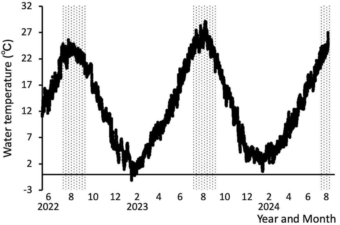
The rearing water temperature fluctuated from 11.9°C on May 17, 2022, at the start of the rearing period, to a peak of 25.7°C on July 30, 2022 (Figure 1). It then decreased, reaching a low of −1.1°C on January 26, 2023. Similar fluctuations in water temperature were observed throughout the remainder of the experiment.
Changes in the water temperature in the rearing tank during the observation period. Hatching indicates the emergence period of aestivating individuals.

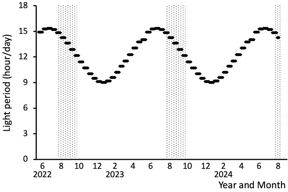
Figure 2 illustrates the changes in day length during the rearing period, expressed as hours of light per day. The maximum light duration was 15.4 hours, recorded from June 16 to June 30, while the minimum duration was 9 h, observed from December 16 to December 31 each year. These variations closely mirrored natural conditions. Observations during measurements indicated that aestivating individuals were consistently present in July, August, and September each year.
Changes in the day length (hours of light per day) in the rearing tank during the observation period. Hatching indicates the emergence period of aestivating individuals.

Changes in intestinal size
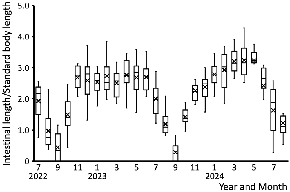
Seasonal changes in relative intestinal length are shown in Figure 3. The relative intestinal length decreased from July 2022, when measurements began, to September 2022, before increasing towards November 2022. It then remained stable until June 2023. Similar seasonal patterns were observed from July 2023 onward.
Seasonal changes in the relative intestinal length of Apostichopus japonicus between July 2022 and August 2024. Cross mark indicates the average value.

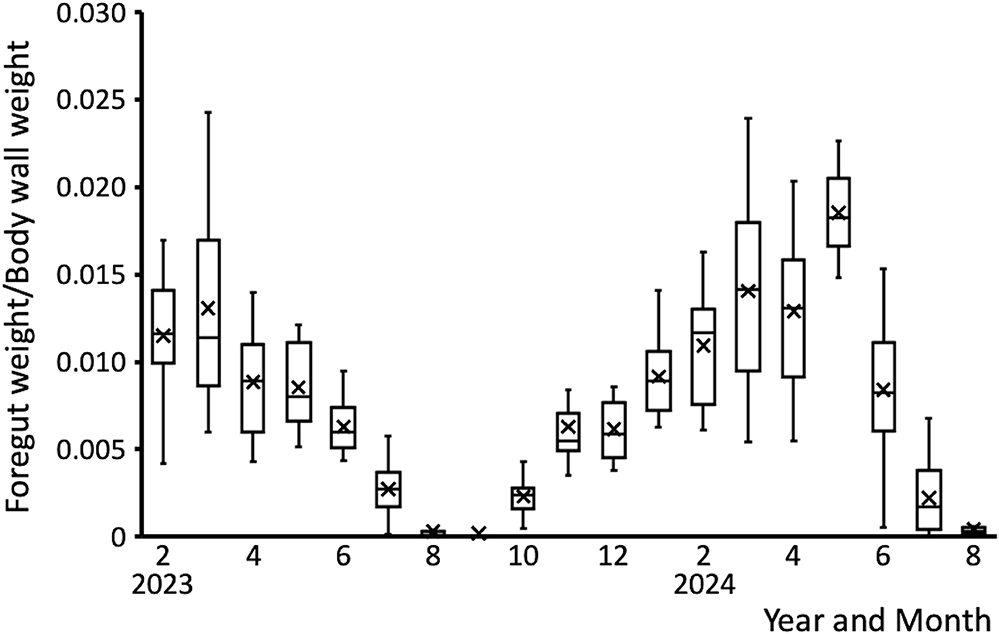
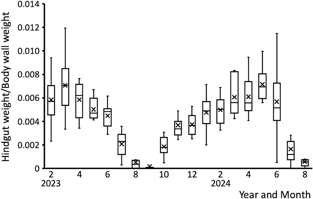
Figures 4 and 5 depict seasonal changes in relative foregut weight and relative hindgut weight, respectively. Relative foregut weight decreased from June 2023 to September 2023, then recovered by January 2024 to the levels observed in May 2023, before the decline. From February 2024 onward, relative foregut weight exhibited similar seasonal trends. Relative hindgut weight decreased from July 2023 to September 2023, followed by an increase toward November 2023. From December 2023 onward, similar seasonal patterns were observed.
Seasonal changes in the relative foregut weight of Apostichopus japonicus between February 2023 and August 2024. Cross mark indicates the average value.

Seasonal changes in the relative hindgut weight of Apostichopus japonicus between February 2023 and August 2024. Cross mark indicates the average value.

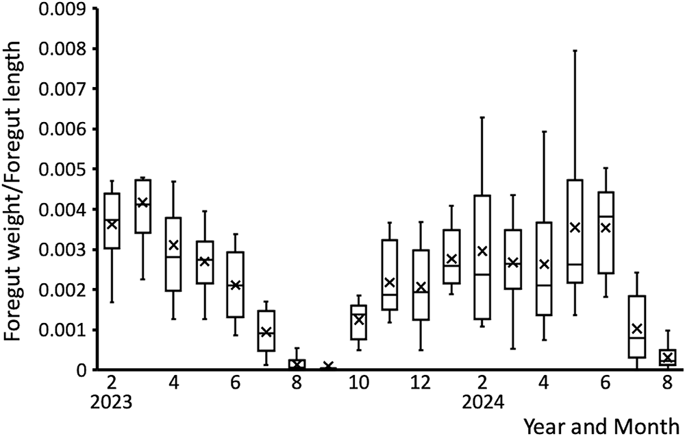
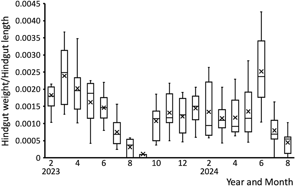
Seasonal changes in the foregut weight/foregut length ratio and hindgut weight/hindgut length ratio are presented in Figures 6 and 7, respectively. The foregut weight/foregut length ratio decreased from July 2023 to September 2023, followed by an increase toward November 2023. It then stabilised and remained consistent until June 2024, with a similar seasonal trend observed from July 2024 onward. The hindgut weight/hindgut length ratio showed a similar pattern, decreasing from July to September, followed by an increase toward November of the same year.
Seasonal changes in the foregut weight-to-foregut length ratio of Apostichopus japonicus specimens between February 2023 and August 2024. Cross mark indicates the average value.

Seasonal changes in the hindgut weight-to-hindgut length ratio of Apostichopus japonicus between February 2023 and August 2024. Cross mark indicates the average value.

Changes in intestinal histology
Histological images of the foregut and hindgut are shown in Figures 8 and 9, respectively. In both regions, the intestinal wall consisted of four layers: the mucosal layer, the submucosal layer, the muscle layer, and the serosa. During the active period, numerous plicae circulares were observed in the mucosal layer of both the foregut and hindgut (Figure 8a–c; Figure 9a, c). In contrast, during the high water temperature period, the mucosal layer showed reduced height (Figure 8d, e; Figure 9d, f), along with partial loss (Figure 8e) or complete disappearance (Figure 9e) in some areas. Additionally, the muscle layer of both the foregut and hindgut during this period was significantly thinner than during the active period (Figure 8d, e; Figure 9e). Although these changes in the mucosal and muscle layers were difficult to quantify, they showed consistent trends across individuals. Lipofuscin-like granules were identified in the submucosal layer of the foregut and both the mucosal and submucosal layers of the hindgut during the high water temperature period (Figure 8d, e; Figure 9e, f). A common feature observed in both the foregut and hindgut during both high and low water temperature periods was the presence of haemolymph-like cells in the submucosal layer, located near the mucosal layer (Figure 8f, g; Figure 9f, g). These haemolymph-like cells were identified morphologically based on previous descriptions of spherical haemolymph cells (5–8 µm in diameter) in the pearl oyster Pinctada fucata, the noble scallop Mimachlamys nobilis, and the sunray surf clam Mactra chinensis (Funahashi Reference Funahashi2000).
Histological images of the foregut of Apostichopus japonicus. (a, b) Intestinal tissue during the active period. Scale bar: 100 µm. (c) Intestinal tissue during the active period. Scale bar: 50 µm. (d) Intestinal tissue during the high water temperature period. The height of the mucosal layer is reduced, and the muscle layer is thinner than during the active period. The black arrow indicates lipofuscin-like granules in the submucosal layer. Scale bar: 50 µm. (e) Intestinal tissue during the high water temperature period. The mucosal layer is partially lost, and the muscle layer is thinner. The black arrow indicates lipofuscin-like granules in the submucosal layer. Scale bar: 100 µm. (f) Intestinal tissue during the high water temperature period. The black arrow indicates haemolymph-like cells in the submucosal layer. Scale bar: 20 µm. (g) Intestinal tissue during the low water temperature period. The black arrow indicates haemolymph-like cells in the submucosal layer. Scale bar: 50 µm. The mucosal layer (ML), submucosal layer (SL), and muscle layer (MUL) are each indicated.

Histological images of the hindgut of Apostichopus japonicus. (a, c) Intestinal tissue during the active period. Scale bar: 100 µm. (b) Intestinal tissue during the active period. Scale bar: 50 µm. (d) Intestinal tissue during the high water temperature period. The height of the mucosal layer is reduced. Scale bar: 100 µm. (e) Intestinal tissue during the high water temperature period. The mucosal and muscular layers are thinner than during the active period. The black arrow indicates lipofuscin-like granules in the submucosal layer. Scale bar: 100 µm. (f) Intestinal tissue during the high water temperature period. The height of the mucosal layer is reduced. Lipofuscin-like granules are present in the mucosal and submucosal layers. The black arrow indicates haemolymph-like cells in the submucosal layer. Scale bar: 50 µm. (g) Intestinal tissue during the low water temperature period. The black arrow indicates haemolymph-like cells in the submucosal layer. Scale bar: 20 µm. The mucosal layer (ML), submucosal layer (SL), and muscle layer (MUL) are each indicated.

Discussion
In this study, aestivating individuals were observed annually in July, August, and September. The aestivation period coincided with the months when water temperatures were high, followed by months when the temperatures began to decline. Day length during these months also marked the onset of the shortening of daily light hours. Although no direct records on the relationship between day length and aestivation were found, the water temperature associated with aestivating individuals followed a similar trend to that reported by Goshima (Reference Goshima, Hirota and Machiguchi2014). It has also been suggested that A. japonicus may undergo hibernation at lower water temperatures (Jeong et al., Reference Jeong, Han and Kang2016; Tazono et al., Reference Tazono, Goda, Nakajima, Sato, Nagata and Motomae2016). However, while a slowdown in activity has been documented, no evidence of true winter dormancy behaviour has been observed. In the present study, no behaviours indicative of winter dormancy were observed. These findings suggest that the winter slowdown in A. japonicus activity is not a result of dormancy. The absence of winter dormancy in A. japonicus may be attributed to its ectothermic physiology. As an ectotherm, the metabolic rate of A. japonicus possibly decreases in response to the lower water temperatures of winter. This reduction in metabolic rate may lead to decreased energy consumption, allowing the species to conserve energy without entering a dormant state. However, the ‘activity slowdown’ defined in this study is not yet supported by measurements of metabolic rate, oxygen consumption, or feeding behaviour. Further physiological and behavioural research is needed to characterise this period more accurately.
In Cyclorana alboguttata, a species of Hylidae, aestivation results in a reduction in the weights of the stomach, colon, and small intestine (Cramp and Franklin, Reference Cramp and Franklin2005). Such responses are commonly observed in long-term aestivating species, where prolonged disuse during aestivation leads to the degeneration of digestive organs, with the rate of cell loss exceeding that of cell gain (Jiang et al., Reference Jiang, Storey, Yang and Sun2023). In the present study, the relative intestinal length, relative hindgut weight, foregut weight/foregut length ratio, and hindgut weight/hindgut length ratio of A. japonicus decreased annually from July to September. Similarly, relative foregut weight declined from June to September each year. These findings are consistent with those of previous research (Jiang et al., Reference Jiang, Storey, Yang and Sun2023). Furthermore, relative intestinal length, relative hindgut weight, foregut weight/foregut length ratio, and hindgut weight/hindgut length ratio increased towards November, suggesting that October–November is the recovery period for intestinal morphology following retraction caused by elevated water temperatures. Interestingly, while the relative hindgut weight returned to pre-decline levels by November, the recovery of relative foregut weight was delayed until January of the following year. This observation suggests that, in comparison to the hindgut, the foregut of A. japonicus is more sensitive to rising water temperatures, shows earlier effects of retraction, and requires a longer time for regeneration. Previous studies have shown that villi in the foregut are more developed than those in the hindgut (Shimizu et al., Reference Shimizu, Mikami and Takahashi1994). Although cells with peptidase- and lipase-positive granules are found throughout the intestine, stronger positive reactions were observed in the foregut (Shimizu et al., Reference Shimizu, Mikami and Takahashi1994). These findings suggest that the foregut has a higher digestive capacity. Consequently, the more complex structure of the foregut may also account for the longer time required for its regeneration.
The histological changes observed in both the foregut and hindgut of A. japonicus were similar. Our finding that the height of the mucosal layer decreases during periods of high water temperature aligns with the results of a previous study, which examined the intestinal tissue of A. japonicus exposed to high water temperatures of 27°C for 6–192 hours (Xu et al., Reference Xu, Sun, Liu, Zhang and Yang2015). The observed partial loss or disappearance of the mucosal layer suggests that the degeneration of this layer is likely an early factor contributing to intestinal regression associated with elevated water temperatures. However, since the sampling interval in this study was once a month, future studies should focus on more detailed tissue observations at shorter intervals to clarify these temporal changes.
This study also confirmed that the muscle layer of the intestine becomes noticeably thinner during periods of high water temperature. Similar changes have been reported in the intestinal tissues of Silurus meridionalis (Zeng et al., Reference Zeng, Li, Li, Cao, Fu and Zhang2012), a catfish species, and Lates calcarifer, Barramundi, during fasting (Purushothaman et al., Reference Purushothaman, Lau, Saju, Sk, Lunny, Vij and Orbán2016). Additionally, A. japonicus exhibits reduced mobility (Tanaka and Sakurai, Reference Tanaka and Sakurai2024) and food intake (Sun et al., Reference Sun, Zhang, Pan, Lin, Wang and Yang2018) at high water temperatures. Feeding cessation has also been observed during aestivation in this species (Yuan et al., Reference Yuan, Yang, Chen and Gao2007). These findings suggest that the thinning of the intestinal muscle layer in A. japonicus may be attributed to fasting or reduced feeding under high water temperature conditions.
Lipofuscin-like granules were identified in the mucosal and submucosal layers of the intestinal tissue of A. japonicus during periods of high water temperature. It has been reported that in turbot (Scophthalmus maximus), abnormalities in lysosomal hydrolytic enzymes in the intestinal epithelium can lead to lipofuscin accumulation, as lipids are less efficiently digested and accumulate in tissues (Lamas et al., Reference Lamas, Novoa and Figueras1996). This phenomenon may be comparable to the accumulation observed in the intestine of A. japonicus. However, in this study, identification of lipofuscin-like granules was based on morphology and staining characteristics in haematoxylin and eosin sections. Future studies should employ biochemical or fluorescence-based methods to confirm the identity of these granules.
A common feature of the A. japonicus intestine during both high and low-temperature periods was the presence of haemolymph-like cells in the submucosal layer, near the mucosal layer. The haemolymph of several marine invertebrates has phagocytic properties (Mori and Nomura, Reference Mori1979). Similarly, haemolymph accumulation at wound sites has been reported in the greater wax moth (Galleria mellonella) during wound healing (Rowley and Ratcliffe, Reference Rowley and Ratcliffe1978), and phagocytic haemolymph cells are commonly found in bivalves and marine mussels (Mitta et al., Reference Mitta, Hubert, Dyrynda, Boudry and Roch2000). These findings suggest that the presence of a large number of haemolymph-like cells in the intestinal tissue of A. japonicus may be a response to tissue damage caused by fasting or reduced food intake during periods of high water temperature. The haemolymph cells possibly phagocytose the damaged cells.
While previous studies have investigated intestinal degeneration using high-frequency sampling at intervals of five days under high-temperature exposure (Xu et al., Reference Xu, Yu, Zhang, Lv, Fu and Wang2018), the novelty of the present study lies in its year-round observation of intestinal changes under rearing conditions closely approximating the natural environment. However, monthly sampling may have limited the ability to capture the detailed progression of tissue changes. Therefore, future studies should consider more frequent sampling, at least once a week, particularly during the aestivation and recovery periods, using A. japonicus reared under similarly naturalistic conditions.
Zeng et al., (Reference Zeng, Li, Li, Cao, Fu and Zhang2012) discussed the tendency of animals to degenerate their digestive organs and reduce their functionality as an energy-conservation strategy. Similarly, Watanabe (Reference Watanabe1985) suggested that the degeneration of the intestinal tissue in two-day starved Chaenogobius annularis (forktongue goby) larvae may be a survival response, with the body catabolising its components when stored nutrients are depleted.
In conclusion, the regression and degeneration of the intestinal tissue in A. japonicus during periods of high water temperatures are likely adaptive responses to conserve energy. These processes allow the organism to tolerate unfavourable conditions for growth and survival, such as high water temperatures and reduced food intake associated with aestivation and slowed activity during the winter. However, the discussion of energy conservation in this study is based solely on morphological evidence. Future research should incorporate metabolomic analyses and measurements of oxygen consumption to clarify the causal relationship between intestinal regression and energy consumption.
Acknowledgements
We want to thank Dr S. Nakamura, a lecturer in the Department of Veterinary Medicine, Faculty of Veterinary Medicine, Okayama University of Science, for his advice on observing intestinal tissue. We also appreciate the cooperation of the staff at Rumoi City Hall and Chief H. Miura of the Academic and Student Affairs Division at Tokai University in collecting and rearing the test specimens. This research was partly funded by the Sasakawa Scientific Research Grant (2024-4005, represented by K. Tanaka). We sincerely thank the individuals mentioned above for their support.
Author contributions
Kai Tanaka mainly conducted the experiments, analysed the results, and wrote the manuscript. Minoru Kihara and Izumi Sakurai established and supervised the experimental methodology. All authors contributed to drafting the manuscript, interpreting the results, and read and approved the final version.
Funding
This research was partly funded by the Sasakawa Scientific Research Grant (2024-4005).
Competing interests
The authors have no competing interests to declare.
Ethical standards
A. japonicus used in the experiment were obtained from a pre-shipment stock population maintained in a storage tank at the Sandomari fishing port in Rumoi, Hokkaido, Japan. As these sea cucumbers were not experimental animals, ethical approval for animal experimentation was not required. Furthermore, throughout the entire rearing process, the standards of the Act on Welfare and Management of Animals (Act No. 105 of October 1, 1973) were met.
Data availability
All relevant data are provided with the article but are not stored in a public repository.