Introduction
Biotite is a typical 2:1 phyllosilicate mineral, constituting ∼8% of the Earth’s crustal mass (Nesbitt and Young, Reference Nesbitt and Young1984). Influenced by the chemical weathering of surface water and microbial activities, biotite undergoes dissolution and transformation, giving rise to secondary minerals, such as hydrobiotite, vermiculite and kaolinite (McMaster et al., Reference McMaster, Smits, Haward, Leake, Banwart and Ragnarsdottir2008; Galán and Ferrell, Reference Galán, R.E, Bergaya and Lagaly2013; Wu et al., Reference Wu, Chen, Wang, Xu, Lin, Liang and Cheng2023), which are significant portions of soil minerals. Moreover, the K+ within the interlayer of biotite is gradually exchanged by H+ or other hydrated metal cations, often accompanied by the oxidation of octahedral Fe2+ and dissolution of other cations (Bray et al., Reference Bray, Oelkers, Bonneville, Wolff-Boenisch, Potts, Fones and Benning2015; Lamarca-Irisarri et al., Reference Lamarca-Irisarri, Van Driessche, Jordan, Cappelli and Huertas2019; Wang et al., Reference Wang, Nurdiana, Sato, Watanabe and Tsuchiya2024). The elements released, such as K, Mg and Fe, serve as crucial nutrients for the biosphere (Bowser and Jones, Reference Bowser and Jones2002; Samantray et al., Reference Samantray, Anand, Dash, Ghosh and Behera2022). Additionally, biotite weathering consumes CO2, acting as a net sink in the global carbon cycle, thereby influencing the global climate (Wilson, Reference Wilson2004; Van Der Kellen et al., Reference Van Der Kellen, Derrien, Ghanbaja and Turpault2022). As such, biotite weathering has an impact on geochemical processes such as soil formation, elemental migration and enrichment, nutrient supply to the biosphere, and the overall carbon balance on Earth.
The dissolution and transformation of biotite are key processes in biotite weathering, which have been proven to be strongly influenced by environmental conditions. For instance, under acidic conditions, a higher concentration of H+ (i.e. lower solution pH) results in an accelerated biotite weathering rate (Pachana et al., Reference Pachana, Zuddas and Censi2012; Cappelli et al., Reference Cappelli, Van Driessche, Cama and Huertas2023; Holgersson et al., Reference Holgersson, Drake, Karlsson and Krall2024). Furthermore, organic acids facilitate mineral dissolution by complexing with metal ions on the mineral surface (Ganor et al., Reference Ganor, Reznik, Rosenberg, Oelkers and Short2009; Haward et al., Reference Haward, Smits, Ragnarsdóttir, Leake, Banwart and McMaster2011). On the other hand, inorganic cations are distributed widely in the surface environment (e.g. soil and groundwater) with various types (e.g. Ca2+, Mg2+, Na+ and K+) and large concentration variations (ranging from μM to M levels) (Breen et al., Reference Breen, Angelo, Masters and Sedam1985; Kharaka and Hanor, Reference Kharaka, Hanor, Holland and Turekian2014), which also have a non-negligible influence on biotite weathering. For example, in experiments simulating the effects of cations on biotite weathering, Na+, Rb+ and Cs+, due to their identical charge and comparable radius to K+, could significantly enhance the release of interlayer K+ in biotite (Kim and Kim, Reference Kim and Kim2015; Wang et al., Reference Wang, Shi, Wu, Ding, Liang, Li and Fan2023). Among them, hydrated Na+ entered the interlayer of biotite and opened the interlayer channels, promoting the dissolution of the layer structures (Kim and Kim, Reference Kim and Kim2015). Min et al. (Reference Min, Kim and Jun2018) studied how Na+ and K+ impact the dissolution/transformation of biotite under acidic hydrothermal conditions. They found that even a small amount of K+ in the solution could inhibit biotite dissolution. A low concentration of Na+ (less than 0.5 mol/L) could promote biotite dissolution, whereas the promoting effect of a high concentration of Na+ (greater than 0.5 mol/L) was reduced, which was attributed to the competitive interaction between Na+ and protons. Moreover, in the presence of Na+, noticeable cracks appeared on the surface of the reacted biotite, and fibrous illite was formed (Hu et al., Reference Hu, Ray and Jun2011). Therefore, metal cations have a great impact on the dissolution process and transformation products of biotite.
Previous research has concentrated primarily on the influence of a certain type of cation (mostly alkali metal cations) on biotite dissolution. However, there are various cations in the terrestrial environment, with significant differences in their properties (e.g. valence, ionic radius and hydration capacity, Supplementary Table S1) (Teppen and Miller, Reference Teppen and Miller2006; Tansel, Reference Tansel2012), which differ in their affinity for biotite. Further research is needed to clarify the influence of the type of cation on the dissolution and transformation of biotite. In addition, in current studies on the interaction between cations and biotite, emphasis is generally on the impact of cations on the dissolution kinetics and mechanisms of biotite, with insufficient research on the accompanying phase transformation process of biotite.
In this work, five typical metal cations (Na+, K+, Ca2+, Mg2+ and Al3+) were selected to study the effect of cation types on the dissolution and transformation of biotite. By combining modern spectroscopic methods and micro-beam characterisation techniques, we focussed on the mineralogical characteristics of the transformation products (e.g. phase composition, microstructure, and surface morphology) and the relevant phase transformation mechanisms. This work sought to clarify the influence of metal cation types on the dissolution and transformation of biotite, providing new information for an in-depth understanding of biotite weathering in the surface environment.
Experimental section
Materials
The pristine biotite (Bt) from Hebei, China, was purified initially through manual selection, light cleaning and magnetic separation. Subsequently, the material was ground into a powder and passed through a 200-mesh sieve, with the undersized particles being reserved for further experiments. The structural chemical formula of biotite was calculated to be (K0.87Na0.04Ca0.02)(Mg1.69Mn0.01Fe0.92Ti0.10Al0.17)(Al1.25Si2.75)O10(F0.06(OH)0.92)2 according to the corresponding chemical composition (Table S2). Sodium chloride (NaCl), potassium chloride (KCl), magnesium chloride (MgCl2), calcium chloride (CaCl2), aluminium chloride hexahydrate (AlCl3·6H2O), and hydrochloric acid (HCl, 37 wt.%) were all procured from Guangzhou Chemical Reagent Factory.
Dissolution experiments
First, solutions of NaCl, KCl, CaCl2, MgCl2 and AlCl3·6H2O with concentrations of 0.60 mol/L were prepared, and the system pH was set to 2 by adding an HCl solution. 50 mL of the above solutions were placed in different reaction vessels (with PTFE liners), followed by adding 0.10 g of biotite. Subsequently, the sealed reaction vessels were transferred to an oven at 150°C to initiate the dissolution/transformation experiment of biotite. The solution pH and reaction temperature settings were designed to accelerate the reaction rate (Cho and Komarneni, Reference Cho and Komarneni2007; Lamarca-Irisarri et al., Reference Lamarca-Irisarri, Van Driessche, Jordan, Cappelli and Huertas2019). Sampling was done at set time points (0 to 90 d), and the post-reaction suspension was centrifuged to collect the supernatant. The concentrations of various cations in the solution were analysed utilising a Thermo iCAP 7000 series inductively coupled plasma-atomic emission spectrometry (ICP-AES). The solid products were washed with distilled water, air-dried at room temperature, and collected for structural and morphological analysis. Based on the type of cation used and the reaction time, the resulting products were labelled as Bt-M-yd (where M represents the cation type and y denotes the sampling days). For instance, Bt-Na-30d signified the product obtained after biotite reacted for 30 d in the NaCl solution. Additionally, the control experiment (blank system) where no cations were added, but all other conditions remained consistent with the above experiment, resulted in products labelled as Bt-H-yd (with y representing the sampling days). For brevity, the systems containing NaCl, KCl, CaCl2, MgCl2 and AlCl3·6H2O solutions were abbreviated as the Na system, K system, Ca system, Mg system and Al system, respectively.
Characterisation methods
Powder X-ray diffraction (XRD) patterns were determined utilising a Bruker AXS D8 ADVANCE X-ray diffractometer fitted with Ni-filtered CuKα radiation (λ = 1.541 Å) at 40 kV and 40 mA. The powdered samples were loaded into the holder and flattened with a glass slide for measurement.
Thermogravimetric (TG) curves were recorded using a NETZSCH STA 409PC synchronous thermal analyser. The sample was heated in a corundum crucible from 30°C to 1000°C under a N2 flow (60 ml min−1) with a ramp rate of 10°C min−1. The derivative thermogravimetry (DTG) curve was generated by differentiating the TG curve.
Fourier transform infrared spectroscopy (FTIR) analysis was conducted using a Bruker Vertex-70 FTIR spectrometer. The mixture of KBr powder and the sample with a mass ratio of 80:0.9 was pressed into moulds to form thin sheets for measurement. The spectra were collected over a range of 400 to 4000 cm−1.
57Fe Mössbauer spectra were obtained at room temperature through transmission mode, employing a Silver Double Limited WSS-10 Mössbauer spectroscopy system with a 57Co/Rh as the Mössbauer source. The velocity-driven sensor functioned in a triangular wave mode within the energy range of ±15 mm/s, and the acquired spectra were performed using MossWinn 4.0 software.
Transmission electron microscopy (TEM) images, high-angle annular dark-field (HAADF) images, and energy-dispersive X-ray spectroscopy (EDS) were obtained using a FEI Talos F200S field-emission transmission electron microscope operating at 200 kV. The sample powder underwent ultrasonic dispersion in anhydrous ethanol, after which a drop of the suspension was deposited on a carbon-coated copper grid and allowed to air-dry before observation. To capture high-resolution transmission electron microscopy (HRTEM) images, a minute sample portion was embedded within an epoxy matrix, cured by heating, and then sliced into thin sections (∼75 nm thick) using the Leica EM UC7 ultramicrotome. These sliced samples were picked up with a copper grid for observation.
Results and discussion
Effect of various cations on biotite dissolution
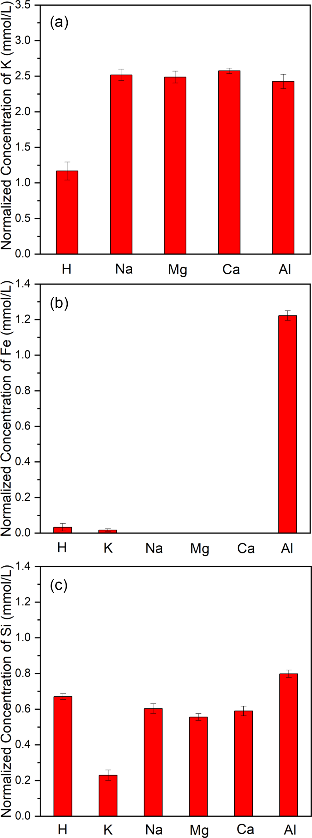
To study the influence of cation types on biotite dissolution, the ion concentrations of K, Fe and Si in the supernatant of different cation systems after 30 d of dissolution experiments were measured. The concentrations of these three elements in the solution can respectively indicate the dissolution of interlayer ions, tetrahedral sheets and octahedral sheets of biotite. The ion concentrations, normalised by the stoichiometry of biotite, revealed that the K concentration in the solution for the Na, Mg and Ca systems was notably higher than in the blank system (Fig. 1a), due to the promotion of interlayer ion exchange of biotite by the metal cations. However, these three systems exhibited no Fe and a lower concentration of Si in the solution compared to the blank system (Fig. 1b, c). In contrast, the solution pH values (3.50–4.00) of the Na, Mg and Ca systems after reactions were higher than that of the blank system (2.53) (Table S3), indicating that more protons were consumed in the cation systems (leading to more intense dissolution of biotite). Hu et al. (Reference Hu, Ray and Jun2013) found through atomic force microscopy that when large-radius hydrated cations entered the interlayer of biotite via ion exchange, the biotite layer underwent significant swelling, which promoted layer bulging and crack formation, and thus increased the chance of proton attack on the layers. In this present study, the dissolution of structural Fe and Si from biotite was expected to be greater in the cation systems compared to the blank system. However, the enhanced dissolution of biotite consumed more protons, and the raised solution pH caused the dissolved Fe to precipitate in the form of hydroxides, hence Fe was almost undetectable in the solution of the cation systems (Fig. 1b). Additionally, the weakly crystalline iron hydroxides formed had a strong adsorption capacity for silicate ions (Carlson and Schwertmann, Reference Carlson and Schwertmann1981; Hiemstra, Reference Hiemstra2018), adsorbing a certain amount of released Si (resulting from the structural degradation of biotite). This led to a lower Si concentration in the cation system. The low Fe content in the solution of the blank system could be linked to the adsorption effect of clay minerals (e.g. hydrobiotite and vermiculite, confirmed later by XRD and TEM) formed during the dissolution and transformation of biotite.
Normalised ion concentrations of K (a), Fe (b), and Si (c) in the supernatant of different cation systems after biotite dissolution for 30 d.

Compared with the blank system, the K system showed lower concentrations of Fe and Si (Fig. 1b, c) and a smaller increase in solution pH after the reaction (Table S3), indicating a lower dissolution degree of biotite in this system. This phenomenon was primarily due to the competitive interaction between K and protons on the biotite surface, leading to a reduced proton attack on the layers. Owing to the hindered dissolution of biotite and the substantial amount of added K+, the alteration in K+ concentration within the solution of the K system resulting from the cation exchange reaction was barely discernible. In the Al system, the K concentration in the solution was comparable to that in the Na, Mg and Ca systems (Fig. 1a), but the concentrations of structural Fe and Si increased greatly (Fig. 1b, c), and the solution pH after the reaction (2.63) was close to that of the blank system (Table S3). This was because the hydrolysis of added Al3+ generated a large number of protons (Cho and Komarneni, Reference Cho and Komarneni2007; Li et al., Reference Li, He, Tao, Zhu, Tan, Ji, Yang and Zhang2020), significantly promoting the dissolution of biotite while preventing a substantial increase in solution pH.
In all the systems, the normalised ion concentrations of K, Si and Fe were asynchronous, indicating a non-stoichiometric dissolution behaviour for biotite. Among them, the normalised ion concentration of K was the highest, followed by Si, and Fe was the lowest (except for the Al system). It was generally believed that under acidic conditions, the dissolution of biotite structural ions was influenced mainly by protons, with octahedral cations such as Mg and Fe dissolving first, followed by tetrahedral Al, and finally Si. This sequence was attributed to the weaker chemical bonds of Fe/Mg–O in the octahedral sheets compared to Al/Si–O in the tetrahedral sheets (Bray et al., Reference Bray, Oelkers, Bonneville, Wolff-Boenisch, Potts, Fones and Benning2015; Masson et al., Reference Masson, Robin, Joussein, Tertre and Baron2024). The normalised ion concentration of Fe in the solution was lower than that of Si in this work, due to the adsorption of dissolved Fe by secondary products (such as clay minerals) (Jolicoeur et al., Reference Jolicoeur, Ildefonse and Bouchard2000) and the formation of iron hydroxides (Tamrat et al., Reference Tamrat, Rose, Grauby, Doelsch, Levard, Chaurand and Basile-Doelsch2018). The non-stoichiometric dissolution of various elements in biotite has been reported frequently in previous studies (Kalinowski and Schweda, Reference Kalinowski and Schweda1996; Bray et al., Reference Bray, Oelkers, Bonneville, Wolff-Boenisch, Potts, Fones and Benning2015; Holgersson et al., Reference Holgersson, Drake, Karlsson and Krall2024). In contrast to the aforementioned systems, the normalised ion concentration of Fe was higher than that of Si in the Al system, as the dissolved Si reacted with Al in the solution to form kaolinite (verified by the phase analysis of the biotite-transformed products below).
Structure of biotite-transformed products in various cation systems
XRD analysis
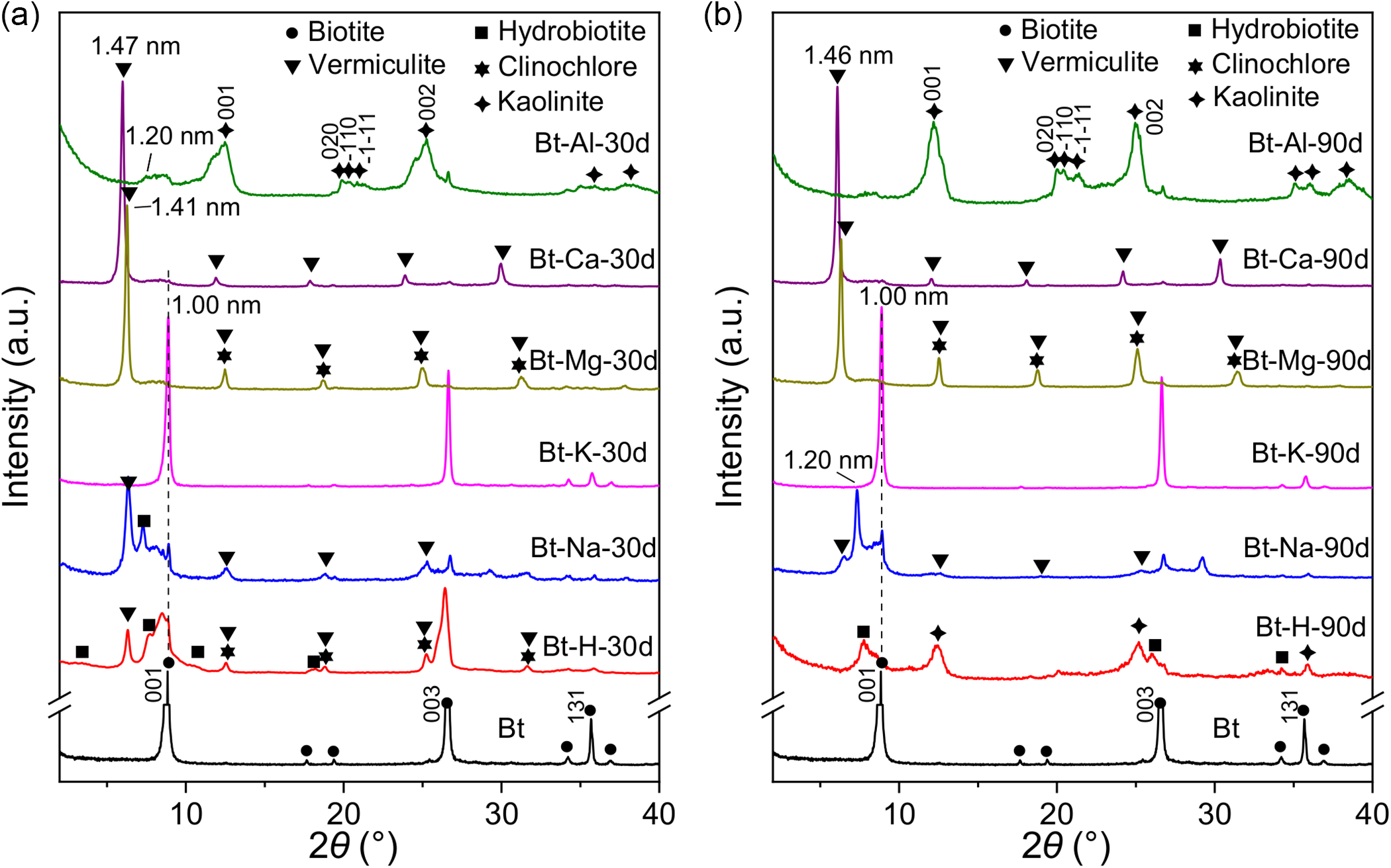
X-ray diffraction patterns of the resulting products following the dissolution reaction of biotite for a certain period in various cation systems were recorded (Fig. 2). The XRD pattern of the original Bt exhibited sharp characteristic reflections of biotite, with no other phase signals (Jeong and Kim, Reference Jeong and Kim2003) (Fig. 2a), indicating the sample’s high purity. For the blank system, the XRD pattern of the product at 30 d showed the characteristic reflections of vermiculite, chlorite and hydrobiotite, while still retaining relatively distinct reflections of biotite (Fig. 2a). Hydrobiotite is a 2:1 regular interlayer clay mineral, with a structure formed by alternating vermiculite layers and biotite layers in a 1:1 arrangement. The products in the Na system exhibited a similar phase composition to the blank system but with stronger reflections of vermiculite and weaker reflections of the original biotite, indicating a higher reaction rate of biotite. According to ion concentration analysis, Na+ could promote the exchange of K+ within the interlayer of biotite. Na+ with a larger hydration capacity than K+ entered the interlayer space from the layer edges, opened the interlayer channels, and allowed more protons to enter the interlayers, facilitating the dissolution of structural ions (Sánchez-Pastor et al., Reference Sánchez-Pastor, Aldushin, Jordan and Schmahl2010). By contrast, the product in the K system presented the same characteristic reflections as the original biotite. In the Ca and Mg systems, vermiculite appeared in the products at 30 d, with basal spacings of 1.47 and 1.41 nm, respectively, attributed to the larger hydration radius of Ca2+ than Mg2+ (Ikeda et al., Reference Ikeda, Boero and Terakura2007). In addition, reflections of chlorite were observed in the XRD pattern of the products in the latter system. For the Al system, the XRD pattern of the products revealed characteristic reflections of kaolinite including (001), (020), ( $\bar 1$10), (
 $\bar 1$
 $\bar 1$1) and (002) planes (He et al., Reference He, Ji, Tao, Zhu, Chen, Liang, Li and Dong2017). Additionally, the interlayer space of 1.20 nm could be derived from a small amount of hydrobiotite in the products.
XRD patterns of the products from biotite in different cation systems after 30 d (a) and 90 d (b).

Subsequently, the reaction time was extended to 90 d. The XRD pattern of Bt-H-90d from the blank system showed the typical reflections of kaolinite and hydrobiotite (Fig. 2b and Fig. S1), indicating the transformation of biotite to kaolinite. Besides the prominent (001) and (002) reflections, kaolinite also exhibited some relatively faint ones within the 2θ range of 20–22°, suggesting weak crystallisation. In the Na, K, Mg and Ca systems, the phase types of the 90 d products were similar to those of the 30 d products. Specifically, the XRD pattern of the Na system’s product exhibited a discernible rise in the intensity of the reflection associated with a d spacing of ∼1.20 nm over time (Fig. 2b). This phenomenon was attributed to greater penetration of hydrated Na+ into the biotite interlayers (i.e., the formation of more Na-containing biotite). In the Al system, with the reaction time extended from 30 to 90 d, the reflections of kaolinite in the products were further strengthened, and the splitting of (020), ( $\bar 1$10) and (
 $\bar 1$
 $\bar 1$1) reflections within the 2θ range of 20–22° became more pronounced, indicating an increase in the crystallinity of kaolinite (Hinckley, Reference Hinckley1962). The above results demonstrated that compared with the blank system, Na+, Mg2+ and Ca2+ could promote the interlayer ion exchange of biotite but inhibit its transformation from biotite to kaolinite; whereas K+ suppressed the dissolution and transformation of biotite, and Al3+ significantly accelerated the process.
TG/DTG analysis
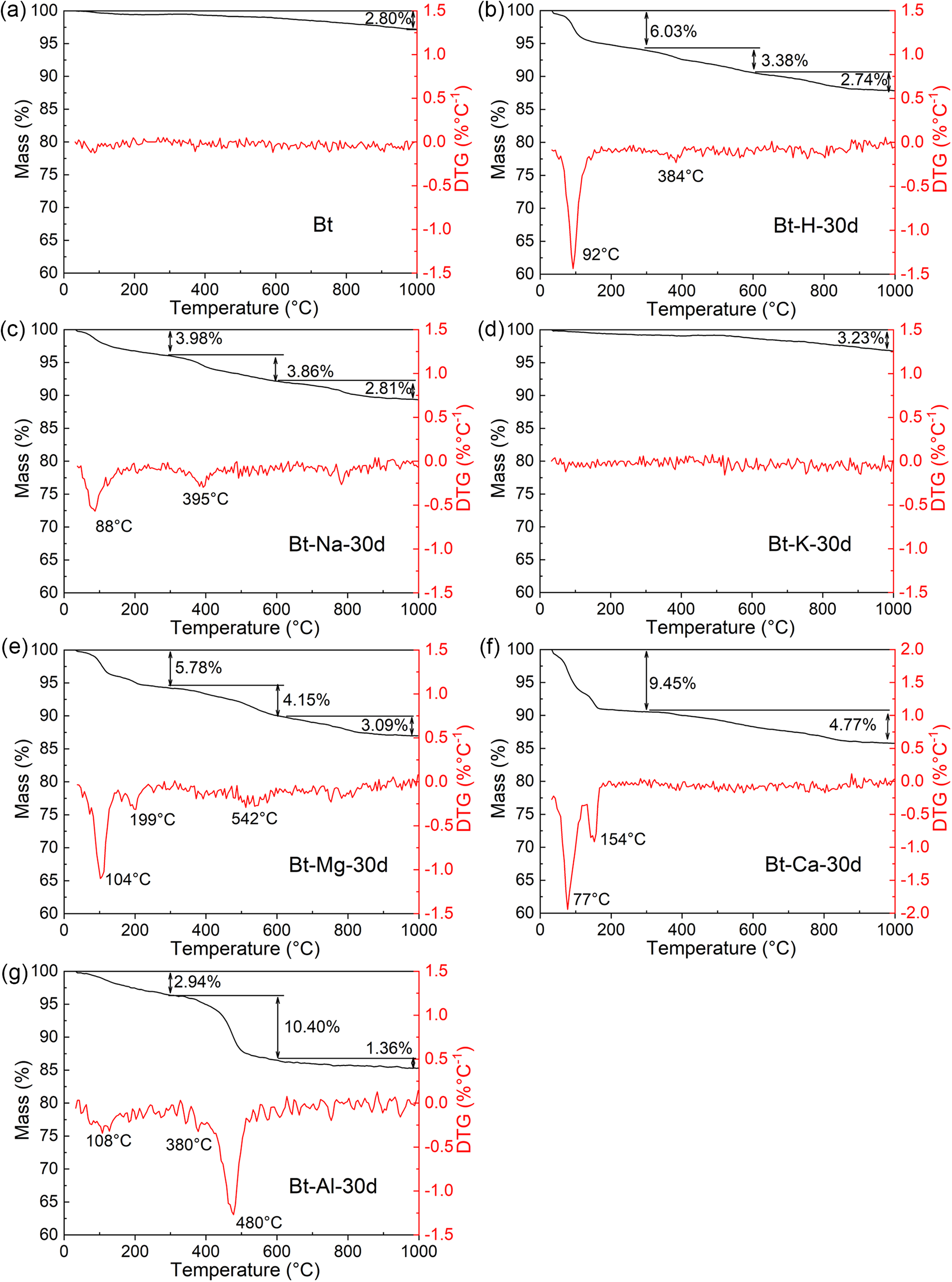
The effect of cation types on the transformation products from biotite was further investigated via TG analysis. The TG/DTG results showed that the original Bt exhibited no prominent DTG peak up to 1000°C because of the lack of adsorbed water and interlayer water in biotite (Fig. 3a), whereas the products Bt-H-30d from the blank system displayed two DTG peaks at 92 and 384°C (Fig. 3b), which were associated with the elimination of interlayer water in clay minerals (e.g. hydrobiotite and vermiculite) and the dehydroxylation of polyhydroxy aluminium cations, respectively (Perez-Rodriguez et al., Reference Perez-Rodriguez, Maqueda, Murafa, Šubrt, Balek, Pulišová and Lančok2011; Chen et al., Reference Chen, Zhu, Ma, Zhou, Zhu and He2017). By contrast, the dehydration temperature (88°C) and the area of the dehydration peak of interlayer water in the products Bt-Na-30d from the Na system decreased (Fig. 3c). In the blank system, the interlayer cations in the products were mainly metal ions (e.g. Mg2+, Al3+ and Fe3+) dissolved from the biotite layers, with a higher charge-to-radius ratio than Na+ (Table S1). These cations exerted a stronger electrostatic attraction to ligand water, thus leading to a higher dehydration temperature of interlayer water. Compared with Na+, the higher hydration enthalpies made these cations more prone to form hydrated ions (Tansel, Reference Tansel2012), bringing more water molecules into the interlayers of biotite, thus leading to greater interlayer water loss. Additionally, Bt-Na-30d showed a larger area of the dehydroxylation peak of polyhydroxy aluminium cations than Bt-H-30d, which could be attributed to more polyhydroxy aluminium cations formed resulting from more intense dissolution of the biotite layers in the Na system. The TG/DTG curve of Bt-K-30d in the K system, similar to that of the original Bt, displayed no significant weight loss up to 1000°C (Fig. 3d), reflecting the inhibitory impact of K+ on the dissolution/transformation of biotite.
TG/DTG curves of the original biotite (a) and the products from biotite in different cation systems after 30 d (b–g).

Unlike the Na system, the products in the Ca and Mg systems exhibited a bimodal peak pattern in interlayer water loss in the DTG curves (Fig. 3e, f), with the dehydration temperature in the Mg system (104 and 199°C) higher than in the Ca system (77 and 154°C), owing to the higher charge-to-radius ratio of Mg2+ (Tansel, Reference Tansel2012). As discussed previously, the higher the hydration enthalpy of the metal cation, the more water molecules it carried into the interlayers, theoretically resulting in a greater amount of interlayer water loss. However, the larger hydration enthalpy of Mg2+ led to less interlayer water loss in the Mg system (5.78% vs. 9.45% in the Ca system). This was because the amount of water loss from clay minerals was also related to the exchange capacity of different cations. Clay minerals typically exhibited a stronger affinity for cations with larger ionic radii (Bergaya and Lagaly, Reference Bergaya, Lagaly, Bergaya and Lagaly2013; Zhu et al., Reference Zhu, Chen, Zhou, Xi, Zhu and He2016), and the larger Ca2+ was more likely to enter the interlayer of clay minerals through an ion exchange reaction, resulting in a larger dehydration amount of the product in the Ca system. In the DTG curve of Bt-Na-30d from the Mg system, a DTG peak at 542°C was assigned to the dehydroxylation of magnesium–oxygen/hydroxide octahedra in chlorite (Okada et al., Reference Okada, Arimitsu, Kameshima, Nakajima and MacKenzie2005), demonstrating the formation of chlorite in this system, consistent with the XRD results (Fig. 2a). It is worth noting that a broad DTG peak appeared at ∼800°C in the DTG curves of the products from the blank, Na, Mg and Ca systems, probably due to vermiculite dehydroxylation (Marcos et al., Reference Marcos, Arango and Rodriguez2009; Ma et al., Reference Ma, Sun, Peng and Zhang2021; Wang et al., Reference Wang, Gainey, Marinelli, Deer, Wang, Mackinnon and Xi2022). For the Al system products, three DTG peaks at 108, 380 and 480°C are indicative of the removal of adsorbed/interlayer water, dehydroxylation of polyhydroxy aluminium cations, and dehydroxylation of kaolinite, respectively (Huertas et al., Reference Huertas, Fiore, Huertas and Linares1999). The TG/DTG results confirm the substantial impact of cation types on the phase composition of the transformation products from biotite.
FTIR analysis
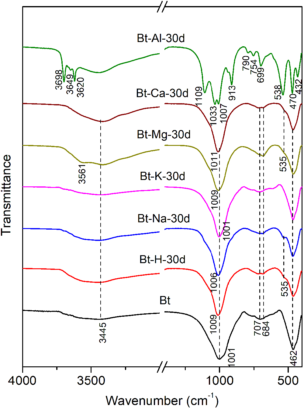
The FTIR spectra of the products from biotite at 30 d in different cation systems were collected (Fig. 4 and Table S4). The products in the K system exhibited similar vibration absorption bands to the original Bt, including the stretching vibration of interlayer water (3445 cm−1), Si–O stretching vibration (1001 cm−1 for the strong absorption region, 707 and 684 cm−1 for the weak absorption regions), and Si–O bending vibration (462 cm−1) (Madejová et al., Reference Madejová, Gates, Petit, Gates, Kloprogge, Madejová and Bergaya2017). Compared to the above two systems, the area of the stretching vibration band of interlayer water increased in the blank, Na, Mg, and Ca systems, and the increase followed the order Bt-Ca-30d > Bt-Mg-30d/Bt-H-30d > Bt-Na-30d, which was consistent with the TG/DTG results. A pronounced shoulder absorption at 3561 cm−1 appeared in the FTIR spectrum of the product from the Mg system, derived from the Mg–OH stretching vibration of chlorite (Madejová et al., Reference Madejová, Gates, Petit, Gates, Kloprogge, Madejová and Bergaya2017). The Si–O stretching vibration absorption bands in the mid-frequency region of the products from the above four systems were blue-shifted (1006–1011 cm−1) and narrowed compared to the original Bt (Fig. 4). This change could be attributed to the reduced Al substitution for Si resulting from the dissolution of Al from the tetrahedra. Moreover, in addition to the Si–O bending vibration absorption band of the original biotite (∼462 cm−1) in the low-frequency region, a weaker shoulder absorption appeared on the high-frequency side of this band for these four products, implying that the current transformation product belonged to a transition phase from trioctahedral to dioctahedral minerals.
FTIR spectra of the products from biotite in different cation systems after 30 d.

Compared to the above systems, the FTIR spectrum of the product from the Al system exhibited the characteristic infrared vibrations of kaolinite, including the structural Al–OH stretching vibrations in the high-frequency region (3698, 3670, 3649 and 3620 cm−1), Si–O stretching vibrations (1109, 1033 and 1007 cm−1) and Al–OH bending vibration (913 cm−1) in the mid-frequency region, as well as the Si–O–Si(AlVI) stretching vibrations (790, 754 and 699 cm−1) and Si–O bending vibrations (538, 470 and 432 cm−1) in the low-frequency region (Fig. 4) (Cuadros et al., Reference Cuadros, Vega and Toscano2015). In particular, the Si–O bending vibration in the low-frequency region split from a single band of biotite (462 cm−1) into three sharp absorption bands, indicating the complete transformation from trioctahedral to dioctahedral minerals. This result also demonstrated that the transformation rate of biotite in the Al system was relatively fast. When the reaction time was extended to 90 d, the phase types did not change significantly for the products in other systems based on the XRD patterns (Fig. 2), except for the blank system. As shown in the FTIR pattern of Bt-H-90d, all the characteristic infrared vibrations of kaolinite appeared (Fig. S2), confirming the formation of kaolinite, consistent with the XRD results.
Mössbauer spectra analysis
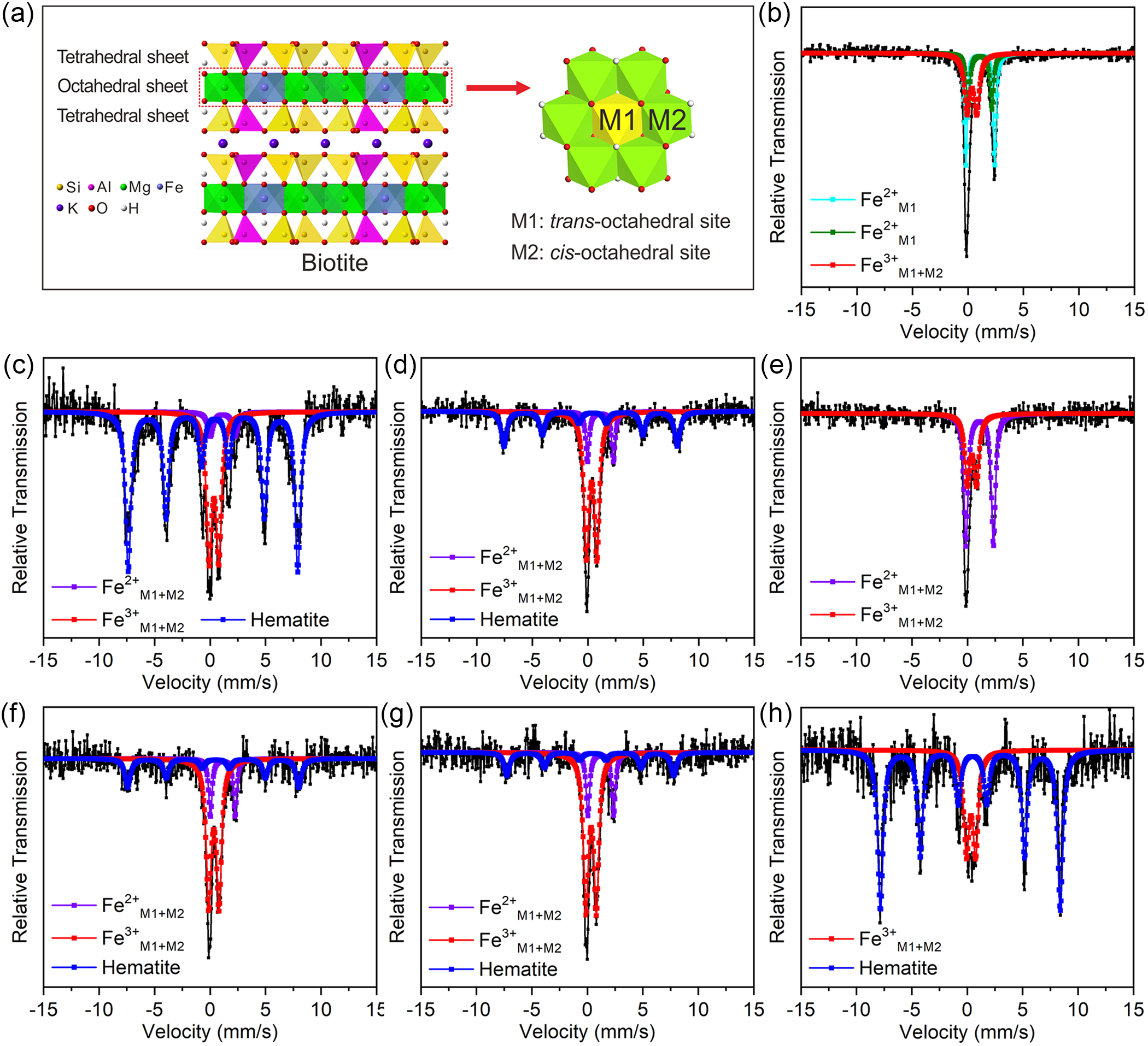
Mössbauer spectra analysis, an important structural analysis method, can be used to analyse the occupancy, valence state, and Fe3+/Fe2+ ratio of iron within clay minerals (Yan et al., Reference Yan, Chen, Yang and Zhu2021). Biotite has two distinct octahedral sites: the trans-octahedral (M1) site and the cis-octahedral (M2) site, with the M1 octahedron being slightly larger and less symmetrical than the M2 octahedron (Fig. 5a). The occupancy and valence state of Fe in the octahedral sites of biotite can be ascertained by examining the quadrupole splitting (QS) and isomer shift (IS) values derived from the Mössbauer spectra. The original Bt exhibited an asymmetric Mössbauer spectrum, which was fitted into three doublets, corresponding to structural Fe2+M1, Fe2+M2, and Fe3+M1+M2 of biotite (Fig. 5b) (Cornell and Schwertmann, Reference Cornell and Schwertmann2003). The fitting results indicated that the biotite sample contained approximately 63% Fe2+ and 37% Fe3+ (Table S5).
(a) Structure diagram of the biotite showing two distinct octahedral sites M1 and M2. Mössbauer spectra of raw biotite and the products from biotite in different cation systems after 90 d recorded at room temperature: (b) Bt, (c) Bt-H-90d, (d) Bt-K-90d, (e) Bt-Na-90d, (f) Bt-Mg-90d, (g) Bt-Ca-90d, and (h) Bt-Al-90d.

Extensive research has proven that the oxidation of structural Fe2+ is a pivotal process in biotite weathering, with the level of oxidation serving as an indicator of the dissolution and transformation stages of biotite (Velde and Meunier, Reference Velde and Meunier2008; Bray et al., Reference Bray, Oelkers, Bonneville, Wolff-Boenisch, Potts, Fones and Benning2015). The Mössbauer spectra of the 90 d products from biotite in various cation systems were measured further (Fig. 5). The Mössbauer spectrum of Bt-H-90d in the blank system was fitted into a sextet and two doublets (Fig. 5c), which were attributed to hematite, Fe2+M1+M2 and Fe3+M1+M2 of the clay minerals (Cornell and Schwertmann, Reference Cornell and Schwertmann2003), with a higher ratio of Fe3+M1+M2 and Fe2+M1+M2 than that of biotite (Table S5). Bt-K-90d from the K system showed a similar Mössbauer spectrum (Fig. 5d) to the original biotite (Fig. 5b), indicating that K inhibited the transformation of biotite. The Mössbauer spectra of the products from the Na, Mg and Ca systems all revealed a sextet and two doublets upon fitting (Fig. 5e–g), similar to the blank system (Fig. 5c). However, the intensity of the sextet was much weaker than that of Bt-H-90d, due to less hematite in the cation systems. The ratios of Fe3+M1+M2/Fe2+M1+M2 in the clay minerals of these systems were also lower than that of the blank one (Table S5). These comparative results indicate that Na+, Mg2+ and Ca2+ inhibited the biotite transformation in the later phases of the reaction. Lemine et al. (Reference Lemine, Sajieddine, Bououdina, Msalam, Mufti and Alyamani2010) studied the Mössbauer spectra of nanoscale hematite and found that a decreased size of hematite would result in a smaller hyperfine magnetic field (BHF). The Mössbauer parameters showed that the BHF values of hematite in the products of the Na, Mg and Ca systems decreased in sequence (Table S5), implying that the size of the hematite produced gradually decreased. Moreover, the ratios of Fe3+M1+M2/Fe2+M1+M2 in the clay minerals in these three systems were 4.15, 3.67 and 3.34, respectively, indicating a decrease in the content of Fe3+M1+M2, which suggested that the inhibitory effect of Na+, Mg2+ and Ca2+ on biotite transformation increased in sequence. As for Bt-Al-90d in the Al system, its Mössbauer spectrum was fitted into a sextet and a doublet (Fig. 6h), corresponding to hematite and Fe3+M1+M2 of the clay mineral. Compared with the blank system, the Fe content in the clay minerals of the Al system product further decreased, while the amount of hematite increased and Fe2+ almost disappeared, indicating that the addition of Al3+ promotes the transformation of biotite.
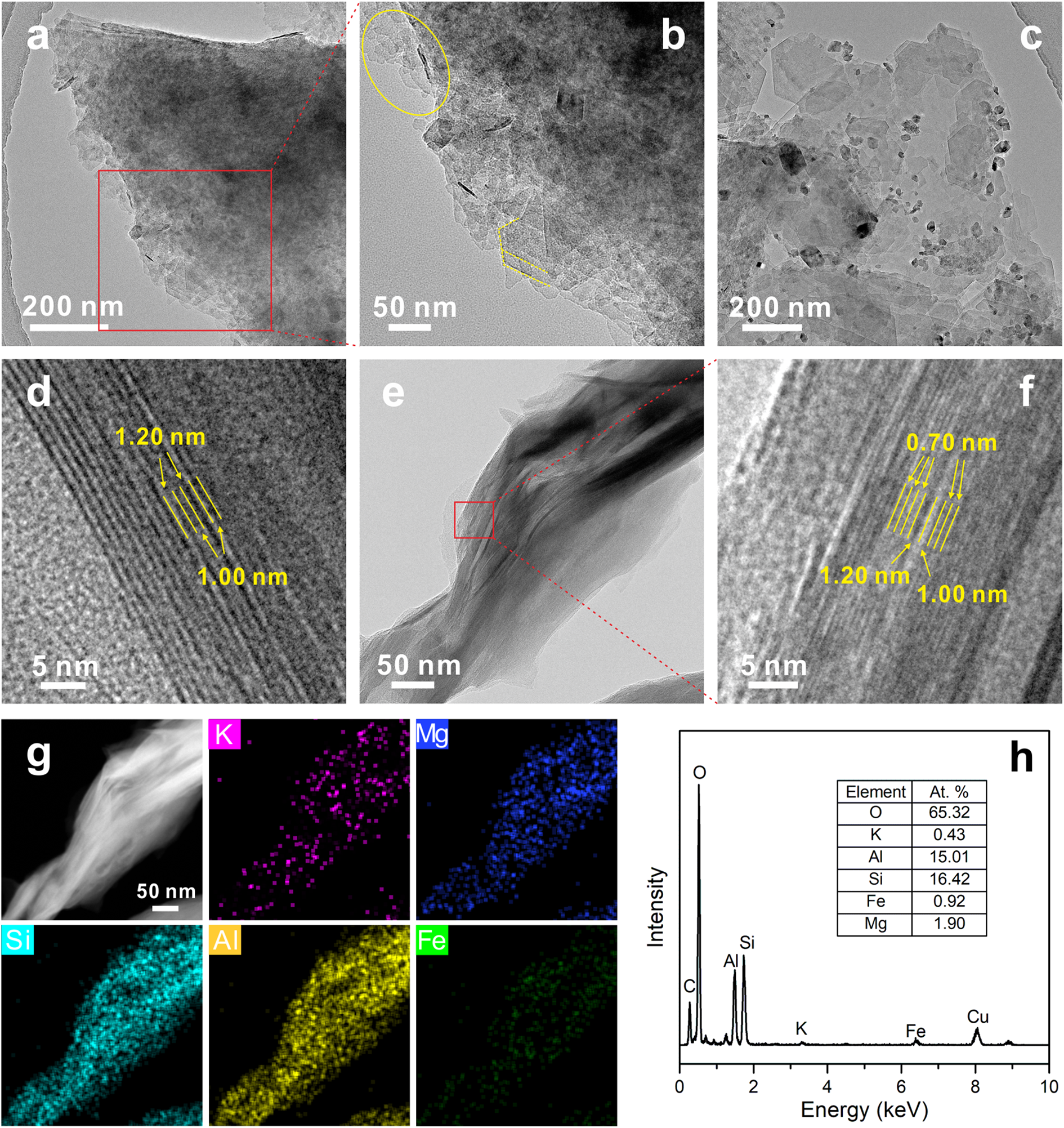
The morphology and composition of Bt-H-90d. (a–c) TEM images of the powdered sample; TEM images (d–f), HAADF image and EDS mapping (g), and EDS pattern (h) of the ultrathin-section specimen.

Morphology of biotite-transformed products in various cation systems
Transmission electron microscopy and EDS results showed that the original biotite possessed large layers with relatively smooth and uniform surfaces and was rich in Si, Al, Mg, Fe and K (Fig. S3). Following the dissolution reaction of biotite for 90 d, the product Bt-H-90d in the blank system exhibited rough edges and surfaces of the layers (Fig. 6a), due to the destruction of the biotite layers by protons. Enlarged TEM images revealed the presence of pseudo-hexagonal kaolinite layers along the edges of the large biotite layers, growing parallel to the biotite layers (Fig. 6b). Additionally, unevenly sized kaolinite particles were observed on the surface of the large layers, with a large amount of nano hematite adsorbed around (Fig. 6c). HRTEM images of the ultrathin-section samples displayed lattice fringes of ∼1.20 and 1.00 nm along the c-axis direction (Fig. 6d). During HRTEM observation, when clay minerals containing interlayer hydrated cations were exposed to a high-energy electron beam, the interlayer water escaped, leading to layer collapse. As such, the lattice fringes of the 2:1 clay mineral along the c-axis direction observed in HRTEM images were generally smaller than those detected by XRD. Therefore, the lattice fringe of ∼1.20 nm corresponded to vermiculite, and the observed structure in this area should be an interstratification of vermiculite and biotite, i.e. hydrobiotite. Furthermore, lattice fringes measuring ∼0.70 nm along the c-axis coexisted with hydrobiotite lattice fringes, with the former corresponding to the (001) plane of kaolinite (Fig. 6e, f). This part of the kaolinite was speculated to be formed by in situ dissolution and recrystallisation of Si and Al from the biotite layers (Banfield and Eggleton, Reference Banfield and Eggleton1988). The corresponding EDS pattern for this area showed a Si/Al ratio close to 1, with K essentially leached out (Fig. 6g, h), further confirming the formation of kaolinite, consistent with the XRD, FTIR and TEM results. In the same area, small amounts of Fe and Mg were still observed, which could be attributed to partially residual hydrobiotite.
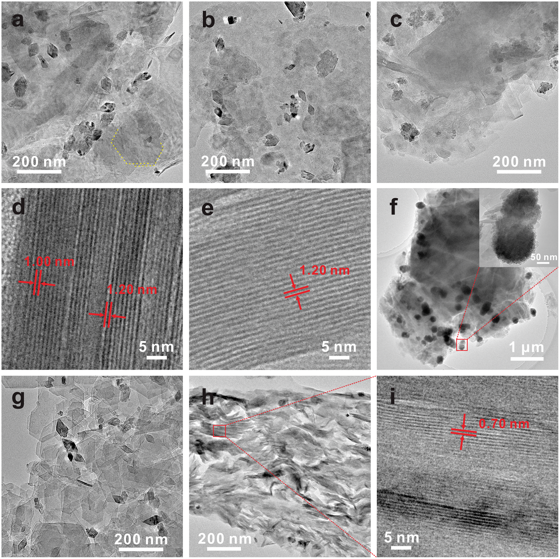
Compared to the products in the blank system, those in the Na, Mg and Ca systems showed more disordered and fragmented layers (Fig. 7a–c), which may be related to the swelling and cracking of the flake layers caused by the entry of large radius hydrated cations into the interlayers of biotite. On the surface of the layers, rhombic-shaped hematite appeared, with particle size decreasing in the sequence of Na, Mg and Ca systems, consistent with the Mössbauer spectra results (Fig. 5). Unlike the blank system, only a small amount of kaolinite was observed at the edge of the layers in the Na system product, with no observation in the Mg and Ca system products. These results further indicate that these cations inhibited the dissolution and transformation of biotite in the later stages of the reactions, with Na+ exhibiting less inhibition than Mg2+ and Ca2+. The HRTEM image of the ultrathin-section sample of the Na system product showed a lattice fringe with a period of ∼1.00 nm (Fig. 7d). Nevertheless, it could not be distinguished whether it corresponded to the original biotite or biotite with interlayer hydrated Na+ (i.e. sodium-bearing biotite), because sodium-bearing biotite lost the interlayer water under high-energy electron beam irradiation, resulting in a reduction of the interlayer spacing from 1.20 to 1.00 nm. In addition, we also observed a lattice stripe of ∼1.20 nm in this sample (Fig. 7d, e), which corresponded to vermiculite, indicating that the structural ions (such as Al) dissolved from biotite entered the interlayer. The TEM image of the K system product shows that the layer structure of biotite remained relatively intact, but that it was covered with more amorphous iron hydroxide aggregates (Fig. 7f). In contrast, the Al system product contained a large amount of well-crystallised pseudo-hexagonal flaky kaolinite, and the hematite particles on the surface were mostly intact rhombi, with a larger size (Fig. 7g). EDS results indicated a Si/Al ratio close to 1, with minor Fe content (Fig. S4). The HRTEM image of the ultrathin-section sample showed clear lattice fringes around 0.70 nm, attributed to the (001) plane of kaolinite (Fig. 7h, i), further revealing the good crystallinity of kaolinite.
The morphology of the products from biotite in different cation systems after 90 d. TEM image of Bt-Na-90d (a), Bt-Mg-90d (b), Bt-Ca-90d (c), and Bt-K-90d (f); HRTEM images (of the specimen prepared via ultrathin-section method) of Bt-Na-90d (d, e); TEM image (g) and HRTEM images (of the specimen prepared via ultrathin-section method) (h, i) of Bt-Al-90d.

Discussion on biotite dissolution and transformation mechanisms
The results above indicated that metal cations can regulate the dissolution and transformation rate of biotite and alter the transformation products, with different metal cations exhibiting different influences on the process (Fig. S5). In the blank system, the products at 30 d consisted mainly of vermiculite, chlorite and hydrobiotite, with a substantial amount of biotite still retained. After 90 d of reaction, poorly crystalline kaolinite appeared along with nano hematite aggregates on its surface. The addition of K+ significantly inhibited the dissolution and transformation of biotite. In the K system, biotite remained the main phase of the products even after 90 d, although protons could still attack the layer edges, leading to the aggregation of dissolved Fe on the biotite surface to form amorphous iron hydroxides. Na+, Mg2+ and Ca2+ could promote biotite dissolution but inhibit its transformation into kaolinite. The products in the Na system were sodium-bearing biotite, vermiculite, hematite, and a small amount of kaolinite, whereas the products in the Mg and Ca systems were mainly vermiculite, chlorite and hematite. Al3+ generated a large number of protons through its hydrolysis, which accelerated the dissolution and transformation of biotite and consequently resulted in well-crystallised kaolinite and hematite as the products.
The TEM images of the 90-d product in the blank system showed that kaolinite appeared at the edges of the biotite layers, with its (001) plane parallel to the basal plane of biotite (Fig. 6b). The HRTEM images of the ultrathin-section sample revealed that the kaolinite layers and hydrobiotite layers were stacked along the c-axis (Fig. 6f). We also observed that kaolinite grew on the surface of the precursor mineral layer in this sample (Fig. 6c). These results all indicated that the newly formed kaolinite had a similar orientation to the precursor mineral, showing a certain inheritability. Banfield and Eggleton (Reference Banfield and Eggleton1988) examined natural biotite weathering samples using TEM. They observed that, unlike vermiculite, which directly inherited the structure and orientation of biotite, kaolinite grew epitaxially on existing layered silicate structures with Al and Si (from solution or amorphous precursors) to obtain a similar orientation to biotite. The dissolution–recrystallisation mechanism emphasised the complete dissolution of precursor minerals followed by the gradual formation of new minerals, making it difficult for the newly formed minerals to inherit the crystal structure characteristics (Dudek et al., Reference Dudek, Cuadros and Fiore2006). Therefore, based on previous studies and our experimental results, the dissolution mechanism of the original biotite layers is speculated as follows: due to the slow dissolution rate, the leached elements (e.g. Al and Si) did not undergo long-distance migration but were adsorbed on the surface of the altered biotite layers, forming an Al and Si supersaturated environment, and gradually crystallising and growing epitaxially into kaolinite (Fig. S5). This dissolution–reprecipitation mechanism occurring at the mineral interface could to some extent maintain the topological structure of the original mineral (Putnis, Reference Putnis2014; Li et al., Reference Li, He, Tao, Xi, Chen, Ji, Zhang, Yang and Zhu2019), which was demonstrated in this study as a similar orientation of kaolinite and biotite. In the Al system, Al3+ hydrolysis produced a large number of protons, accelerating the dissolution of biotite (Bickmore et al., Reference Bickmore, Bosbach, Hochella M.F. and Rufe2001). Moreover, Al in the solution combined with leached Si to form a silicon–aluminium complex (Li et al., Reference Li, He, Tao, Zhu, Tan, Ji, Yang and Zhang2020), accelerating the diffusion of leached Si from the mineral surface into the solution. The rapid dissolution of biotite provided a large source of Si, which combined with Al in the solution and eventually crystallised into kaolinite (Fig. S5). In this dissolution–reprecipitation process, the precursor mineral layers were completely dissolved into ions or ion clusters that easily migrated and diffused in the solution. The mineral layers were formed by recrystallisation, making it difficult to maintain the orientation of the precursor minerals. The TEM image of Bt-Al-90d in the Al system showed that kaolinite was well crystallised and disordered in stacking (Fig. 7g–i), once again demonstrating the formation of kaolinite through the dissolution–recrystallisation mechanism. The results indicated that metal cations altered the generation mechanism of kaolinite by changing the dissolution process of biotite.
Previous studies showed that the dissolution of clay minerals in acidic environments primarily occurred at the edges because the silicon–oxygen bonds in the basal plane were charge-saturated and highly stable (Kaviratna and Pinnavaia, Reference Kaviratna and Pinnavaia1994; Li et al., Reference Li, He, Tao, Zhu, Tan, Ji, Yang and Zhang2020); in acidic solutions (pH 1.08), the rate of dissolution at the edges of biotite layers was ∼250 times greater than that at the basal plane (Turpault and Trotignon, Reference Turpault and Trotignon1994). For the Na, Mg and Ca systems, cations first entered the interlayers of biotite through ion exchange, causing internal stress that promoted the buckling and cracking of the layers (Hu et al., Reference Hu, Ray and Jun2013; Bray et al., Reference Bray, Oelkers, Bonneville, Wolff-Boenisch, Potts, Fones and Benning2015). The development of cracks results in the formation of more edges, thus accelerating the dissolution of biotite layers. However, Na+, Mg2+ and Ca2+ inhibited the subsequent phase transformation. Min and Jun (Reference Min and Jun2016) found that Na+ and Ca2+ could hinder plagioclase (CaAl2Si2O8) dissolution by vying with protons for adsorption on the plagioclase surface, thereby reducing the proton attack on its structure. Similarly, cations would rival protons for access to the surface and interlayer sites of biotite, inhibiting proton attacks on the layers. Therefore, we believe that the effect of cations on biotite dissolution was simultaneously constrained by the cation–K+ exchange reaction (enhancing effect) and the cation–proton competitive adsorption (inhibitory effect). During the initial phase of the reaction, a large number of protons in the solution resulted in rapid layer dissolution and the intense cationic exchange reaction led to more layer cracks, so the overall effect was that cations enhanced the dissolution of biotite. As Na+, Mg2+ and Ca2+ entered the interlayers, the enhancing effect gradually decreased due to diffusion limitations of the cation–K+ exchange reaction, and as the reaction proceeded, protons were gradually consumed. In the later stages of the reaction, with fewer protons in the solution, the dissolution rate of the layers slowed down, and the inhibitory effect produced by the competition between cations and protons became dominant, leading to the inhibition of phase transformation. Due to the small ionic radii and high charges of Mg2+ and Ca2+, the mineral (e.g., biotite) surface had a stronger adsorption effect on these two cations than on Na+ (McDonald et al., Reference McDonald, Evangelou, Chappell and Hillel2005; Hu et al., Reference Hu, Ray and Jun2013). Therefore, the competition between protons and Na+ on the mineral surface was less than the competition between protons and Mg2+/Ca2+, in other words, the chance of protons attacking the layers in the Mg and Ca systems was smaller, leading to a stronger inhibitory effect of Mg2+ and Ca2+ on the biotite transformation. Mössbauer spectroscopy and TEM results both illustrated this point well. Similarly, in the K system, adding K+ prevents the outward diffusion of K+ within the interlayers of biotite, and the competitive effect between K+ and protons leads to a reduced proton attack on the layers, thereby inhibiting the dissolution and transformation of biotite.
Conclusion
In summary, we selected five typical metal cations (Na+, K+, Ca2+, Mg2+ and Al3+) as representatives to study the influence of various metal cations on the dissolution and transformation process of biotite. The results indicated that the type of metal cations could significantly regulate the dissolution and transformation of biotite. Compared with the blank system, K+ impeded the dissolution and transformation of biotite, leading to the formation of amorphous iron hydroxides on the biotite surface; Na+, Mg2+ and Ca2+ promoted the dissolution of biotite but inhibited its transformation into kaolinite. In the Na system, the products included sodium-bearing biotite, vermiculite, hematite, and a small amount of kaolinite; whereas in the Mg and Ca systems, the products consisted primarily of vermiculite, chlorite and hematite. Al3+ notably accelerated the dissolution and transformation of biotite, resulting in the formation of well-crystallised kaolinite and hematite. Furthermore, metal cations could alter the dissolution rate of biotite, thereby changing the formation mechanism of kaolinite. In the blank system, the dissolution rate of biotite was slow, and the released elements such as Al and Si accumulated on the surface of biotite, growing epitaxially into kaolinite; while in the Al system, the rapid dissolution of biotite provided a large source of Si, which combined with Al in the solution to form kaolinite through a dissolution–recrystallisation mechanism. In addition, the exchange reaction of metal cations with K+ and the competitive adsorption between metal cations and protons simultaneously affected the dissolution process of biotite. This work offers a theoretical basis for a deeper comprehension of the factors influencing the weathering of silicate minerals and new insights into the evolution of clay minerals in terrestrial surface environments.
Supplementary material
To view supplementary material for this article, please visit https://doi.org/10.1180/mgm.2024.67.
Acknowledgements
This study was supported by the Key-Area Research and Development Program of Guangdong Province (2020B0101370003), National Natural Science Foundation of China (42225203, 42272045, 41921003, 41825003), Tuguangchi Award for Excellent Young Scholar GIG, CAS (TGC202302), Youth Innovation Promotion Association CAS (2020347), Natural Science Foundation for Distinguished Young Scientists of Guangdong Province (2023B1515020006), and Science and Technology Planning of Guangdong Province, China (2023B1212060048). This is contribution No.IS-3615 from GIGCAS.
Competing interest. The authors declare that there are no competing financial interests.