Introduction
Retraction of the tympanic membrane, in its pars tensa and/or pars flaccida portions, is associated with nearly all clinical cases of otitis media. Except for the positively pressurised purulence of acute suppurative otitis media, low pressure in the various regions of the middle ear, specifically the mesotympanum for pars tensa retraction and Prussak's space for pars flaccida retraction, is a hallmark of otitis media.Reference Sade, Fuchs and Luntz1 Small mastoid size is a lifelong hallmark of ongoing childhood otitis media.Reference Luntz, Teszler, Shpak, Feiglin and Farah-Sima'an2 Therefore, a correlation between retracted pars flaccida and small mastoid size would be expected, as Sade et al. Reference Sade, Fuchs and Luntz1 found in adults attending an otology clinic in Israel. The position of the pars flaccida in well pneumatised ears differed from that found in ears with poorly or non-pneumatised mastoids, ‘irrespective of whether these ears are associated with some chronic inflammatory process’.Reference Sade, Fuchs and Luntz1 In the same patient series, these authors also found that ‘well pneumatised mastoids are rarely associated with pars flaccida retraction’.Reference Sade, Fuchs and Luntz1
Kobayashi et al. used computed tomography (CT) to study epitympanic aeration in patients with pars flaccida retraction.Reference Kobayashi, Toshima, Yaginuma, Ishidoya, Suetake and Takasaka3 In both paediatric and adult patients attending a Japanese otology clinic, most (22 of 28) ears with attic (pars flaccida) retraction pockets had CT evidence of aeration of both the epitympanum and mastoid antrum. In contrast, a minority (five of 25) of ears with attic cholesteatoma had CT evidence of aeration of both the epitympanum and mastoid antrum. Kobayashi et al. did not consider their findings to ‘verify the hypothesis that the obstruction of the aeration route between the mesotympanum and the attic is a contributing factor in the pathogenesis of attic retraction pockets and cholesteatomas’.Reference Kobayashi, Toshima, Yaginuma, Ishidoya, Suetake and Takasaka3 To summarize succinctly, the clinical literature on the pathogenesis of pars flaccida retractions is confusing. The present study addressed the hypothesis that childhood otitis media, identifiable in adults by small mastoid size, correlates with retraction of the pars flaccida.
Materials and methods
Specimens
Forty-one adult crania were provided by the anatomy department. Institutional review board approval was not applicable to these post-mortem specimens. All were from elderly humans who had bequeathed their bodies to science; most were white, the remainder black. No specific age, sex, or racial information was available. Although no subject had died of ear disease, no specific ear historical data were available.
The specimens, which had not been fixed, were kept in a freezer at −2°C. Most soft tissues, except the tympanic membranes, were removed.
Temporal bone pneumatisation
Law lateral plain mastoid radiographs were used to describe the extent of pneumatisation. Temporal bone pneumatisation is considered to be unchanging in adults, and the strong correlation between childhood otitis media and a small mastoid air cell system is ‘beyond debate’.Reference Luntz, Teszler, Shpak, Feiglin and Farah-Sima'an2 The correlation of Law mastoid area versus CT mastoid volumetry is good (r=0.74, N=30, 95 per cent confidence interval 0.52–0.88). For the Law radiographs, the central X-ray beam projected through the external ear canal; the source to target distance was 210 cm, so magnification was negligible. The outline of pneumatisation of each radiographic image was traced (by the author) onto paper, on two separate, independent occasions. In contrast to Diamant's method of excluding the antrum region on the tracings, this region was included.Reference Todd, Pitts, Braun and Heindel4 Care was taken to avoid knowledge of the size of the contralateral mastoid. Areas were measured by planimetry (K & E model 620005, serial number 82305, West Germany).
Pars flaccida retraction
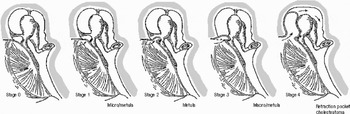
The retraction of each pars flaccida was determined, via an operating microscope, using the categorisation of Sade (Figure 1).Reference Sade, Fuchs and Luntz1 To assess whether the pars flaccida was stuck to the neck of the malleus, direct palpation with a tiny wax curette was performed.
Sade's schematic representation of the various degrees of pars flaccida (PF) retraction. Stage 0 is normal, without retraction, the PF overlaying a gas-filled Prussak space. Stage 1 is a dimpled PF, not touching the neck of the malleus (‘metula’ means cone- or pyramid-shaped). Stage 2 is the PF reclining on the neck of the malleus. Stage 3 involves partial destruction of the scutum. Stage 4 is keratin debris in a pocket (i.e. an attic cholesteatoma). Reproduced with permission.Reference Sade, Fuchs and Luntz1 © (1997) American Medical Association. All Rights Reserved.

Epitympani assessed by dissection and computed tomography
The epitympani of all 82 temporal bones were dissected, with removal of the tegmina tympani, and viewed with an operating microscope via the middle cranial fossa approach.
In addition, 10 crania (five with the smallest mastoid pneumatisation areas and five with the largest areas) were imaged by high resolution CT with a prospective bone algorithm (GE Light Speed Ultra multi-detector scanner, GE Medical Systems, Waukesha, Wisconsin, USA). (Because of financial constraints, the remaining 31 crania were not studied by CT.) Direct axial and coronal images, and coronal reformations from the axial data set, were obtained with each specimen in a non-metallic cephalostat. The 1.2 mm, non-helical images, parallel and perpendicular to the Frankfort horizontal plane, were prepared with a 200 mA, 120 kVp, scan field of view 25 cm, and edge-enhancing algorithm.
Statistics
Both parameters (mastoid area and pars flaccida categorisation) were independently determined on both sides of each specimen, twice, on separate occasions.
The statistical unit was each cranium's right or left ear, as specified. For statistical assessment of a relationship between variables, the non-parametric Spearman rank correlation rs was computed; rs was always rounded toward zero. The non-parametric Wilcoxon rank-sum test was used to compare the mastoid areas of specimens with a normal versus a retracted pars flaccida. Bland–Altman plots were constructed for the mastoid area determinations. Any p values much smaller than 0.001 were nevertheless expressed as <0.001. No Bonferroni correction was applied.
Results
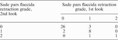
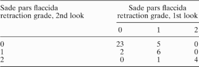
The repeatability (i.e. the reliability) of measurements and categorisations was good (at least). For right mastoid areas, rs=0.89, p<0.001; for left mastoid areas, rs=0.92, p<0.001. For the repeatability of left pars flaccida Sade category, rs=0.79, p<0.001 (Table I). For right pars flaccida repeatability, rs=0.82, p<0.001 (Table II). The frequency distribution of mastoid areas approximated a bell shape: for the right sides, the five-number summary was 2.4, 5.7, 9.6, 12.4 and 14.2 cm2; for the left sides, 2.0, 7.5, 10.0, 11.6 and 18.0 cm2. In contrast, the frequency distribution of pars flaccida categorisations was quite asymmetric (i.e. skewed); the majority of pars flaccida, for both the left and right sides, was normal, i.e. Sade grade zero.
Repeatability of pars flaccida retraction categorisation, left side*
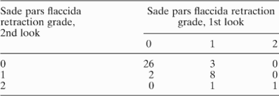
* Forty-one ears. The degree of repeatability relationship was excellent: rs=0.82, p<0.001. The second look yielded, at most, a one-grade difference from the first look. PFR=pars flaccida retraction
Repeatability of pars flaccida retraction categorisation, right side*
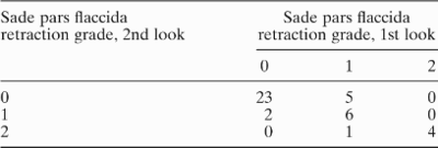
* Forty-one ears. The degree of repeatability relationship was excellent: rs=0.79, p<0.001. The second look yielded, at most, a one-grade difference from the first look. PFR=pars flaccida retraction
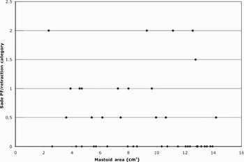
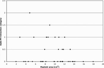
No pars flaccida was categorised as worse than Sade grade two (i.e. retracted onto the neck of the malleus). No association of pars flaccida retractions versus small mastoids was apparent: for right sides, rs=−0.13, p=0.38; for left sides, rs=0.00, p=0.98 (Figures 2 and 3, respectively). On comparing the mastoid areas of specimens with normal pars flaccida versus those with retracted pars flaccida, no significant difference was found: for left sides, p<0.69; for right sides, p<0.13. Of the 82 tympanic membranes of the 41 crania, every pars tensa was in a normal position.
Right ear; areas of pneumatisation and extent of pars flaccida (PF) retraction, for the 41 crania. No association is apparent: rs=−0.13, p=0.38.

Left ear; areas of pneumatisation and extent of pars flaccida (PF) retraction, for the 41 crania. No association is apparent: rs=0.00, p=0.98.

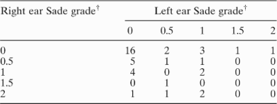
Bilateral symmetry was apparent for both mastoid size and pars flaccida retraction: respectively, rs=0.68, p<0.001; rs=0.35, p<0.03 (Table III).
Bilateral symmetry* of pars flaccida retraction, Sade categorisationReference Sade, Fuchs and Luntz1
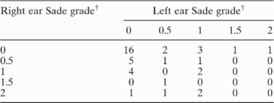
* Right versus left. †Mean of first and second looks. See Figure 1. For the 41 cranial specimens, the right–left degree of relationship was fair: rs=0.35, p<0.03.
All 82 epitympani were found to be normal on anatomical dissection via the tegmina tympani, as were the 20 studied by CT. Specifically, there was no suggestion of cholesteatoma, tympanosclerosis or small volume of any epitympanum.
Discussion
Both clinicalReference Sade, Fuchs and Luntz5 and experimentalReference Larsson, von Unge, Dirckx, Decraemer and Bagger-Sjoback6 evidence substantiates the fact that the pars flaccida commonly and easily collapses in Prussak's space. This evidence is consistent with the findings of this study and the work of Kobayashi et al.,Reference Kobayashi, Toshima, Yaginuma, Ishidoya, Suetake and Takasaka3 Sade et al. Reference Sade, Fuchs and Luntz1 and Palva et al.,Reference Palva, Ramsey and Northrop5if pars flaccida retractions are considered a prolonged consequence of comparatively short-lived episodes of sub-atmospheric pressure in Prussak's space. Such may well be the pathogenesis of the condition, if ventilation routes into Prussak's space are tiny, indeed, tinier than the ventilation channels at the tympanic isthmus and the aditus ad antrum. Otherwise stated, Prussak's space may be more vulnerable to collapse due both to the lack of collagenous fibres involving the pars flaccida (in contrast to the pars tensa) and to the tiny ventilation channels most susceptible to obstruction by inflammation. This study's finding of greater bilateral symmetry of mastoid size, compared with pars flaccida retraction symmetry, is consistent with this concept. The concept can be checked by clinical observation studies, e.g. long-term observation of the pars flaccida of patients whose childhood otitis media courses were prospectively documented.
Some may argue that the pars flaccida retractions did not progress because the mastoids and non-Prussak's compartments of the epitympani were sufficiently well pneumatised by a route independent of the anterior (i.e. major) tympanic isthmus – i.e. ventilated via the so-called ‘posterior isthmus’ behind the tip of the incus. However, Palva et al. considered the ‘posterior isthmus’ to be an ‘inconsistent aeration pathway via the incudal fossa’.Reference Palva, Ramsey and Northrop5 This author's anecdotal observations (both in the operative suite and the dissection laboratory) were that the ‘posterior isthmus’ was indeed inconsistently present, tiny, and an unlikely ventilation route. Because of the vulnerability of the major anterior tympanic isthmus (also termed the epitympanic diaphragm or interatticotympanic diaphragm) to inflammation-induced blockage, the aeration (and drainage) of the epitympanum and mastoid are often (at least transiently) compromised. Prussak's space would be concomitantly affected. It is important to note that Prussak's space is an ‘independent unit’Reference Palva, Ramsey and Northrop5 of the epitympanum, and ‘can be blocked or obliterated without any influence as such on the workings of the major compartments superior to the epitympanic diaphragm, the anterior and posterior epitympanum, aditus ad antrum, and the mastoid air cell system’.Reference Palva, Ramsey and Northrop5 Such a statement is consistent with the findings in the present study's 82 clinically normal specimens. Also consistent with this study's findings is the report by Kobayashi et al. Reference Kobayashi, Toshima, Yaginuma, Ishidoya, Suetake and Takasaka3; their data do not support the classic ‘hypothesis that the obstruction of the aeration route between the mesotympanum and the attic is a contributing factor in the pathogenesis of retraction pockets and cholestatomas’.Reference Kobayashi, Toshima, Yaginuma, Ishidoya, Suetake and Takasaka3
An exception to the correlation of childhood otitis media with a small mastoid air cell system must be discussed. Children with cholesteatoma are reported to ‘typically’ have large mastoids.Reference Shohet and de Jong7 Such cases, in addition to being difficult and extensive surgical challenges, beg aetiologic explanations other than otitis media. One explanation is congenital cholesteatoma, which by definition does (or did) not involve otitis media. Another explanation may be minimal or clinically silent otitis which resolved without affecting the mastoid or epitympanum – except for retraction into the comparatively vulnerable Prussak's space, or postero-superior portion of the pars tensa in children. Such retraction could eventuate in a pocket with keratin debris (i.e. cholesteatoma) into a generously sized mastoid. The natural history of pars flaccid retraction is, however, more complex. Retractions in three- to six-year-old children with persistent middle-ear effusion (which involve the pars tensa about twice as often as the pars flaccida (involving 9 and 5 per cent of ears, respectively)) were found to resolve in two-thirds of cases over a 12-week period of ‘no potentially effective management’, without prior ear or adenoid surgery.8 However, over the 12 weeks, the otitis media with effusion had resolved in only about 15 per cent of the ears. So, one may presume, the very few children with acquired cholesteatoma (the annual incidence of all cholesteatoma types being only about three per 100 000 children)Reference Dornelles, Costa, Meurer and Schweiger9 and a large mastoid had experienced otitis media that resolved, but the retraction pockets advanced to cholesteatoma. The rapid growth of acquired paediatric cholesteatomas may be explained by their higher rate of keratinocyte proliferation,Reference Bujia, Holly, Antoli-Candela, Tapia and Kastenbauer10 and an increase in the number of perimatrix inflammatory mononuclear elements with increased collagenase activity.Reference Quaranta, Resta and Santangelo11
• The relationship of pars flaccida retraction with epitympanic aeration and mastoid size is ill-defined
• This paper studied, in clinically normal specimens, the relationship of pars flaccida retraction with mastoid pneumatisation and epitympanic aeration
• The study involved post-mortem anatomical dissection of 41 bequeathed adult crania without clinical otitis
• The findings of this study do not support the hypothesis that, in clinically normal temporal bone specimens, there is a correlation between small mastoid size and retraction of the pars flaccida
The limitations of this study included the lack of specific information about the age, sex, race and otologic history of the specimens. The delimitations of this study included, at least, the fact that CT was performed on only one-fourth of the specimens (because of cost), plus the fact that the appearance of the pars tensa may have influenced categorisation of the pars flaccida retraction. Otherwise stated, as the observer knew of both (1) the fact that scarring of the pars tensa correlates with small mastoids,Reference Todd12 and (2) the report of Sade et al. Reference Sade, Fuchs and Luntz1 associating pars flaccida retraction with small mastoid size, observer bias may thus have occurred. Erroneous or inaccurate categorisations of the pars flaccida, and post-mortem changes in the specimens, are unlikely. Law lateral radiographs assessing pneumatisation have a good correlation with pneumatisation volume determined by CT. Thus, there is no cause for concern that predominantly antero-posterior pneumatisation overestimates mastoid size, or, conversely, that predominantly medial-lateral pneumatisation underestimates mastoid size.Reference Todd, Pitts, Braun and Heindel4
Conclusion
The findings of this study do not support the hypothesis that, in clinically normal temporal bone specimens, there is a correlation between small mastoid size and retraction of the pars flaccida. Enduring pars flaccida retractions may be explained by comparatively short-term, inflammation-induced constriction of Prussak's space.
Acknowledgements
M Katherine Thurman prepared one set of planimetry determinations of mastoid pneumatisation. Patricia A Hudgins MD facilitated obtaining the CTs. She, Richard T Jackson PhD and John H Per-Lee MD critiqued a draft of the manuscript.