Right aortic arch with an isolated left subclavian artery from the left pulmonary artery is a rare congenital arch anomaly in which the left subclavian artery arises from the left pulmonary artery via a patent arterial duct or is connected to it through an arterial ligament.Reference Edwards 1 Nearly 60% of patients have associated intracardiac anomalies that influence clinical presentation. Some patients may have symptoms associated to subclavian or pulmonary steal.Reference Carano, Piazza and Agnetti 2 Unlike right aortic arch with an aberrant left subclavian artery and a patent arterial duct, which is commonly associated with a vascular ring, right aortic arch with an isolated left subclavian artery has never been associated with airway obstruction or vascular ring in any previously published reports to the best of our knowledge.
Case 1
A 5-year-old boy was diagnosed with tetralogy of Fallot at 2 months of age. His parents reported a history of noisy breathing since early infancy and multiple admissions for pneumonia associated with severe bronchospasm requiring mechanical ventilation. On physical examination, he had pectus carinatum, was not tachypnoeic despite having inspiratory stridor with mild cyanosis, and his oxygen saturations were 88–91% on room air. His heart rate was 97 bpm with reduced pulse volume in the left arm. His blood pressure was 123/46 mmHg in the right arm and 80/50 mmHg in the left arm. The cardiovascular examination revealed an ejection systolic murmur grade IV/VI at the left sternal border. His respiratory examination was unremarkable.
A chest radiograph revealed a right aortic arch with narrowing of the tracheal shadow. Echocardiography confirmed tetralogy of Fallot but the pulmonary trunk did not bifurcate at the usual location, and there was a low velocity flow at the origin of the left pulmonary artery representing the origin of the left subclavian artery. There were bilateral superior caval veins with left-sided draining into the coronary sinus.
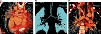
Cardiac catheterisation demonstrated a pulmonary artery sling, right aortic arch, and late retrograde filling of the left vertebral artery with subsequent filling of the left subclavian artery and left pulmonary artery via a patent arterial duct, suggestive of isolated left subclavian artery. In view of the pulmonary artery sling and to further delineate his airway, a chest CT was performed. It showed moderate tracheal stenosis with bilateral left-sided bronchi (Fig 1). Subsequently, he underwent re-implantation of the left pulmonary artery to the pulmonary trunk and of the left subclavian artery to the left common carotid artery, as well as tetralogy of Fallot repair with preservation of the pulmonary valve. The tracheal stenosis was not considered to be severe enough to warrant intervention. The bypass time was 89 minutes with cross-clamp time of 57 minutes. There were no major intraoperative or postoperative complications; he was extubated on postoperative day 1 and discharged well on postoperative day 5. At his sixth-month follow-up evaluation, he remained well with no respiratory symptoms and symmetrical pulses in the arms.
(a) Reconstructed CT- scan shows pulmonary artery sling, the left pulmonary artery (LPA) arises from the right pulmonary artery (RPA), curves posteriorly behind the lower trachea to the left, encircling the trachea. (b) Reconstructed CT-scan of the airways reveals long segment narrowing of the trachea above the carina and bilateral left- sided bronchi. (c) From posterior view shows the isolated left Subclavian artery (LSCA) from the left pulmonary artery (LPA) and its relationship with the trachea (T).

Case 2
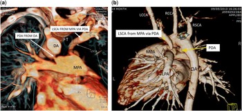
A 1-year-old girl with a history of laryngomalacia and global developmental delay secondary to perinatal asphyxia was diagnosed with a moderate-sized patent arterial duct, patent foramen ovale, and a tiny perimembranous ventricular septal defect. Initially, she had signs of pulmonary over-circulation, but this resolved spontaneously within a few months. Her most recent echocardiogram revealed a small patent arterial duct, a right aortic arch, and an abnormal vessel, which was thought to be an aberrant left subclavian artery. A vascular ring was suspected as she had a persistent stridor in the presence of a right aortic arch. Clinically, she was pink and small for age as all growth measurements were below the 3rd percentile, with the presence of inspiratory stridor and suprasternal retractions. Her blood pressure was 91/40 mmHg in the right arm and 63/33 mmHg in the left arm, along with a reduced pulse volume in the left arm. A soft systolic grade II/VI murmur was heard at the upper left sternal border. A thorax CT revealed a right aortic arch, a right patent arterial duct, and an isolated left subclavian artery from the pulmonary artery with mild compression of the trachea (Fig 2). Her parents were counselled regarding the findings and the need for arterial duct division and re-implantation of left subclavian artery to the aorta but they refused treatment.
(a) Reconstructed CT scan reveals isolated left subclavian artery (LSCA) from pulmonary trunk via a left patent arterial duct (PDA) and the right PDA from descending aorta (DA) and connected to proximal right Pulmonary artery (RPA). The relationship of both arterial ducts and the aorta to the trachea (T) and mild compression of the trachea from both sides are demonstrated. (b) Volume rendered technique CT scan shows the left subclavian artery (LSCA) arises from pulmonary trunk via left PDA and the right PDA from DA.

Discussion
The first case had an extremely rare combination of lesions: tetralogy of Fallot, pulmonary artery sling, isolated left subclavian artery from the left pulmonary artery, right aortic arch, bilateral superior caval veins, and left bronchial isomerism suggesting heterotaxy syndrome; however, there was no left atrial appendage isomerism, polysplenia, or intestinal malrotation noted in this case. From the literature review, this is the first reported case with such a combination of cardiac lesions. The association of bronchial isomerism with pulmonary artery sling was discussed by Landing et alReference Landing and Dixon 3 . Bricker described a case of tetralogy of Fallot, pulmonary artery sling, and isolated left subclavian artery from the pulmonary artery in a newborn baby with a left-hand deformity. He did not provide any clear description of evaluation of the airway of this patient who subsequently died of severe respiratory distress.Reference Bricker, Nihill and McNamara 4
The upper airway obstruction symptoms in case 1 occurred as a result of tracheal stenosis caused by the pulmonary artery sling. The presence of an isolated left subclavian artery might not have contributed significantly to the airway obstruction as demonstrated by the CT-scan, and the patent arterial duct did not form a complete ring, as it was not connected to the descending aorta. Tracheobronchial tree anomalies including complete cartilaginous rings and hypoplasia of the distal trachea and bronchi are seen in 50% of pulmonary artery sling cases.Reference Sade, Rosenthal and Fellows 5 These intrinsic airway abnormalities may also play an important role in respiratory symptoms.
In the second case, a very mild compression of the distal end of the trachea was seen due to bilateral arterial ducts and possibly by the transverse arch. A chest CT-scan showed that the compression occurred at both sides. This is possible as the patient had a right aortic arch with bilateral arterial ducts. Stephen et alReference Miller, Campbell and Barker 6 noted that the vascular ring was not formed, as the ducts were ipsilateral to the subclavian artery and not connected to the aorta. We believe this very mild compression might not cause symptomatic airway obstruction by itself, but may be responsible for the development of secondary tracheomalacia. Cartilage destruction and malacia as a result of vascular compression have been observed even within a couple of weeks.Reference Filler and de Fraga 7
Both patients were diagnosed only after multiple visits to the clinic. This could be due to the rare association of tetralogy of Fallot with pulmonary artery sling, as reviewed by Gikanyo et alReference Gikonyo, Jue and Edwards 8 , who described only 3 (2.6%) cases of tetralogy of Fallot among 130 cases of pulmonary artery sling. It is important to investigate 22q11 deletion in patients with tetralogy of Fallot and aortic arch anomaly, as they are 3.14 times more likely to have a 22q11del. In addition, 22q11del is seen in almost 30% of patients with perimembranous ventricular septal defect with arch anomaly compared with ~5% of those with a normal aortic arch anatomy.Reference Peyvandi, Lupo and Garbarini 9
The isolated subclavian artery cannot be diagnosed by echocardiography alone. Further imaging is required to confirm the anatomy, by means of CT-scan, MRI, or cardiac catheterisation.Reference Van Son, Julsrud and Hagler 10 For these patients who need further airway assessment, we found that CT-scan is an excellent technique to delineate the vascular and airway anatomy accurately as well as the effect of vascular compression on the airway.
The management involves surgical re-implantation of the left subclavian artery and the left pulmonary artery along with correction of the associated intracardiac anomalies. To our knowledge, this is the first reported case of single-stage operation for repair of tetralogy of Fallot, re-implantation of the left pulmonary artery to the main pulmonary artery, and re-implantation of the left subclavian artery to the left common carotid artery. Even though the operation was successful, this patient will require long-term follow-up by both cardiology and respiratory teams for assessment of late complications related to the operation and to the airway abnormalities.
In conclusion, right aortic arch with isolated left subclavian artery may not cause symptomatic upper airway obstruction by itself, but it can be associated with airway compression in the presence of an associated lesion-like pulmonary artery sling or bilateral arterial ducts with intrinsic airway abnormalities. A high index of clinical suspicion of this rare but surgically correctable condition is of paramount importance to avoid delay in diagnosis. Whenever symptoms are not readily explained by the obvious CHD, a careful examination of the pulmonary anatomy, the aortic arch, and its branching pattern should be performed to rule out a lesion that may be causing or be associated with airway compressions.
Acknowledgements
The authors are grateful to Mr Sivakumar Sivalingam who did the successful operation, Dr Hasri Samion, the head of Pediatric Congenital Heart Center and to all dedicated National Heart Institute staffs who are taking care of these patients.
Financial Support
This case report received no specific grant from any funding agency or from commercial or not-for-profit sectors.
Conflicts of Interest
None.
Ethical Standards
These case reports comply with the ethical standards and was accepted by the National Heart Institute Ethics Committee.