INTRODUCTION
Trichomonads are defined as microorganisms possessing the general structural characteristics of the order Trichomonadida. Trichomonas species are commonly known to be pathogenic to humans (Honigberg and Brugerolle, Reference Honigberg, Brugerolle and Honigberg1990). In particular, Trichomonas tenax is a protozoan that inhabits the human oral cavity in and around diseased teeth and gums (Roberts and Schmidt, Reference Roberts and Schmidt2009). Although its prevalence in the mouth ranges from 4 to 53%, it is believed to be commensal (Ghabanchi et al. Reference Ghabanchi, Zibaei, Afkar and Sarbazie2010). While its pathogenic role remains relatively unclear (Honigberg and Brugerolle, Reference Honigberg, Brugerolle and Honigberg1990), T. tenax appears implicated in various infections outside the limits of the oral cavity, such as lung diseases (Maritz et al. Reference Maritz, Land, Carlton and Hirt2014); however, a role in the periodontitis process has not yet been demonstrated. This protozoan is classified in the same genus as Trichomonas vaginalis (Yamamoto et al. Reference Yamamoto, Asaga, Nagao, Igarashi and Goto2000), the pathogenicity of which has been extensively studied. It is not typical for dentists to carry out a systematic search for oral parasites in cases of gum disease; however, here we report finding protozoans morphologically similar to Trichomonas species, in a child with periodontal disease.
CASE REPORT
An 8-year-old boy was referred by his physician for gum bleeding. In the preceding 2 months, the patient had undergone surgical excision of an odontoma that was followed by antibiotic therapy (amoxicillin for 6 days). He presented no other pathological conditions.
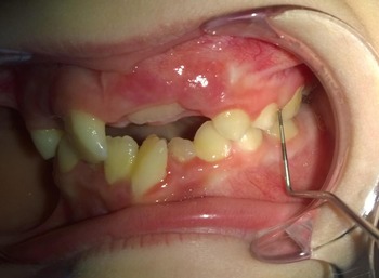
The medical history highlighted gum bleeding during toothbrushing and meals, plus mild oral pain. The clinical examination showed a generalized moderate gum inflammation, with vivid red gingiva that bled easily (Fig. 1). The Community Periodontal Index (CPI) demonstrated the presence of periodontal pockets (4 mm depth in two sites) (Clerehugh and Tugnait, Reference Clerehugh and Tugnait2001). The radiographic examination (periapical technique) revealed no bone destruction. Both the pocket depth and inflammation signalled the diagnosis of acute periodontitis and a microbiological examination was subsequently pursued. A dental plaque sample was collected in the gingival sulcus area by a sterile periodontal probe in four different sites. The smears taken were immediately spread onto a glass slide with physiological serum to avoid the loss of organism motility. A direct examination, using phase-contrast optical microscopy with a magnification X1000, was performed in order to search for motile flagellate Trichomonas species trophozoites (Ponce de Leon et al. Reference Ponce De Leon, Zdero, Vasconi, Nocito, Lucca and Perez2001). This method was employed to enable the use of the X100 oil immersion objective before fixing the cover glass over the slide, thereby avoiding dryness or alteration of parasitic structures (Abdel-Hamid, Reference Abdel-Hamid1997). This procedure revealed a large number of non-motile and motile bacteria, including numerous widespread motile flagellated protozoa. The shapes of the organisms were either ellipsoidal or ovoidal, along with four anterior flagella of unequal length. This observation corresponded to Trichomonas species (Benchimol, Reference Benchimol2004) (Fig. 2).
Clinical view of gum inflammation using a sterile probe in a periodontal pocket.

(A) Numerous flagellated motile protozoa (phase-contrast optical microscopy X1000) and (B) isolated specimen.

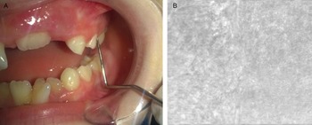
Patient management included the cleansing of gums with a hydrogen peroxide mouthwash (DENTEX®, Colgate, USA) applied with a sterile compress over a 2 week period. The patient was also treated with Metronidazole (300 mg three times daily for 10 days), following the recommendation of the American Association of Pediatric Dentistry (2014) (AAPD guideline, 2014). A non-surgical removal of infected crevicular epithelium and debridement of root surfaces were performed. Notably, 2 months following this intervention, a renewed measurement of the CPI revealed the absence of both gum inflammation and periodontal pockets. Microscopic examination following the same protocol as before treatment, revealed no flagellated protozoa (Fig. 3). A clinical examination of the patient's parents showed that they both suffered from chronic periodontitis. Interestingly, parent-to-child transmission of oral microbials has previously been documented (Nelson-Filho et al. Reference Nelson Filho, Macari, Faria, Assed and Ito2000).
(A) Absence of gum inflammation after anti-parasitic treatment and (B) normalization of flora.

Concluding remarks
Although Trichomonas species are often considered to be non-pathogenic in periodontal diseases, numerous authors have described their presence in disease cases and have thus suggested a relationship between this protozoan and periodontal pocket depth (Kurnatowska et al. Reference Kurnatowska, Dudko and Kurnatowski2004; Abualqomsaan et al. Reference Abualqomsaan, Töz, Yolasiğmaz and Turgay2010). One study demonstrated the proteinase activity of T. tenax (El Sibaei et al. Reference El Sibaei, Abdel-Fattah, Ahmed and Abou-Seri2012). The same species was shown more recently to fulfil the requisites of a parasite – causing damage to different mammalian cells and behaving similarly to T. vaginalis when in contact with target cells in vitro (Ribeiro et al. Reference Ribeiro, Santos and Benchimol2015). The direct microscopic examination described in our report is not the most efficient method to demonstrate the presence of Trichomonas species. This is primarily due to the inability of this technique to identify different species. It also requires a microbiologically trained operator as well as carefully treated samples. A more suitable approach to demonstrate the presence of parasites in periodontal diseases would be the use of molecular data, such as in the polymerase chain reaction method (Bonner et al. Reference Bonner, Amard, Bar-Pinatel, Charpentier, Chatard, Desmuyck, Ihler, Rochet, Roux de La Tribouille, Saladin, Verdy, Gironès, Fresno and Santi-Rocca2014). Nevertheless, provided it is performed by an experienced operator, it remains a simple, cost-effective and non-invasive procedure that is appropriate for an initial chair-side diagnosis. In paediatric dentistry, correct practice must ensure the care of both patients and their parents. A direct microscopic observation of patients has the potential to increase the acceptance and compliance of treatment (Marty, Reference Marty2012). This case report describes the first direct observation of Trichomonas species at the dental office in children with acute periodontitis in Europe. The successful patient response to a treatment known to target trichomonads, which led to the loss of the parasite and associated symptoms, indicates the role of protozoa in the periodontitis process. Therefore, this underlines the importance for the scientific community to further investigate oral protozoan infection. An additional study has been planned, focusing on the use of molecular data to detect T. tenax in children with periodontal diseases.
ACKNOWLEDGEMENTS
The authors would like to thank the members of the clinical paediatric dentistry department of Toulouse University Hospital, Pr Isabelle Bailleul-Forestier, Dr Emmanuelle Noirrit-Esclassan and Dr Philippe Kemoun. They also would to thank Dr Pierre Amiot for his support.
FINANCIAL SUPPORT
This research did not receive specific grants from any funding agencies, commercial or not-for-profit sectors.