


Introduction
In insects, juvenile hormone (JH) is biosynthesised and excreted by the corpus allatum. JH functions as an important growth hormone and gonadotropin and has roles in the growth, development, metamorphosis, and reproduction of insects (Riddiford, Reference Riddiford1994; Li et al., Reference Li, Jiang and Cao2004; Sugahara et al., Reference Sugahara, Tanaka and Shiotsuki2017; Nouzova et al., Reference Nouzova, Edwardsc, Michalkova, Ramirez, Ruiz, Areiza and DeGennaro2021; Zhang et al., Reference Zhang, Li and Liu2022). JH titres in insects are maintained and balanced by the synthesis and catabolism of JH (Li et al., Reference Li, Falabella, Kuriachan, Vinson, Borst, Malva and Pennacchio2003; Kayukawa et al., Reference Kayukawa, Furuta, Nagamine, Shinoda and Okabe2020), and the degradation of JH is catalysed by juvenile hormone epoxide hydrolase (JHEH) and juvenile hormone esterase (JHE) (Share and Roe, Reference Share and Roe1988; Maxwell et al., Reference Maxwell, Weleh and Horodyski2002). JHEH and JHE encode degradative enzymes that regulate JH titres (Li et al., Reference Li, Jiang and Cao2004; Wang et al., Reference Wang, Liang, Cheng and Zhu2020). JHEH is responsible for irreversibly opening the epoxy ring of JH and hydrolysing JH to produce JH diol (Li et al., Reference Li, Jiang and Cao2004; Newman et al., Reference Newman, Morisseau and Hammock2005; Lü et al., Reference Lü, Fu, Guo and Li2015). JHE is a carboxylesterase that hydrolyses the methyl ester group of JH converting it into JH acid (JHA) that can be converted back into JH by JHA methyl transferase (Tan et al., Reference Tan, Tanaka, Tamura and Shiotsuki2005; Borovsky et al., Reference Borovsky, Ekert, Buytaert, Peeters and Rouge2023). JHEH and JHE have important regulatory roles in insect growth, development, reproduction, and diapause (EI-Sheikh et al., Reference EI-Sheikh, Kamita and Hammock2016; Liu et al., Reference Liu, Wang, Wang, Gao, Zhang, Li, Zang and Zhang2019; Nouzova et al., Reference Nouzova, Edwardsc, Michalkova, Ramirez, Ruiz, Areiza and DeGennaro2021). For example, JHEH impacted the growth and development of Mythimna separata by regulating JH content (Li et al., Reference Li, Yang, Zhang, Zhang, Hu and Fan2021). The activity, content, and distribution of JHEH and JHE vary among insect species, tissues, organs, and developmental stages (Li et al., Reference Li, Jiang and Cao2004; Anand et al., Reference Anand, Crone and Zera2008; Liu et al., Reference Liu, Li, Prasifka, Jurenka and Bonning2008; Wang et al., Reference Wang, Liang, Cheng and Zhu2020). JHE is primarily distributed in the haemolymph, midgut, and adipose tissues, and it can degrade JH into JH acid and causes changes in JH levels and impacts moulting, pupation, and reproduction (EI-Sheikh et al., Reference EI-Sheikh, Kamita and Hammock2016).
Studies have confirmed that JH regulates the downstream transcription factor Krüppel homolog1 (Kr-hl) via the JH receptor Met (methoprene-tolerant), and this results in various physiological responses (Jin and Lin, Reference Jin and Lin2014). Both Met and the downstream transcription factor Kr-hl play important roles in the JH-mediated regulation of female reproduction and vitellogenesis (Roy et al., Reference Roy, Saha, Zou and Raikhel2018; Wu et al., Reference Wu, Yang, He and Zhou2021; Han et al., Reference Han, Feng, Zhang and He2022). For example, RNA interference studies demonstrated that Met modulates ovary development and egg production in C. septempunctata (Cheng et al., Reference Cheng, Zhou, Li and Jin2024b).
The ladybird beetle Coccinella septempunctata L. (Coleoptera: Coccinellidae) is the dominant natural enemy of aphids, whiteflies, leafhoppers, leaf mites, and other important pests (Pang, Reference Pang1996; Honek, Reference Honek2010; Zhou et al., Reference Zhou, Cheng, Jin, Li and Li2017). When C. septempunctata was supplied with artificial diets, the egg-laying capacity and hatching rates were reduced, thus restricting large-scale reproduction (Zeng and Chen, Reference Zeng and Chen2009). In preliminary studies, we discovered that egg production and hatching rates of adult ladybirds supplied with JH-amended diets were 3–4 times higher than those supplied with diets lacking JH. These findings were consistent with the prior results showing that JH plays an important role in ladybird reproduction (Chen et al., Reference Chen, Qin, Fan and Li1984). Unfortunately, the effects of JH titre and dosages on Met and the genes that degrade JH are unclear, and it is difficult to determine how much JH should be added to artificial diets to achieve optimal reproduction of ladybirds. In this study, we used ultra-high-performance liquid chromatography with tandem mass spectrometry (UPLC-MS/MS) to determine the JH titres in ladybird beetles. The transcription of JHEH, JHE, and Met in various reproductive stages were studied in ladybirds by quantitative real-time PCR (RT-qPCR). Our results provide a reference for further elucidating how JHEH, JHE, and Met regulate JH levels, which will contribute to further improvement of ladybird artificial diets.
Materials and methods
Insects
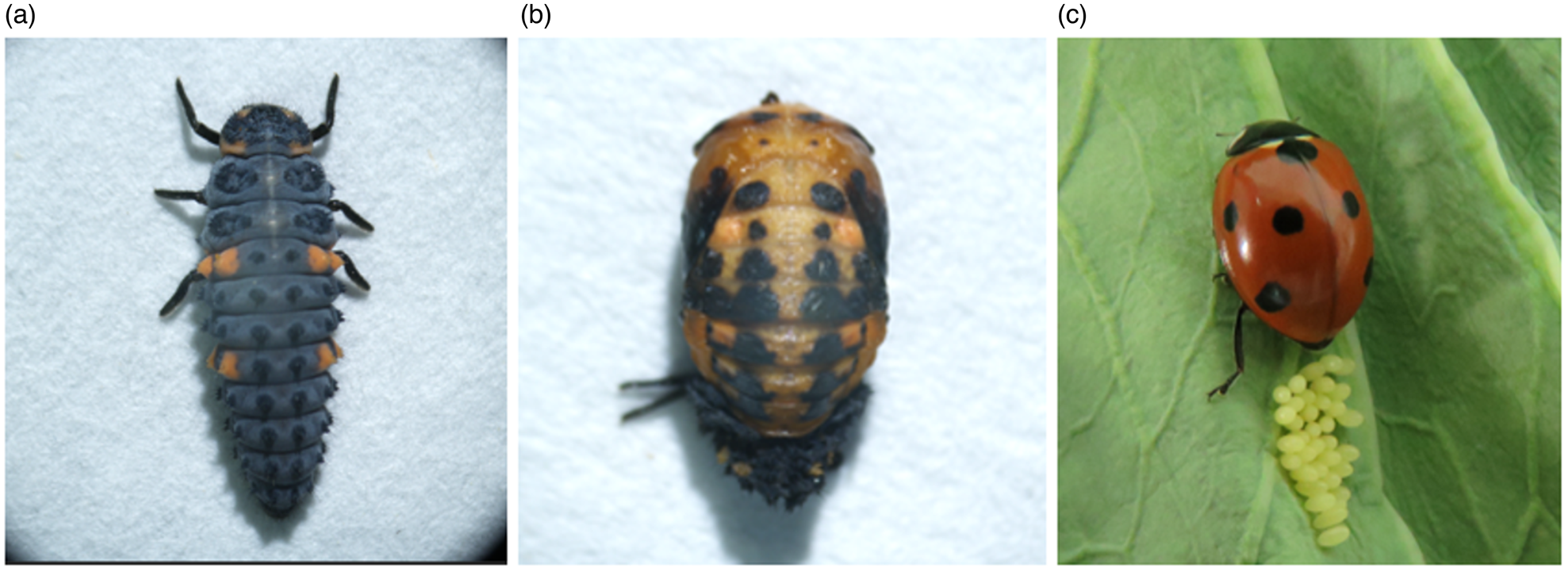
C. septempunctata were collected in wheat fields and reared indoors on Aphis craccivora as described previously (Cheng et al., Reference Cheng, Zhou and Li2024a, Reference Cheng, Zhou, Li and Jin2024b) (fig. 1). Studies were conducted in growth chambers maintained at 70 ± 5% RH, 25 ± 1°C, with a 16:8 h light:dark photoperiod.
A generation of photographs of Coccinella septempunctata. (a) Larva, (b) pupa, (c) adult and eggs.

Artificial diet
The ingredients of the artificial diet and the procedure for preparing it have been described previously (Cheng et al., Reference Cheng, Zhou, Ran and Li2023). Juvenile hormone III (JHШ, 65.0%, Toronto Research Chemicals) was added to the artificial diet (582.2 g) at 0, 1, 3, and 5 μl and mixed. An aphid diet consisting of A. craccivora served as a control (CK).
Feeding protocols
Male and female adults were paired at 24 h after emergence. Five pairs were placed in 500 ml plastic bottles (10 × 8 cm) and supplied with the artificial diet as described (Cheng et al., Reference Cheng, Zhou, Ran and Li2023). Female adults were collected 5, 10, 15, 20, 25, and 30 days after feeding and stored at −80°C until they were used in experiments. A single replication consisted of five adults, and three replications were used in each treatment.
Determination of JH titres
Six female adults were weighed and transferred to 5 ml centrifuge tubes; methanol (4 ml, 98%, Macklin Co.) was then added and mixtures were homogenised at 4°C for 120 s, 60 Hz for a total of three times. Solutions were sonicated for 30 min at 4°C, and supernatants were transferred to 15 ml centrifuge tubes. The sonication was repeated three times and solutions were combined, volatilised at low temperature until nearly dry, and then dissolved with 50% methanol to 0.5 ml. Solutions were gently agitated for 1 min and centrifuged at 4°C 13,000 rpm for 20 min; supernatants were subjected to filtration through a 0.22 μm membrane, and JH titres were determined by UPLC-MS/MS with a Waters BEH amide chromatographic column (2.1 × 100 mm, 1.7 μm). Compounds were separated at 40°C with a flow rate of 0.3 ml min−1 in an aqueous solution of 0.1% formic acid (solvent A) and 0.1% formic acid in methanol (solvent B).
RNA extraction and cDNA synthesis
Female adults were ground to a powder in liquid nitrogen and transferred to 1.5 ml RNase-free microcentrifuge tubes. Total RNA was extracted from samples according to the protocols provided with the Eastep® Super Total RNA Isolation Kit (Promega Co.). RNA integrity and purity were assessed by spectrophotometry and 1.2% agarose gel electrophoresis. One microgram of RNA was used as a template, and cDNA was synthesised with the Revert Aid First Strand cDNA Synthesis Kit (Fermentas Co.). Samples were stored at −20°C until needed.
Quantitative gene-expression analysis
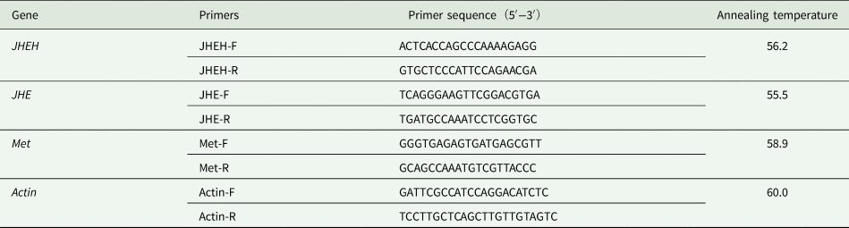
The sequences of JHEH (GI: 2125268544), JHE (GI:1027077779) and Met (OR135688) were obtained from the National Center for Biotechnology Information (https://www.ncbi.nlm.nih.gov/). ClustalX and Primer Premier v. 5.0 were used to design primers (table 1), which were synthesised by Sangon Biotech Co. (Shanghai). The cDNA obtained by reverse transcription was used as the template, and Actin (Liu et al., Reference Liu, Wang, Wang, Gao, Zhang, Li, Zang and Zhang2019) was used as an internal reference. RT-qPCR was performed using the iTaq Universal SYBR Green Supermix Kit (Bio-Rad). RT-qPCR was conducted in 20 μl reaction volumes containing upstream and downstream primers, 2 μl each; cDNA template, 2 μl; Sso Advanced Universal SYBR Green Supermix, 10 μl; and ddH2O, 4 μl. The RT-qPCR conditions consisted of 95°C for 2 min (pre-denaturation); 95°C for 5 s (denaturation), and incubation for 30 s at the annealing temperature for each primer for a total of 39 cycles. The melting curve conditions were 65–95°C in 0.5°C increment at 2–5 s step−1. Relative expression levels were calculated using the 2− ΔΔ Ct method (Pfaffl, Reference Pfaffl2001).
The primer sequences used to amplify juvenile hormone epoxide hydrolase (JHEH), juvenile hormone esterase (JHE), the JH receptor (Met), and Actin in Coccinella septempunctata

Statistical analysis
Data Processing System v. 19.0 was used for statistical analysis (Tang and Zhang, Reference Tang and Zhang2013), and the relative expression of JHEH and JHE were evaluated using the Student's t test. The effects of JH on expression of JHEH, JHE, and Met were analysed by one-way ANOVA, and significance was determined by the LSD method.
Results
JH titres in female adults reared on the aphid diet
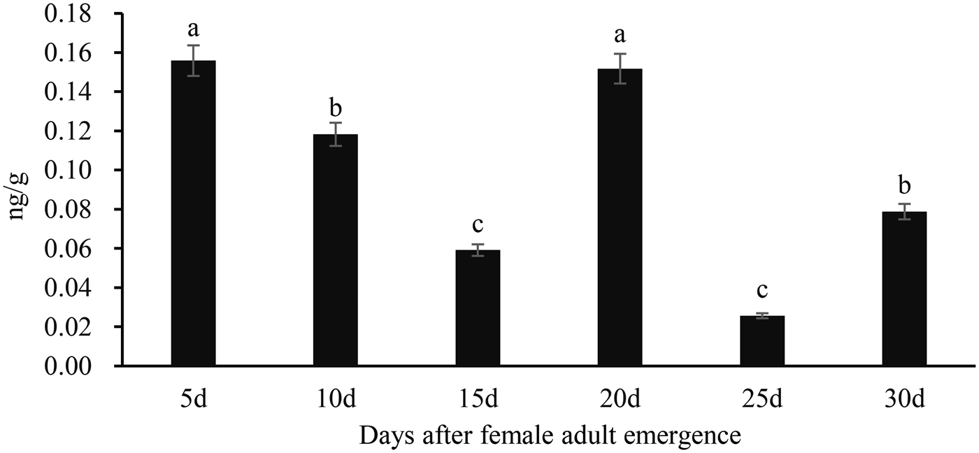
JH titres in female adults reared on the aphid diet ranged from 0.03 to 0.16 ng g−1 (fig. 2). JH levels were the highest (0.16 ng g−1) in 5-day-old female adults and were lowest (0.03 ng g−1) in 25-day-old female adults. JH titres gradually decreased in 5-, 10-, to 15-day-old female adults, and the titres were 0.16, 0.12, and 0.06 ng g−1, respectively. A notable increase in JH was apparent in 20-day-old female adults where titres increased to 0.15 ng g−1; however, in 25- and 30-day-old female adults, the titre value dropped to 0.03 and 0.08 ng g−1, respectively.
JH titres in days after female adult reared on the aphid diet emergence of Coccinella septempunctata. Data points represent means ± SE, and columns labelled with different letters are significantly different at P < 0.05.

Expression of JHEH, JHE, and Met in female adults reared on the aphid diet
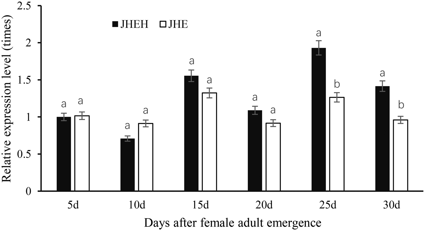
In female adults reared on the aphid diet, the relative expression of JHEH in 5-day-old adults was set to a value of 1 (fig. 3). JHEH expression was the highest in 25-day-old adults and was lowest in 10-day-old female adults, which were 1.93- and 0.71-fold of the values observed for 5-day-old female adults, respectively. JHE expression was highest in 15-day-old adults and lowest in 10-day-old female adults, which were 1.32- and 0.91-fold that of 5-day-old female adults, respectively. The expression levels of JHEH in 5-day-old (F = 1.31, P = 0.9550) and 10-day-old (F = 7.13, P = 0.2610) female adults were lower than JHE transcript levels, and the differences were not significant. The expression of JHEH in 25- (F = 173.06, P = 0.0115) and 30-day-old (F = 2.29, P = 0.0285) female adults was significantly higher than JHE transcript levels. Although JHEH expression was higher than JHE in 15- and 20-day-old female adults, the differences were not significant.
Relative expression of genes encoding juvenile hormone epoxide hydrolase (JHEH) and juvenile hormone esterase (JHE) in Coccinella septempunctata female adults reared on the aphid. Data points represent means ± SE, and columns labelled with different lowercase letters are significantly different at P < 0.05.

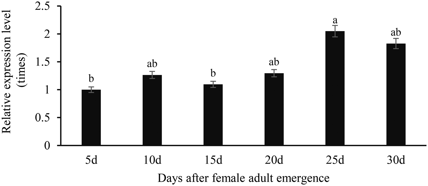
Met transcript levels in 5-day-old female adults reared on the aphid were set to 1 (fig. 4). Met expression in 5- to 20-day-old female adults was lower than levels observed for 25 to 30-day-old adults. Met expression was the highest in 25-day-old female adults where levels were 2.05-fold greater than levels in 5-day-old females; furthermore, this difference was significant (F = 25.97, P = 0.0364).
Relative expression levels of Met in Coccinella septempunctata female adults reared on the aphid. Data points represent means ± SE. Different lowercase letters indicate significance at P < 0.05.

Gene expression in female adults reared on artificial diets containing different doses of JH
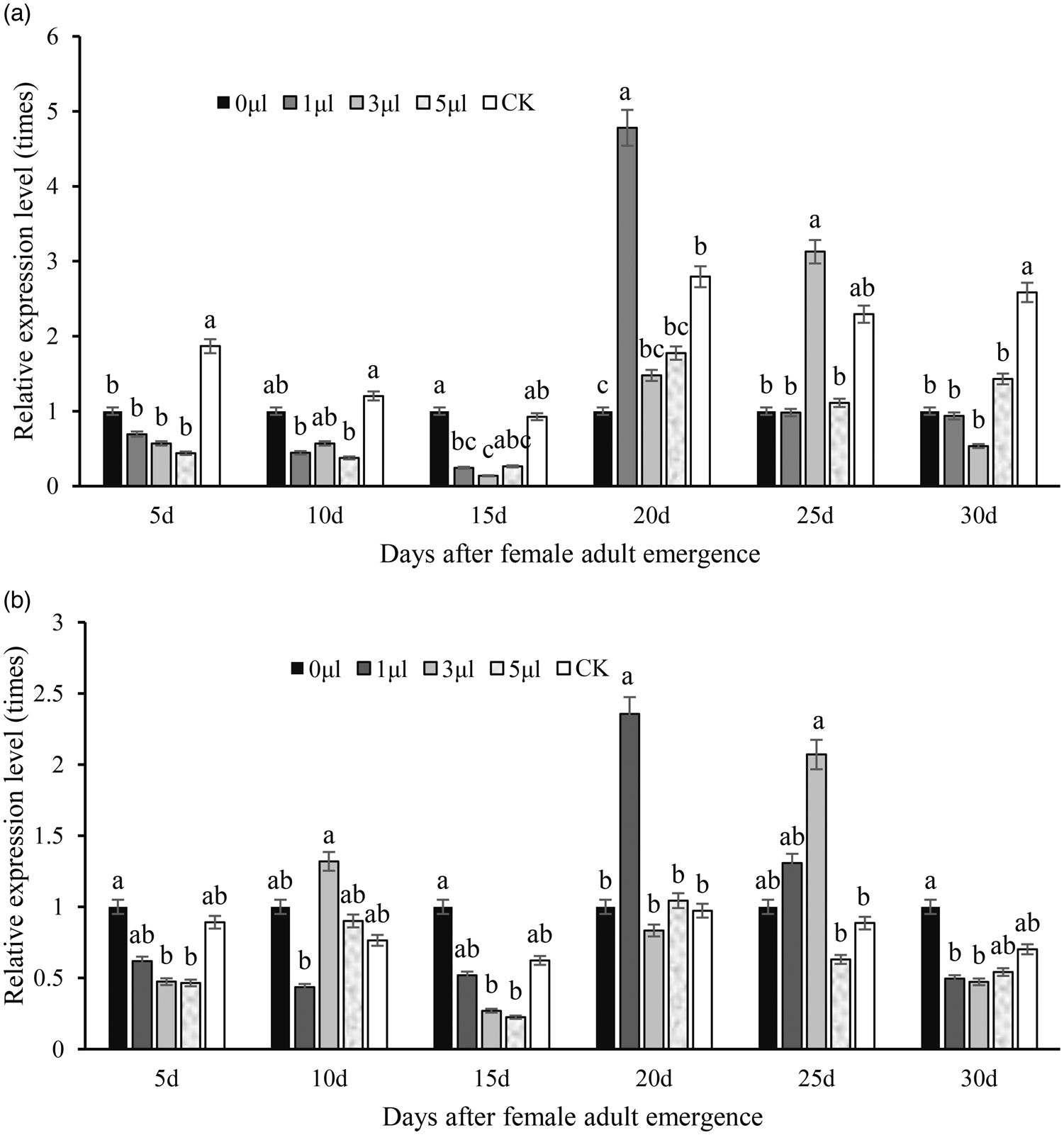
The statistical analysis was compared between different doses at the same time point (fig. 5A, B). The relative expression of JHEH in female adults reared on the 0 μl JH diets was set to a value of 1 (fig. 5A). In 5-day-old female adults, the expression of JHEH was highest when ladybirds were reared on the aphid diet (CK) (fig. 5A) (F = 7.88, P = 0.0070); the expression level of JHEH gradually decreased in response to 0, 1, 3, and 5 μl JH, which were 1, 0.69, 0.57, and 0.44, respectively, of the levels observed on the 0 μl JH diet. JHEH expression levels in 10- and 15-day-old female adults reared on the aphid diet (CK) were similar to adults that received no JH artificial diets (0 μl). In 10- to 15-day-old females, JHEH expression levels in beetles supplied with 0 μl JH or the aphid diet were higher than beetles fed with 1, 3 or 5 μl JH. JHEH expression in 20-day-old female adults supplied with 1 μl JH was higher than that in adults supplied with 0, 3, and 5 μl JH and the aphid diet. In 25-day-old female adults supplied with 3 μl, JH had higher JHEH expression than adults supplied with 0, 1, and 5 μl JH and the aphid diet. In 30-day-old female adults, the expression of JHEH supplied with aphid diet was highest and was lowest in adults supplied with 3 μl JH.
Effects of different JH levels on the expression of JHEH and JHE in Coccinella septempunctata female adults at the same time point. Panels show expression levels for (a) JHEH and (b) JHE. The control (CK) shows expression levels in ladybird beetles supplied with an aphid diet. Data represent means ± SE. Different lowercase letters indicate significant differences at P < 0.05.

In 5-day-old ladybird beetles, JHE expression was lower when diets contained 1, 3, or 5 μl JH as compared to expression levels in diets lacking JH (0 μl) or the aphid diet (fig. 5B). JHE expression in 10-day-old female adults supplied with 1 μl JH was lower than expression levels in beetles supplied with the aphid diet or with 0, 3, or 5 μl JH; furthermore, the artificial diet amended with 3 μl led to significantly higher JHE expression. In 15-day-old females, JHE expression levels in beetles supplied with 0 μl JH diet was higher than beetles fed with 1, 3, 5 μl JH diet or with aphid. In 20-day-old female adults supplied with 1 μl JH, JHE expression was significantly higher than levels in adults fed with aphids or with 0, 3, or 5 μl JH (F = 9.94, P = 0.0034). In 25-day-old female adults fed with an artificial diet containing 3 μl JH, JHE expression was higher than adults fed with aphids or with 0, 1, or 5 μl JH. In 30-day-old females, JHE expression levels in beetles supplied with 0 μl JH was higher than beetles fed with 1, 3, and 5 μl JH or with aphids, and there were no significant difference in expression when beetles were fed with 1, 3, or 5 μl JH (F = 4.74, P = 0.0295).
Met expression in female adults fed artificial diets supplemented with different JH doses
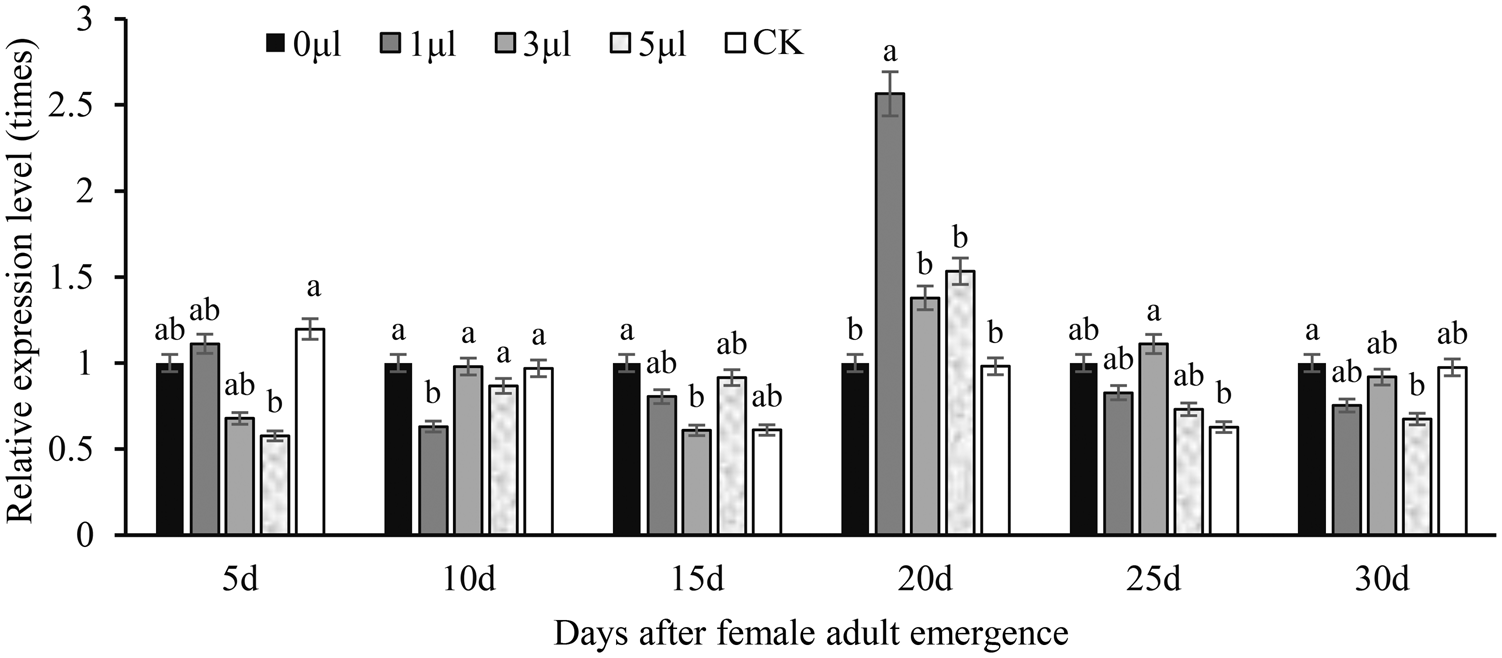
The statistical analysis was compared between different doses at the same time point (fig. 6). The relative expression of Met in female adults reared on the 0 μl JH diets was set to a value of 1. In 5-day-old female adults, Met expression was highest when ladybird beetles were supplied with the aphid diet. In 10-day-old females, Met expression was significantly lower when ladybirds were supplied with 1 μl JH as compared to diets containing 0, 3, or 5 μl JH or the aphid diet (F = 7.66, P = 0.0077). In 15-day-old females, Met expression was significantly higher when diets contained no JH (0 μl) as compared to diets containing 3 μl JH artificial diet. In 20-day-old females, Met expression in adults supplied with 1 μl JH was significantly higher than expression levels in beetles supplied with the other diets (F = 6.80, P = 0.0109). In 25-day-old females, Met expression levels in response to artificial diets amended with 3 μl JH were significantly higher than the levels observed for females supplied with the aphid diet (F = 20.79, P = 0.0449). In 30-day-old female adults, Met expression levels in diets containing no JH (0 μl) were significantly higher than levels observed for females supplied with 5 μl JH (F = 59.48, P = 0.0164). Among the diets amended with different levels of JH, Met expression in females supplemented with 3 μl JH was most similar to expression levels in response to the aphid diet.
Effects of different JH levels on Met expression in Coccinella septempunctata female adults at the same time point. The control (CK) shows Met expression levels in ladybird beetles supplied with an aphid diet. Data represent means ± SD. Columns labelled with different letters indicate significance at P < 0.05 using the LSD test.

Discussion
In this study, JH titres varied among female adults reared on the aphid of different ages (fig. 2). In 5- to 15-day-old adult females, JH titres decreased; however, titres increased at 20 days and then fluctuated at 25 and 30 d. Interestingly, JHEH and JHE expression levels were highest in females at 15- and 25-day-old and this resulted in JH degradation and a drop in JH titre. JHEH and JHE expression levels were lower in 5- and 20-day-old adults, and this correlated with higher levels of JH (fig. 3). This supports the fact that JH titres in female insects are affected by JHEH and JHE activity. We observed higher expression levels for JHEH as compared to JHE, which indicates that the former enzyme may have a greater role in JH degradation (fig. 3). JHEH belonged to the microsomal epoxide hydrolase family and appears to be more potent than JHE in terms of function as these are non-secreted enzymes, active in various organs and tissues and catalyse JH degradation reaction in an irreversible way (Seino et al., Reference Seino, Ogura, Tsubota, Shimomura, Nakakura, Tan, Mita, Shinoda, Nakagawa and Shiotsuki2010). Thus, JHEH holds a great importance in JH regulation in insect body compared to JHE (Sarkar and Kalia, Reference Sarkar and Kalia2023). The degradation of JH by JHEH has been studied in Manduca sexta, Tribolium castaneum, and Nilaparvata lugens (Tsubota et al., Reference Tsubota, Nakakura and Shiotsuki2010; Zhao et al., Reference Zhao, Zhou, Li, Cai and Hua2017). Wang et al., (Reference Wang, Liang, Cheng and Zhu2020) reported that JHEH has a crucial role in JH degradation during metamorphosis in Sitodiplosis mosellana. Knocking down of JHEH in adult female Aedes aegypti using double-stranded RNA (dsRNA) did not affect egg development or caused adult mortality (Borovsky et al., Reference Borovsky, Ekert, Buytaert, Peeters and Rouge2023). In addition, injecting JHE into Manduca sexta can reduce the titre of JH in the haemolymph (Bonning et al., Reference Bonning, Ward, Van Meer, Booth and Hammock1997).
The expression level of JHEH in C. septempunctata females fed on diets lacking JH was significantly lower than those supplied with the aphid diet (fig. 5A), and the transcription of JHEH in females was affected by the diets. Studies of adding JH to artificial diet have shown that (fig. 5A-B), expression levels of JHEH and JHE in 5- to 15-day-old females supplied with JH were lower than levels in females that were not supplemented with JH. Due to the addition of JH in artificial diets, ladybugs need to consume a portion of the proteins JHEH and JHE to degrade and metabolise JH, resulting in decreased expression of JHEH and JHE. In 20- to 30-day-old adult females, JHEH expression increased when ladybugs were fed on artificial diets supplemented with JH. In 20- to 30-day-old ladybugs, which are the optimum age for females to lay eggs, the titre of JH decreases, reducing the degradation of JH by JHEH and JHE and causing increased expression of JHEH and JHE. However, JHEH expression levels were lower than those in females supplied with the aphid diet, and the feeding of ladybugs on artificial diets reduced the expression of JHEH. We previously documented the effects of different JH analogues on the biological characteristics of C. septempunctata and showed that adult mortality increased at higher quantity of JH (Cheng et al., Reference Cheng, Zhou, Ran and Li2023). We speculated that the expression of JHEH and JHE decreased after the addition of JH in the artificial diet, resulting in excessive accumulation of JH and death. However, knockdown of JHEH in Aedes aegypti females by RNAi did not affect egg development or lead to death (Borovsky et al., Reference Borovsky, Ekert, Buytaert, Peeters and Rouge2023).
In our study, Met expression levels in females reared on the aphid were stable in the early stages of oviposition (5- to 20-day-old adults); however, expression levels increased in 25- to 30-day-old adults (fig. 4). High levels of Met expression in ladybird adults may help regulate reproductive development and life span (Smykal et al., Reference Smykal, Bajgar, Provaznik, Fexova, Buricova, Takaki, Hodkova, Jindra and Dolezel2014; Hejnikova et al., Reference Hejnikova, Paroulek and Hodkova2016). We show that Met expression in female adult ladybirds varied when JH was supplied at different doses (fig. 6). Met expression levels in females reared without JH (except for 5-day-old ladybugs) were higher than levels reared on aphids, but the difference was not significant (P < 0.05). Therefore, Met transcription in females reared without JH was not affected; however, adding different doses of JH to artificial diet resulted in variable Met expression after feeding, which may be due to the addition of JH to artificial diet. Ladybugs require more protein Met to participate in the JH metabolic pathway to regulate reproductive function and reduce Met expression. After feeding on JH diet, Met expression increased in different stages and doses. When female adults were supplied with 3 μl JH, Met expression levels were similar to those in females supplied with an aphid diet. In a related study focusing on the biological characteristics of C. septempunctata, after feeding on 1, 3, and 5 μl of the JH-amended artificial diet, ladybugs produced 186, 426, and 260 eggs in 50 days, with hatching rates of 45, 60 and 63%, respectively (Cheng et al., Reference Cheng, Zhou, Ran and Li2023). This suggests that the 3 μl dose of JH was the most suitable for the reproductive regulation of C. septempunctata.
Our results indicated that JH titres varied among C. septempunctata females of different ages and suggested that JHEH was the dominant JH-degrading enzyme. The artificial diet reduced JHEH and JHE expression and led to unfavourably high JH titres, which may result in increased mortality. Adding JH to the artificial diet requires more protein JHEH and JHE to degrade and metabolise JH, leading to a further decrease in the expression of JHEH and JHE in ladybugs. The transcription level of Met in ladybugs fed without JH artificial diet was not affected. Met expression patterns in ladybirds supplied with 3 μl JH were similar to those observed with the aphid diet, and egg production and hatching rates of ladybug were higher, indicating that the 582.2 g artificial diet with 3 μl JH was the most suitable concentration for female reproduction. It remains unclear why JH supplementation of the artificial diet resulted in decreased expression of JHEH and JHE, and this warrants further research and exploration. This study provides a theoretical basis for artificial diet improvement and breeding of ladybugs.