INTRODUCTION
Strongyloidiasis infection affects 30–100 million people in 70 different countries (Genta, Reference Genta1989; Siddiqui and Berk, Reference Siddiqui and Berk2003). The disease usually propagates by penetration of filiform larvae (L3) through the skin, but ingested parasites may also transform into the filiform infective stage inside the gut and may cause autoinfection (Siddiqui and Berk, Reference Siddiqui and Berk2001, Reference Siddiqui and Berk2003). Most of the patients infected by S. stercoralis are asymptomatic, but uncontrolled hyperinfection may occur in immunosuppressed patients, as in T-cell leukaemia virus type-1 (HTLV-1) infection and in long-term corticosteroid treatment (Siddiqui and Berk, Reference Siddiqui and Berk2001; Carvalho and Porto, Reference Carvalho and Porto2004). S. venezuelensis larvae have an obligatory migration through the lungs of the host before they become established in the duodenal mucosa (Takamure, Reference Takamure1995), similar to S. stercoralis infection in humans. In experimental murine infection adult worms are spontaneously eliminated after 10 to 14 days of infection (Sato and Toma, Reference Sato and Toma1990). Type-2 immune responses have been associated with host protection against nematode infections. These are represented by high IL-4, IL-5, IL-9 and IL-13 secretion and strong IgE production in infected mice with high accumulations of eosinophil and mast cells at the site of infection. In experimental infections with S. venezuelensis mice lacking the IL-4R chain or the STAT6 transcriptor factor (Negrão-Corrêa and Teixeira, Reference Negrão-Corrêa and Teixeira2006; Negrão-Corrêa et al. Reference Negrão-Corrêa, Pinho, Souza, Pereira, Fernandes, Scheuermann, Souza and Teixeira2006) showed severe impairment of parasite expulsion, stressing the importance of type-2 immune responses for host protection.
Conversely, in infections induced by mycobacteria such as Mycobacterium tuberculosis, the protective immune response is associated with IFN-γ production by Th1 cells (Flynn and Chan, Reference Flynn and Chan2001). Cells producing IL-10, although playing a detrimental role by down-regulating the Th1 protective immune response against Mycobacterium avium, reduce inflammation and thereby play a role minimizing pathology (Couper et al. Reference Couper, Blount and Riley2008). It is important to note that an increased incidence of tuberculosis in tropical and subtropical regions usually coincides with high prevalence of gastrointestinal parasitic infections. This may suggest a modulatory effect of helminthic infections on mycobacterial disease and on the response to BCG vaccination (Ferreira et al. Reference Ferreira, Aguiar, Fava, Corrêa, Teixeira and Teixeira2002; Elias et al. Reference Elias, Akuffo, Pawlowski, Haile, Schön and Britton2005; Talaat et al. Reference Talaat, Bonawitz, Domenech and Nutman2006). However, the effect of mycobacterial infections on the immune response to helminths has not received proper attention.
The aim of the present study was to evaluate the interference of M. bovis infection in the development of the Th2 protective immune response induced by S. venezuelensis at 2 important time-points: at 7 days post-infection when the number of worms in the intestine and eggs in the feces is highest, and at 10 days post-infection when this number has greatly diminished (Fernandes et al. Reference Fernandes, Pereira, Eschenazi, Schilter, Sousa, Teixeira and Negrão-Corrêa2008). The production of IL-4, IFN-γ, IL-10 and the expression of the costimulatory molecules CD80, CD86, CTLA-4 e CD28 on pulmonary cells as well as IL-4 and IL-13 production on intestinal homogenate from BALB/c mice were investigated.
MATERIALS AND METHODS
Animals
Six to eight-week-old male BALB/c mice were obtained from the animal facility of the Federal University of Juiz de Fora (UFJF). Animals were maintained in micro-isolators, fed laboratory chow and given tap water to drink ad libitum. All procedures were performed in accordance with the principles of the Brazilian Code for the Use of Laboratory Animals. This project was approved by the Ethics Committee of UFJF on the use of laboratory animals (034/2006).
Strongyloides venezuelensis infection
S. venezuelensis isolated from Rattus norvegicus and maintained by serial passage in Wistar rats (Department of Parasitology, Federal University of Minas Gerais) was used. For the experimental infections, S. venezuelensis infective filiform larvae (L3) were obtained from charcoal culture of infected-rat feces using a Baermann apparatus. Recovered larvae were washed in phosphate-buffered saline (PBS), the number of viable infective larvae were counted and adjusted to 7000 L3/ml in PBS. Each mouse was subcutaneously inoculated with 700 L3/0·1 ml PBS in the abdominal region (Sv group; n=5).
M. bovis infection
M. bovis 19274 strain was obtained from the American Type Culture Collection (ATCC) in lyophilized form. M. bovis was cultured in Lowenstein-Jensen medium for 21 days. Colonies were harvested at mid-log phase and stirred vigorously with sterile glass beads for 5 min and resuspended in phosphate-buffered saline (PBS). To further disrupt clumps, the bacterial suspensions were sonicated (20 watts for 5 sec). The viable counts of bacteria were determined by serial dilutions and plating into 6-well plates containing Lowenstein-Jensen. Then, it was resuspended in 0·2 ml of PBS containing 1×10⁶ colony-forming units (CFU). BALB/c mice were infected by intravenous bolus injection of 5×10⁶ CFU (Mb group; n=5).
Co-infection
At 3 days after infection, S. venezuelensis-infected mice were i.v. co-infected with M. bovis (CI group; n=5). Non-infected animals were used as control (C group; n=5). At 7 and 10 days after S. venezuelensis infection (or at 4 and 7 days after M. bovis infection), animals were sacrificed by an overdose of anaesthesia (0·9% NaCl, 2% xylazine, 5% ketamine) and lungs and intestines were removed for evaluation of parasite burden, cytokines and cell marker expression.
Parasite burden
Infectivity rates were determined by assessing fecal egg counts and numbers of worms recovered from the small intestine at day 7 and day 10 post-infection. For worm recovery from the small intestine, the upper half of the small intestine was removed after sacrifice, washed, cut open longitudinally, and incubated in PBS at 37°C for 4 h. Worms that emerged from the organ were quantified by stereomicroscopy. To determine the number of nematode eggs eliminated in the feces, well-formed fecal pellets were obtained from the rectum of each infected mouse, weighed, and homogenized in a known volume of PBS. For each infected mouse, 2 fecal samples (100 μl) were examined by light microscopy, and the total number of parasite eggs was determined. The number of S. venezuelensis eggs was expressed per gramme of mouse feces.
Cytokine production in lungs and intestines
Tissues were homogenized in extraction solution (100 mg of tissue per ml), containing: 0·4 M NaCl, 0·05% Tween 20 (Merck & Co., Inc., Whitehouse Station, USA), 0·5% bovine serum albumine (BSA), 0·1 M phenylmethylsulphonyl fluoride (PMSF), 0·1 M benzethonium chloride, 10 mM ethylenediaminetetracetic acid (EDTA) and 20 kIU/ml aprotinin (Sigma, St Louis, MO, USA), using Ultra-Turrax (IKA Works, Wilmington, USA). Tissue homogenates were centrifuged at 3000 g for 15 min at 4°C and supernatants were collected and stored at −70°C. The concentrations of IFN-γ, IL-10, IL-13 and IL-4 in supernatants of tissues extracted, at 1:2 dilution in 1% PBS-BSA, were assayed in an ELISA set-up using commercially available antibodies according to the procedures supplied by the manufacturer (BD Biosciences Pharmingen, San Diego, CA, USA).
Flow cytometry
Lungs were perfused by gently infusing 10 ml of PBS into the right ventricle of the heart to minimize contamination with blood. Perfused lungs were aseptically removed and cut into small pieces with a scalpel. Dissected tissue was incubated at 37°C for 30–45 min in 5 ml of RPMI medium containing type I collagenase (0·5 mg/ml, GIBCO, Grand Island, NY, USA). After incubation, the enzymatic activity was stopped by adding 10 ml of RPMI medium containing 10% FBS. The digested lungs were further disrupted by gently pushing the tissue through a nylon screen (70 μm). The single-cell suspension was then washed and centrifuged for 10 min. To lyse contaminating red blood cells (RBC) the cellular preparations were resuspended in RBCs lyse buffer (containing 10 μM Tris-HCl and 0·83% ammonium cloride) and incubated at room temperature for 5 min. After centrifugation, pelleted cells were then washed with PBS containing 0·1% BSA and further incubated with combinations of fluorescent antibodies anti-CD11b)/anti-CD80, anti-CD11b/anti-CD86, anti-CD3/CTLA-4, anti-CD3/CD28 for 45 min (BD Pharmingen). After incubation, 70 μl of FBS were added to each well and cell suspensions were washed 3 times, and then resuspended in PBS with 0·5% of paraformaldehyde. All samples were analysed by cytometry using FACScalibur (BD software CellQuest, Becton Dickinson, San Jose, CA, USA).
Statistical analysis
Data are reported in quintuplicate as the mean±S.E.M. and were analysed by using D'Agostino-Pearson normality test as well ANOVA and Dunns as a post-hoc test (GraphPad Prism 5.00). The differences were considered significant at P<0·05. Results are representative of 2 independent experiments.
RESULTS
Kinetics of S. venezuelensis infection
The number of S. venezuelensis worms recovered from small intestine and the number of eggs from feces were evaluated at day 7 and day 10 post-infection in mice infected with S. venezuelensis (Sv) and co-infected with M. bovis (CI) (Fig. 1). At day 7, the number of worms in the CI group was significantly higher than in the Sv group (Fig. 1A; P<0·01). At days 7 and 10, the number of eggs (Fig. 1B) was higher in the CI group in comparison to the Sv group (P<0·01 and 0·001, respectively). In addition, there was a 20-fold reduction in the number of eggs in the Sv group (35,194±4·2 to 1,775±0·3) as well as a 7-fold decrease in the number of eggs in the CI group (75,190±2·9 to 9,733±0·8) from day 7 to day 10 (Fig. 1B).
Parasite burden. Number of worms (A) and eggs (B) in Strongyloides venezuelensis-infected mice (Sv) and Mycobacterium bovis-co-infected mice (CI). The determination of the number of worms and eggs was performed in the small intestine and feces, respectively, at day 7 and day 10 post-infection with S. venezuelensis. Each bar represents the arithmetic mean±S.E.M. of 5 mice per group, and results are representative of 2 independent experiments. ** P<<0·01; *** P<<0·001.

Cytokine in the lungs and intestine
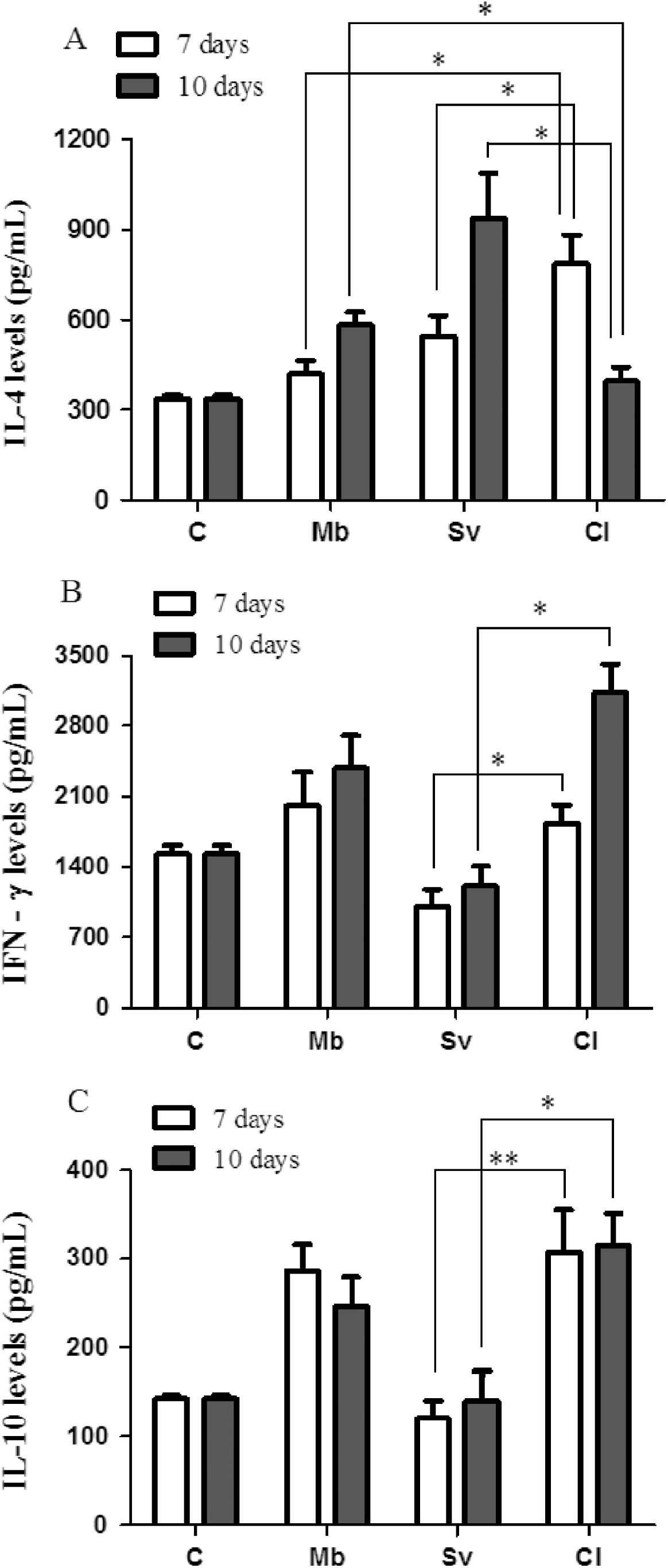
IL-4, IFN-γ and IL-10 were measured in lung supernatants at days 7 and 10 after infection (Fig. 2). Higher IL-4 production was observed at day 7 post-infection in the CI group when compared with the Mb and Sv groups (Fig. 2A; P<0·05). At day 10 post-infection a significant decrease in IL-4 production was detected only in the CI group (P<0·05). IFN-γ production was higher in the CI group when compared to the Sv group at both day 7 and day 10 post-infection (Fig. 2B; P<0·05). Likewise, IL-10 levels in lung supernatants in the CI group were higher when compared to the Sv group at both time-points (Fig. 2C; P<0·05). Fig. 3 shows the production of IL-4 and IL-13 in intestinal homogenates of infected mice. Higher levels of both cytokines were found in the Sv group in comparison with the CI group at day 7 post-infection (Fig. 3A and 3B).
Production of cytokines in pulmonary cells. (A) Interleukin-4 (IL-4); (B) interferon-gamma (IFN-γ); (C) interleukin-10 (IL-10). Lungs from non-infected (C) or Mycobacterium bovis-infected (Mb) or Strongyloides venezuelensis-infected (Sv) or co-infected (CI) mice were collected at day 7 and day 10 post-infection with S. venezuelensis and homogenized in extraction solution. Cytokine levels were determined by ELISA. Each bar represents the arithmetic mean±S.E.M. of 5 mice per group, and results are representative of 2 independent experiments. * P<0·05; ** P<0·01.

Production of cytokines in intestinal cells. (A) Interleukin-4 (IL-4); (B) interleukin-13 (IL-13). Small intestine from non-infected (C) or Mycobacterium bovis-infected (Mb) or Strongyloides venezuelensis-infected (Sv) or co-infected (CI) mice were collected at day 7 and day 10 post-infection with S. venezuelensis and homogenized in extraction solution. Cytokine levels were determined by ELISA. Each bar represents the arithmetic mean±S.E.M. of 5 mice per group, and results are representative of 2 independent experiments. ** P<0·01.

Expression of costimulatory molecules on pulmonary cells
Figure 4A shows that at day 7 post-infection CD80 expression was higher in the Mb group in comparison with the CI group (P<0·05). At day 10 post-infection, pulmonary cells from the Sv group showed higher CD80 expression in comparison to the CI group (P<0·05). In reference to CD86 expression, no difference was detected between the groups at day 7 post-infection. However, CD86 expression was higher in the CI group when compared to the Mb group at day 10 after infection (Fig. 4B; P<0·05). CTLA-4 expression was very low in all groups analysed, but its expression in the CI group was higher in comparison to Sv and Mb groups at day 10 post-infection (Fig. 5A; P<0·05). CD28 expression augmented more in the Mb group at day 10 post-infection in relation to the CI group (Fig. 5B; P<0·05).
Expression of CD80 (A) and CD86 (B) co-stimulatory molecules on CD11b+ pulmmonary cells. Lungs from non-infected (C) or Mycobacterium bovis-infected (Mb) or Strongyloides venezuelensis-infected (Sv) or co-infected (CI) mice were collected at day 7 and day 10 post-infection with S. venezuelensis, homogenized in collagenase I. Flow cytometry was performed by FACS. Each bar represents the arithmetic mean±S.E.M. of 5 mice per group, and results are representative of 2 independent experiments. Gates are representative of 1 animal in its respective group. * P<0·05.

Expression of CTLA-4 (A) and CD28 (B) co-stimulatory molecules on CD3+ pulmmonary cells. Lungs from non-infected (C) or Mycobacterium bovis-infected (Mb) or Strongyloides venezuelensis-infected (Sv) or co-infected (CI) mice were collected at day 7 and day 10 post-infection with S. venezuelensis, homogenized in collagenase I. Flow cytometry was performed by FACS. Each bar represents the arithmetic mean±S.E.M. of 5 mice per group, and results are representative of 2 independent experiments. Gates are representative of 1 animal in its respective group. * P<0·05.

DISCUSSION
In the present report, the interference of M. bovis infection in the development of a Th2 protective immune response induced by S. venezuelensis was evaluated. It was observed that co-infection with M. bovis influenced S. venezuelensis infection progression in a direct manner, resulting in an increase of the parasite burden in co-infected animals.
Interestingly, increased IFN-γ production was detected in animals infected only with M. bovis and in the co-infected group, indicating the mycobacteria as the major inducer of this Th1 cytokine. The down regulation of the Th2 immune response to S. venezuelensis observed in the co-infected group was correlated to increased levels of IFN-γ production and to increased parasite burden. Similarly, patients co-infected with HTLV-1 and S. stercoralis presented reduced Th2 cytokines and elevated virus-induced IFN-γ production (Porto et al. Reference Porto, Neva, Bittencourt, Lisboa, Thompson, Alcântara and Carvalho2001).
The Th2-dominant immune response is characteristic of helminthic infections. This work shows that levels of IL-4 and IL-13 in intestines decrease progressively after co-infection, suggesting that IFN-γ production in response to M. bovis infection may lead to down-modulation of this Th2 cytokine response incrementing the number of worms and eggs in the CI group. Recent data have shown that Th2 cytokines are of crucial importance in the expulsion of intestinal helminths such as S. venezuelensis (Negrão-Corrêa et al. Reference Negrão-Corrêa, Pinho, Souza, Pereira, Fernandes, Scheuermann, Souza and Teixeira2006; Ferreira et al. Reference Ferreira, Pereira, Souza, Cassali, Souza, Lemos, Teixeira and Negrão-Corrêa2007; Machado et al. Reference Machado, Ueta, Lourenço, Aníbal, Roque-Barreira and Faccioli2007). Furthermore, previous studies using IL-4R-deficient mice infected with S. venezuelensis demonstrated that augmented levels of IL-4 in the intestine coincided with the decline of egg production and worm elimination (Negrão-Corrêa et al. Reference Negrão-Corrêa, Pinho, Souza, Pereira, Fernandes, Scheuermann, Souza and Teixeira2006).
Experimental infections of S. venezuelensis larvae have an obligatory migration through the lungs of the host between the 2nd and 5th day before they become established in the duodenal mucosa, similar to S. stercoralis infection in humans (Takamure, Reference Takamure1995; Negrão-Corrêa and Teixeira, Reference Negrão-Corrêa and Teixeira2006, Fernandes et al. Reference Fernandes, Pereira, Eschenazi, Schilter, Sousa, Teixeira and Negrão-Corrêa2008). In this experimental model adult worms are spontaneously eliminated after 10 to 14 days of infection (Sato and Toma, Reference Sato and Toma1990). Moreover, in experimental murine M. bovis infection, the lungs are the major site of this mycobacteria and it is found in this tissue from the 1st day after intravenous infection (Jung et al. Reference Jung, LaCourse, Ryan and North2008).
The increased level of IFN-γ stimulated by M. bovis infection could inhibit the protective Th2 response to S. venezuelensis in co-infected animals which is dependent on IL-4 production. Our results suggest that the increase of IFN-γ levels from day 7 to day 10 could increment the number of worms and eggs in the CI group, which is correlated to decreased IL-4 levels and the progression of infection. Although the increased number of worms at day 10 post-infection was not statistically significant as was the number of eggs, it suggests not only higher worm infectivity but also lower host immune response activity on worm fecundity (Baek et al. Reference Baek, Islam, Kim, Lim, Oluoch, Kim and Kakoma2003).
It is noteworthy that the increased production of IL-4 in the lungs of the Sv group from day 7 to day 10 could be explained by the induction of a memory immune response against antigens secreted by the worm during its passage through the lungs. Fernandes et al. (Reference Fernandes, Pereira, Eschenazi, Schilter, Sousa, Teixeira and Negrão-Corrêa2008) observed that S. venezuelensis antigen-immunized mice had more IL-4 production and long-lasting protection. However, the levels of IL-4 detected in the lungs of CI animals showed a decrease from 7 to 10 days after infection, suggesting that production of IFN-γ and IL-10 detected in response to M. bovis may have modulated the activity of IL-4, resulting in its decline. Previous studies have demonstrated the role of IL-10 in regulating the production and activity of IL-4 during S. venezuelensis infection (Ferreira et al. Reference Ferreira, Pereira, Souza, Cassali, Souza, Lemos, Teixeira and Negrão-Corrêa2007; Negrão-Côrrea and Teixeira, Reference Negrão-Corrêa and Teixeira2006). So the elevated production of IL-10 from the 7th day after infection could reduce the activity of IL-4 detected in the CI group, which explains the elevated number of worms at this point of infection.
IL-10 can even be produced by regulatory T cells, which might hinder pathogen clearance and also reduce the tissue damage (Couper et al. Reference Couper, Blount and Riley2008). Previous studies have shown that overproduction of IL-10 by T-cells is associated with a suppression of immunity and an increase of susceptibility to mycobacterial infection (Kursar et al. Reference Kursar, Koch, Mittruker, Nouailles, Bonhagen, Kamradt and Kaufmann2007). Besides, another work demonstrated that IL-10 led to a recruitment error in Th1 differentiation from naïve into effector cells in mycobacterial infections (Demangel et al. Reference Demangel, Bertolino and Britton2002). In addition, IL-10 produced by regulatory T cells is part of an anti-inflammatory network that ensures that Th2 and the downstream effectors are kept under control (Yazdanbakhsh et al. Reference Yazdanbakhsh, Kremsner and Ree2002).
The expression of CD80 in the Mb group was higher at day 7 post-infection while its expression in the Sv group was greater on day 10. However, M. bovis co-infection led to a significant decrease in CD80 expression in pulmonary cells. In contrast, CD86 was markedly expressed at day 10 post-infection not only in the CI group but in Sv group as well. Therefore, M. bovis co-infection did not interfere with CD86 expression here. Previous reports showed that co-stimulatory molecule expression such as CD80 and CD86 can influence the helminth-induced cytokine profile (Mueller, Reference Mueller2000; Liu et al. Reference Liu, Liu, Pesce, Whitmire, Ekkens, Foster, VanNoy, Sharpe, Urban and Gause2002). According to studies involving Heligmosomoides polygyrus, the blockage of CD80/CD86 co-stimulatory molecules led to inhibition of the primary Th2 response to parasitic infection (Lu et al. Reference Lu, Zhou, Chen, Moorman, Morris, Finkelman, Linsley, Urban and Gause1994). In Nippostrongylus brasiliensis-infected BALB/c mice, the anti-CD86 mAb treatment decreased the IL-4 production and did not change the IFN-γ production (Nakajima et al. Reference Nakajima, Watanabe, Yoshino, Yagita, Okumura and Azuma1997). The reciprocal balance of CD28 and CTLA-4 is pivotal in modulating T cell responses (Lenschow et al. Reference Lenschow, Su, Zuckerman, Nabavi, Jellis, Gray, Miller and Bluestone1993; Linsley et al. Reference Linsley, Bradshaw, Urnes, Grosmaire and Ledbetter1993; Manzotti, Reference Manzotti, Tipping, Perry, Mead, Blair, Zheng and Sansom2002). Also, it has been demonstrated that CTLA-4 expression inhibition in helminthic infections induces an increased Th2 immune response. Thus, our results agree with such work because not only was there a reduction in CD80 expression but also an increased CTLA-4 and a diminished CD28 expression was noted in co-infected animals.
This report suggests that the production of IFN-γ and IL-10 in response to M. bovis can decrease CD80 expression by antigen presenting cells and increase CTLA-4 expression by T lymphocytes leading to reduced IL-4 and IL-13 production. Consequently, this immune response makes the host more susceptible to S. venezuelensis infection, increasing parasite burden. It may be important in subtropical regions where M. tuberculosis and S. venezuelensis are endemic. The possibility that the Th1 immune responses induced by M. bovis after vaccination interfere in the host response against intestinal parasites deserves more study.
This work was partly supported by grants from Fundação de Amparo à Pesquisa do Estado de Minas Gerais (FAPEMIG- CBB 1231/2005), Conselho Nacional de Desenvolvimento Científico e Tecnológico (CNPq) and CAPES-PQI 070. Michelle A. Vicentini was recipient of a scholarship from FAPEMIG.