Brady- and tachyarrhythmias are a common finding in patients with congenital heart disease. The two lesions highlighted in this series are no exception. Arrhythmias occur in the peri-operative period or can develop years after surgery. Many patients with congenital heart disease do not tolerate arrhythmias over a long period of time, although they may not be immediately symptomatic. Therefore, arrhythmia surveillance should be a standard part of the long-term management strategy for these patients. This chapter will discuss strategies regarding the management of arrhythmias in the peri-operative period, as well as long-term management.
Pre-operative period
Both common arterial trunk and d-transposition of the great arteries are cyanotic congenital lesions that require early neonatal surgery. This makes these patients vulnerable to arrhythmias. It is therefore important to monitor these patients immediately after birth and identify those infants who are at high risk of developing arrhythmias. The initial goal following birth is to maintain a stable physiologic state with adequate systemic blood flow (Qs) with little to no metabolic acidosis and adequate end-organ perfusion.
Once resuscitation of the newborn occurs and the infant is stabilised, he should be placed on a continuous telemetry system. This provides the best opportunity to not miss any signs that the patient may be at risk for post-operative arrhythmias. A baseline 12- or 15-lead electrocardiogram should be obtained. It is becoming more common that these lesions are diagnosed in utero by foetal echocardiography. However, a confirmatory transthoracic echocardiogram should be obtained, not only to confirm the prenatal diagnosis, but to evaluate the cardiac function and identify any lesions that may be haemodynamically significant and therefore precipitate arrhythmias, such as valvar insufficiency and chamber dilation. These diagnostic tests can help identify infants at higher risk of developing arrhythmias, such as the presence of ventricular pre-excitation on a baseline electrocardiogram, atrioventricular node dysfunction, or the presence of heterotaxy syndrome, although uncommon in common arterial trunk and d-transposition of the great arteries.
Central line placement, especially umbilical catheters, is a mainstay in the management of these patients. Both atrial and ventricular arrhythmias can be triggered by irritation to the myocardium when the lines are intracardiac. Therefore, a chest X-ray should be obtained if arrhythmias develop, in order to determine the location of the tip of the catheters. Atrial flutter has been associated with umbilical line placement,Reference Texter, Kertesz, Friedman and Fenrich 1 and once sinus rhythm is restored and the line is pulled out of the heart the patient may not be at risk for further arrhythmias and treatment should be withheld unless subsequent arrhythmias arise.
Infants in the pre-operative period can have both atrial and ventricular ectopy, as well as tachy- and bradyarrhythmias. Although ectopy is generally considered benign and does not warrant antiarrhythmic treatment, they can be a marker for the development of sustained arrhythmias and closer surveillance is warranted. Furthermore, the presence of any concerning pre-operative arrhythmia may impart useful information to the cardiovascular surgeon and anesthesiologist during surgery and increase awareness for similar post-operative arrhythmias, which may be less well tolerated. Strategies to minimise the arrhythmia risk should be undertaken, such as correcting electrolyte derangements and minimising acidosis. The decision to treat an arrhythmia should be based on its potentially negative haemodynamic effects – hypotension, loss of atrioventricular synchrony – and not just on “cleaning up the arrhythmias on telemetry” for the sake of “making it look better”. Similarly, an arrhythmia may signify a worsening haemodynamic milieu and a less balanced pulmonary/systemic blood flow ratio or overall systemic output. Treatment of a particular arrhythmia in neonates with d-transposition or a common arterial trunk should also be considered pre-operatively if it is deemed that non-intervention will complicate the intra-operative or post-operative period.
Maintenance of sinus rhythm and atrioventricular synchrony with a narrow QRS complex generally provides the best haemodynamics. Both automatic tachycardias (focal atrial tachycardias and ventricular tachycardias) and reentrant tachycardias (accessory pathway mediated tachycardia) may not be as well tolerated in infants with marginal hemodynamics due to their congenital heart disease as opposed to infants without congenital heart disease. Occasionally, correction of electrolyte disturbances, respiratory optimisation, or repositioning intra-thoracic lines will resolve an arrhythmia. However, when these measures fail and it is deemed necessary to control the arrhythmia, antiarrhythmic therapy should be initiated. All antiarrhythmic agents in the pre-operative period should be used with caution because of the potential for developing bradyarrhythmias, as temporary pacing is not readily available at this juncture. Once treatment has been decided, the drug and route must be decided. Generally, both oral and intravenous antiarrhythmic therapies are available. Neonates who cannot take enteral medications should be placed on continuous drips. Esmolol, procainamide, and amiodarone are the most commonly used intravenous antiarrhythmics in the neonatal period. Although there are some concerns about the negative inotropic effects of many of these, they are generally well tolerated. Nonetheless, a discussion regarding the use of antiarrhythmics should involve the entire team.
Esmolol as an intravenous beta-blocker has a short half-life – 3–10 minutes.Reference Trippel, Wiest and Gillette 2 It is effective for both atrial and ventricular reentrant and automatic tachycardias. However, it should be used with caution, especially in the setting of ventricular dysfunction. Its short half-life is advantageous in the peri-operative period; if the haemodynamics do not tolerate it, its effects subside promptly. Procainamide is a sodium channel blocker that is also effective for atrial and ventricular arrhythmias. It causes less bradycardia than esmolol. Levels of procainamide, as well as its active metabolite N-acetyl-procainamide-acetate, need to be monitored during the infusion in a timely manner to avoid toxicity. Amiodarone is also effective for both atrial and ventricular arrhythmias, and is more likely to control arrhythmias in a shorter time for patients who have significant ventricular dysfunction or poor haemodynamics as a result of the arrhythmia. The side-effect profile of amiodarone makes it less than ideal in the long term. It has a long half-life (45 days).
If the patient will tolerate enteral medications, it may be preferable to minimise the iatrogenic risks of intravenous use, such as extravasations and infections. Propranolol, digoxin, amiodarone, and sotalol are all commonly used in the newborn period for peri-operative arrhythmias. Both amiodarone and sotalol act by prolonging phase III of the myocyte action potential – prolong repolarisation – and careful scrutiny of the QT interval should be assessed to avoid a negative pro-arrhythmic effects. Digoxin should be used with caution as it has a narrow therapeutic window and may be less effective than other antiarrhythmics in preventing recurrences.
Post-operative period
The operative period exposes the neonate with congenital heart disease to a vast array of physiologic and non-physiologic machinations that further compound the proclivity for arrhythmias. The neonate with a history of arrhythmias in the pre-operative period may no longer have the benefit of antiarrhythmic protection. Cardiopulmonary bypass will sequester the majority of these medications from the infant's system. Therefore, the post-operative neonate continues to be at risk for arrhythmia development. Peri-operative arrhythmias have been shown to be associated with an increased mortality.Reference Shamszad, Cabrera and Kim 3 In addition, the pressure and volume changes that occur following repair of the congenital heart disease put these patients at higher risk. Myocardial protection is extremely important during cardiopulmonary bypass, and if the myocardium is not well protected this increases the potential for arrhythmias. In addition, most infants will return from the operating room with intracardiac catheters for pressure monitoring, as well as for medication infusion. Trauma from these catheters can cause tachyarrhythmias, and if there is trauma to the atrioventricular node varying degrees of heart block can occur.Reference Perry, Fenrich, Hulse, Triedman, Friedman and Lamberti 4 Many of the medications to help support inotropy and lusitropy can cause tachyarrhythmias. In addition, pain or a low cardiac output state can increase sympathetic activity, increase the sinus rate, and further aggravate the haemodynamic balance, limit cardiac filling, and increase certain arrhythmias, such as junctional ectopic tachycardia.
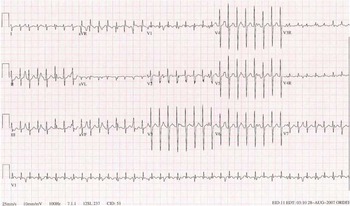
One of the more common arrhythmias in neonates following reparative transposition of the great arteries with a complex ventricular septal defect or common arterial trunk is junctional ectopic tachycardia (Fig 1). It occurs following ventricular septal defect repair or when there is traction applied close to the atrioventricular node and generally leads to haemodynamic compromise. It is the result of irritation to the atrioventricular node and its associated conduction tissue. Patients who undergo repair of complex ventricular septal defects, especially those that involve the conotruncus and require prolonged bypass times, are at risk for junctional tachycardia.Reference Hoffman, Bush and Wernovsly 5 Patients can have such profound haemodynamic compromise that might even require extreme mechanical support as rescue, such as extracorporeal membrane oxygenation. Junctional ectopic tachycardia generally occurs in the first 24 to 48 hours after surgery, and aggressive management is important. Weaning inotropes and cooling the patient are helpful. As a general approach, cooling below 35.5°C is avoided. Overdrive atrial pacing is often attempted to restore atrioventricular synchrony, which is successful as long as the junctional rate can be slowed with medications and other measures. Correcting acidosis and electrolytes are key strategies. Magnesium in particular should be given if tolerated. If these strategies do not work, then intravenous amiodarone should be promptly initiated. In a neonate with already compromised haemodynamics, care must be given when administering intravenous amiodarone. It should be given in doses of 5 mg/kg and run over 20–60 minutes. Pre-medicating infants with a bolus of calcium chloride often prevents the hypotension that can be seen with rapid amiodarone administration. Procainamide is a second agent that can be used to treat junctional tachycardia. It is less potent than amiodarone and therefore not used as commonly, but may be better tolerated.Reference Madapati, Byrum and Kavey 6 There is increasing evidence that dexmedetomidine may be useful in post-operative junctional ectopic tachycardia.Reference Chrysstomou, Beerman and Shiderly 7 Typically, once sinus rhythm is restored and post-surgical inflammation resolves, this arrhythmia subsides and does not require long-term treatment.
This electrocardiogram demonstrated junctional ectopic tachycardia 12 hours after repair of d-transposition and a ventricular septal defect. Note the atrioventricular dissociation.

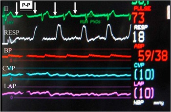
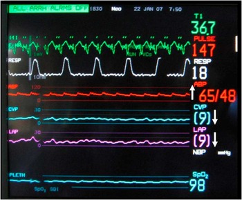
Atrioventricular block can also occur following surgery owing to mechanical injury to the atrioventricular node, especially during closure of the ventricular septal defect in common arterial trunk or in d-transposition with a complex ventricular septal defect (Figs 2 and 3). Atrioventricular demand pacing should be performed using temporary pacing wires, and atrioventricular conduction should be monitored on a daily basis. If atrioventricular conduction does not return by post-operative day 10, a permanent epicardial pacing system should be placed.Reference Epstein, DiMarco and Ellenbogen 8 It must be remembered that arrhythmias may be a sign of an electrolyte malady, ventricular dysfunction, or significant residual post-operative lesions such as moderate to severe atrioventricular valve or semi-lunar valve insufficiency. Often, the electrophysiologist is asked whether to treat an arrhythmia, but the following questions should always be addressed first:
Bedside monitor of a 9-day-old infant after repair of a common arterial trunk with post-operative atrioventricular block with demonstration of atrioventricular dyssynchrony – P–P interval marked with arrows and dissociated from the QRS complex. Note the cannon waves on the CVP and LAP tracings. BP = blood pressure; CVP = central venous pressure; LAP = left atrial pressure; NBP = noninvasive blood pressure measurement; RESP = respiration.

Bedside monitor of the same 9-day-old infant after repair of a common arterial trunk with post-operative atrioventricular block with atrioventricular sequential pacing improving the blood pressure and lowering both the CVP and LAP. ABP = arterial blood pressure; CVP = central venous pressure; LAP = left atrial pressure; NBP = noninvasive blood pressure measurement; PLETH = raw fingertip plethysmogram outputs; RESP = respiration.

-
• “What is the status of the repair?”
-
• “What are the findings on the echocardiogram and how do they relate to either the current haemodynamic state or the arrhythmia itself?”
Treatment for arrhythmias outside of the post-operative state will vary from institution to institution. Most arrhythmias seem to resolve within the first 6 months of surgery. Consideration should be made to wean off medication within 6–12 months of therapy and continue to monitor for arrhythmia recurrence. Immediate post-operative arrhythmias do not always predict recurrence later in life, with perhaps the exception of reentrant arrhythmias due to accessory pathways, especially in the setting of Wolff–Parkinson–White syndrome. These patients are at higher risk of recurrence even in the setting of a structurally normal heart.Reference Perry and Garson 9
Long-term arrhythmia surveillance and follow-up
While patients are on antiarrhythmic therapy as an outpatient, surveillance tools, such as electrocardiogram, and 24-hour ambulatory Holter monitoring should be performed on a routine basis. The QRS duration and QTc interval should be checked on each electrocardiogram if a patient is on a medication that will affect conduction or repolarisation. The Holter will detect non-sustained arrhythmias that may otherwise go undetected in an asymptomatic infant, and help assess for ventricular pauses or other conduction abnormalities. Patients on amiodarone should have thyroid testing performed every 3–6 months, as well as liver function testing and complete blood counts. Yearly chest X-rays, thyroid studies, and ophthalmologic exams should also be performed for older children on chronic amiodarone therapy. Older children should undergo yearly formal pulmonary function testing.Reference Perry, Fenrich, Hulse, Triedman, Friedman and Lamberti 4 It is reasonable to wean off amiodarone therapy after 6–12 months, and if the arrhythmia recurs a different antiarrhythmic agent with fewer side effects should be tried before reinitiating amiodarone.
Arrhythmia surveillance becomes particularly important as these children become older. Patients who are status post common arterial trunk repair will continue to have residual pressure and volume loading lesions that will predispose them to both atrial and ventricular arrhythmias. These residual haemodynamic lesions should be addressed in the operating room or cardiac catheterisation laboratory. Patients who experience a deterioration in ventricular function, especially over a relatively short period of time, should be aggressively evaluated for arrhythmias as the possible causative factor.
The most common reasons adults with congenital heart disease die is secondary to sudden death, presumably from arrhythmias, and congestive heart failure, where arrhythmias are prevalent.Reference Oeschlin, Harrison, Connelly, Webb and Siu 10 In addition, studies have shown increased morbidity and mortality in adults with congenital heart disease who have pre-existing arrhythmias, including atrial arrhythmias.Reference Yap, Harris and Chauhan 11 This underscores the need for close monitoring and aggressive arrhythmia management for these patients, which may include medical therapy, catheter ablation, or insertion of a pacemaker or internal cardioverter defibrillator.
Few studies have studied arrhythmia burden and prognosis specifically in common arterial trunk. Several long-term surgical studies are available from the surgical literature, but they do not characterise arrhythmia burden.Reference Tlaskal, Chaloupecky and Hucin 12 – Reference Mair, Sim, Danielson and Puga 14 It is generally assumed that, given the nature of the repair, the risk of arrhythmias and sudden death is similar to those with tetralogy of Fallot, plus any increased risk if the truncal valve is abnormal. It should be remembered that diagnostic dysfunction, which is equally seen in patients with common arterial trunk or complex transposition, may be causative for arrhythmias and can be difficult to manage medically. Both ventricular systolic dysfunction and abnormal diastology can have negative long-term effects and may be pro-arrhythmic.
Arrhythmias in adults with atrial switch operations – Senning or Mustard procedure – are much more prevalent than in adults who underwent the arterial switch operation.Reference Rhodes, Wernovsky and Keane 15 Nevertheless, the potential risk of late arrhythmias cannot be overlooked in d-transposition after an arterial switch. Factors that contribute to the arrhythmia risk include the coronary manipulation during the initial surgery. If a ventricular septal defect is present and needs to be patched, there is risk of damage to the atrioventricular node or fascicles, which over time can result in atrioventricular or ventriculo-ventricular dyssynchrony and consequent cardiac failure.
When symptoms are present, such as palpitations, dizziness, or syncope, aggressive evaluation is necessary. The use of both non-invasive and invasive testing may be required. These tools include exercise testing, 24-hour Holter monitoring, event monitors, implantable loop recorders, and electrophysiology studies.
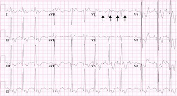
It is important to remember that not all tachyarrhythmias have rapid ventricular conduction. Intra-atrial reentrant tachycardia, which is also known as scar flutter, is a macroreentrant atrial tachyarrhythmia. See the electrocardiogram in Figure 4. It occurs usually around an atriotomy scar, where the myocardium is damaged, resulting in an area of slowed conduction. This slowed conduction is the set-up for reentry.Reference Kannankeril and Fish 16 Varying degrees of atrioventricular node conduction may result in a slower than expected ventricular rate. In such a situation, the patient may be asymptomatic or have minimal symptoms, and the presence of intra-atrial reentrant tachycardia may be missed if careful attention is not paid to resting electrocardiograms. Symptoms of palpitations, out of proportion to the heart rate may be one clue to this diagnosis. In addition, older patients may be at risk for atrial fibrillation, which will have variable conduction through the atrioventricular node. Further testing with Holter monitoring or more invasive electrophysiology studies should be performed when clinical suspicion arises.
This is a 12-lead electrocardiogram of a 12-year-old boy status post common arterial trunk repair in intra-atrial reentrant tachycardia. Although the ventricular rate is 110 beats per minute, the atrial rate is much faster, as indicated by the arrows.

Exercise testing is a useful diagnostic outpatient tool to assess for chronotropic incompetence, and ischaemic changes, which may be a sign of a coronary hypoperfusion or myocardial scarring. Aerobic capacity can also be quantified, and is useful to follow if symptoms of fatigue or exercise intolerance occur. Pasquali et al Reference Pasquali, Marino, McBride, Wernovsky and Paridon 17 recently demonstrated that variant coronary patterns had more chronotropic incompetence on long-term evaluation than the typical pattern for d-tranposition of the great arteries. Other studies have shown ischaemic changes, which can be due to residual haemodynamic lesions,Reference Mahle, McBride and Paridon 18 , Reference Giardini, Khambadkone and Rizzo 19 even in asymptomatic patients.Reference Mahle, McBride and Paridon 18 Therefore, exercise testing should be routinely performed, especially in patients status post the arterial switch operation, even in the absence of symptoms.
Depending on the individual risk factors, 24-hour ambulatory Holter monitoring should be done on a routine basis. They are useful at looking at average heart rates in addition to detecting tachyarrhythmias and may help detect the presence of sinus node dysfunction and bradyarrhythmias.
When paroxysmal symptoms are present, portable event monitors may help detect arrhythmias. These are particularly useful when they occur randomly. Two general types are available. Hand-held monitors can be carried and applied during symptoms for episodes that last for several minutes. For shorter episodes, a looping event monitor that is worn for 15 to 30 days at a time is likely more effective.
Patients who are status post the arterial switch operation are prone to chronotropic incompetence.Reference Giardini, Khambadkone and Rizzo 19 , Reference Hui, Chau, Leung, Chiu and Cheung 20 Symptoms of chronotropic incompetence, such as fatigue, hypersomnolence, and exercise intolerance, should be evaluated with exercise testing, and consideration should be made for permanent pacemaker placement when indicated. Owing to the fact that both patients with a common arterial trunk and d-transposition have biventricular circulations, transvenous pacemaker implantation is possible, provided there is no residual intracardiac shunting. Anatomical variations, such as bilateral superior caval veins and/or no left subclavian vein, should be identified before device placement.
Implantable cardioverter defibrillator placement should be considered in patients who are at risk for lethal ventricular arrhythmias and sudden cardiac death. Left ventricular dysfunction, left-sided obstructive lesions, and residual coronary artery disease are particularly associated with sudden death, and defibrillator therapy needs to be offered when the risk of sudden death is high.
Electrophysiology studies with ventricular extrastimulus protocols – ventricular tachycardia study – may help risk stratify patients with non-sustained ventricular arrhythmias or unexplained syncope, especially in patients with a common arterial trunk, as their haemodynamic substrate is similar to patients with repaired tetralogy of Fallot.Reference Khairy, Landzberg and Gatzoulis 21 There are no data looking at ventricular tachycardia studies in high-risk patients with d-transposition, but their disease may be considered similar to adults with ischaemic myocardium and those guidelines may be helpful to determine appropriate therapy.
In addition to device therapy, catheter ablation with radiofrequency or cryoablation is a useful tool, especially with the current three-dimensional mapping systems, in arrhythmia treatment. These tools allow for very precise and effective lesions to be placed, eliminating atrial flutter/fibrillation, focal atrial and ventricular tachycardias, and reentrant arrhythmias. Correction of residual lesions that result in increased wall stress, such as residual right ventricular outflow tract obstruction or truncal valve stenosis, should be addressed either in the catheterisation laboratory or in the operating room, especially in the setting of potentially dangerous arrhythmias. Surgical arrhythmia surgery – Cox-Maze – can also be considered when patients meet other indications for surgery.Reference Giamberti, Chessa and Abella 22
Long-term survival of patients with congenital heart disease who undergo surgical repair in the neonatal period is excellent, but these patients do require lifetime arrhythmia surveillance. Antiarrhythmic therapy, electrophysiology studies, catheter ablation, and device therapy should be part of the armamentarium in the management of these patients.