Introduction
During thyroid surgery, injury to the recurrent laryngeal nerve (RLN) is the major iatrogenic cause of vocal fold paralysis. In order to decrease the incidence of this injury, the RLN should always be identified; an awareness of RLN anatomical variations and meticulous surgical technique are necessary.
The left RLN usually runs deeply in a straightforward course in the neck, and the right RLN runs superficially and may be more variable. There is always a possibility of anatomical variations in the course of the RLN, and the surgeon must carefully identify the RLN during surgery.Reference Jatzko, Lisborg, Müller and Wette1–Reference Steinberg, Khane, Fernandes and Nel3 One of the major variants is the non-RLN, which originates from an aberrant origin of the subclavian artery.
The first discovery of non-RLNs was in cadavers. StedmanReference Stedman4 reported a right-sided non-RLN in 1823. The incidence of non-RLNs is much greater on the right side than on the left side, and varies from 0.4 to 2.4 per cent.Reference Sanders, Uyeda and Karlan5–Reference Friedman, Toriumi, Grybauskas and Katz7 BerlinReference Berlin8 was the first to report finding a non-RLN on the left side in 1935. Left-sided non-RLNs are extremely rare, only occurring in cases of situs inversus combined with an aberrant left subclavian artery and a right-sided ligamentum arteriosum; an incidence of 0.04 per cent has been reported.Reference Jatzko, Lisborg, Müller and Wette1
The course of non-RLNs may vary. Two main types of course have been described by Cagnol et al.Reference Cagnol, Santini and Demard9 The high type of non-RLNs (type I) arise perpendicularly from the vagal nerve trunk to join the laryngotracheal junction transversally via a short route. The low type of non-RLNs (type II) originate from the vagal nerve trunk and display a supero-external concavity before reaching the tracheoesophageal groove. In such an instance, the nerve may meet the inferior thyroid artery rather than passing underneath it, as it usually does.Reference Page, Monet, Peltier, Bonnaire and Strunski10 Abboud and AouadReference Abboud and Aouad11 reported only three cases of non-RLNs and classified the three types as horizontal, transverse and downward direction.
The course of non-RLNs from the vagus nerve to the larynx is very variable. To avoid injury to the non-RLNs, thyroid surgeons are warned about their existence; an intimate knowledge of their anatomical course is required. Here, we report 15 cases of non-RLNs, and discuss their incidence and travelling patterns, which may well represent variations of non-RLNs for thyroid surgeons.
Materials and methods
A retrospective review of the patients who underwent thyroidectomy over a 15-year period was performed.
From January 1997 to July 2012, 2658 patients (2179 females and 479 males) underwent thyroid surgery in our head and neck surgery department: 1674 total thyroidectomies and 984 unilateral lobectomies were performed (513 right lobectomies). A total of 2187 right laryngeal nerves were exposed.
The pathological findings were as follows: thyroid cancer (68 per cent), nodular goitre (21 per cent) and other benign disease (11 per cent).
Most of the patients (n = 2366, 89 per cent) had undergone pre-operative computed tomography (CT) of the neck. We evaluated abnormalities of the subclavian artery that led to an aberrant origin of the RLNs (Figure 1), particularly those of a retroesophageal origin.
(a) Axial computed tomography (CT) image through the upper mediastinum and lower neck showing the right subclavian artery (arrow) arising from the dorsal part of the aortic arch, and (b) volume-rendered, three-dimensional CT image of the aortic arch showing the right subclavian artery (arrow) arising from the aortic artery.

During surgery, the course of the non-RLN from the vagus nerve trunk was evaluated. Cases were classified according to the following four non-RLN travelling pattern types: descending, vertical, ascending, and V-shaped with a descending–ascending pattern (Figures 2 and 3).
Intra-operative views showing the four patterns of non-recurrent laryngeal nerves (arrows) arising directly from vagus nerve and entering the larynx in: (a) a descending, (b) a vertical, (c) an ascending and (d) a V-shaped manner.
Schematic presentations of the four types of non-recurrent laryngeal nerve course: (a) descending – the nerve descends from the vagus nerve trunk; (b) vertical – the nerve runs vertically to the cricothyroid joint; (c) ascending – the nerve runs upward to the cricothyroid joint; and (d) V-shaped – the nerve takes a downward course and runs upward to the cricothyroid joint.
Results
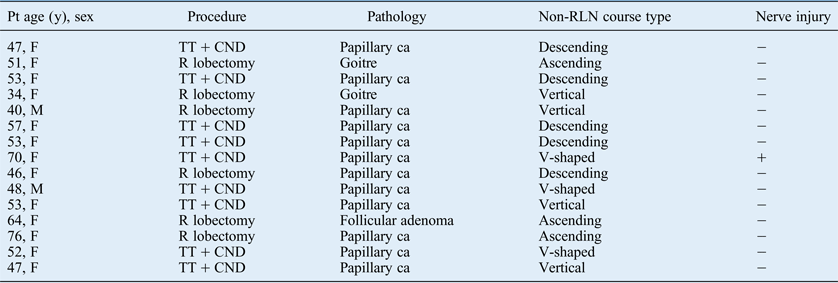
Of the 2187 right laryngeal nerves exposed during thyroid surgery, we identified 15 (0.68 per cent) patients with non-RLNs on the right side (Table I). There were 13 females and 2 males, with a mean age of 52.7 years. Nine patients underwent total thyroidectomy with central neck dissection, and six patients underwent right hemithyroidectomy. Pathologically, 12 cases had papillary carcinoma and 3 cases had goitre or follicular adenoma (Table I). With regard to clinical symptoms (identified via a retrospective chart review), four patients (27 per cent) complained of mild dysphagia but denied other clinical symptoms such as dysphonia. There were pre-operative neck CT images for 14 of the 15 patients, all of which showed a retroesophageal aberrant subclavian artery that arose directly from the aortic arch (Figure 1).
Patient data summary
Pt = patient; y = years; non-RLN = non-recurrent laryngeal nerve; F = female; TT = total thyroidectomy; CND = central neck dissection; ca = carcinoma; R = right; M = male
When classifying the travelling patterns of the non-RLNs, we found five cases of the descending type, four of the vertical type, three of the ascending type and three of the V-shaped type (Table I, Figure 2). Non-RLNs arose from various levels of the vagus nerve, and the levels of the inferior thyroid artery were also very variable. However, we could not compare the relationship between non-RLNs and the inferior thyroid artery because of the variable location of the inferior thyroid artery. With regard to nerve injury among the 15 cases, 1 non-RLN was damaged during thyroid surgery; this non-RLN was classified as a V-shaped type with a descending–ascending pattern. The affected patient received end-to-end anastomosis of the injured nerve, but vocal fold paralysis was noted after surgery.
Discussion
Careful dissection of the RLN is necessary to avoid nerve injury during thyroidectomy. The existence of anatomical variations in the RLN course is an additional factor contributing to its injury during thyroid surgery.Reference Jatzko, Lisborg, Müller and Wette1, Reference Wax and Simpson GT12 One of the potential variants is a non-RLN; this variant originates from an aberrant embryological origin of the subclavian artery.
Embryologically, both RLNs supply the sixth branchial arches.Reference Mra and Wax13 These nerves pass beneath the sixth aortic arch and ascend to the larynx with the descent of the heart. On the right side, the distal portion of the sixth aortic arch, and the fifth aortic arch, disappear, and the RLN moves up to lie beneath the fourth arch. However, the right fourth aortic arch and proximal right dorsal aorta are occasionally obliterated, and the origin of the subclavian artery becomes anomalous. Its final origin is just below that of the left subclavian artery, and it reaches the right side by crossing the midline behind the oesophagus. This anomalous subclavian artery allows the inferior laryngeal nerve to be non-recurrent from the vagus nerve and to rise higher in the neck. This non-RLN then runs directly to the larynx, usually at the level of the thyroid gland upper lobe on the right side. Non-RLNs have very rarely been reported on the left side too.Reference Rustad and Morrison14 This latter anomaly is invariably associated with situs inversus, a right-sided ligamentum arteriosum and a left retroesophageal subclavian artery.Reference Bowden15
The overall reported incidence of non-RLNs varies between 0.4 and 2.4 per cent on the right side.Reference Katz and Nemiroff2, Reference Wax and Simpson GT12 Henry et al.Reference Henry, Audiffret, Denizot and Plan6 observed 31 cases of non-RLNs in 4921 neck dissections on the right side during thyroid and parathyroid excision within a 10-year period. Katz and Nemiroff Reference Katz and Nemiroff2 noted the RLN to arise directly from the vagus in the neck in 11 of 719 patients over 10 years. Sanders et al.Reference Sanders, Uyeda and Karlan5 identified 7 cases of a non-RLN on the right side during 1000 thyroidectomies over a 20-year period. HumphreyReference Humphrey16 reported a 0.68 per cent incidence of this variation. The incidence of an aberrant subclavian artery identified on CT images in our study population is typical. The incidence of surgically identified non-RLNs in our study (0.68 per cent) is somewhat similar to that usually reported.
Generally, there are no reliable symptoms or signs that indicate the possibility of a non-RLN pre-operatively.Reference Bowden15 Some patients in this study complained of mild dysphagia and a foreign body sensation. Before surgery, the diagnosis of non-RLN may be made only if an aberrant subclavian artery is suspected. This aberrant artery is almost always associated with a non-RLN; however, the identification of an aberrant subclavian artery would be tantamount to identifying a non-RLN before surgery. Freed and LowReference Freed and Low17 have suggested that barium oesophagography be included in pre-operative evaluation, especially for dysphagic patients. Relevant findings on oesophagography include an impression from the left to the upper right of the oesophagus, and on anterior plain chest X-ray include an anomalous origin of the right subclavian artery at the right peak of the aortic arch. A diagnosis of an aberrant subclavian artery with neck CT images is sound, as it is based on the direct identification of an aberrant artery passing behind the oesophagus.Reference Page, Monet, Peltier, Bonnaire and Strunski10 Many patients undergo CT study of the neck before thyroid surgery to evaluate the anatomical relationships between the thyroid gland and the surrounding structures, especially the lateral neck lymph nodes. Thus, the identification of an aberrant subclavian artery can be reliably established using only CT images.Reference Materazzi, Berti, Iacconi and Miccoli18, Reference Nagayama, Okabe, Katoh and Furukawa19 The existence of non-RLNs can also be evaluated using intra-operative nerve monitoring. Surgeons should be aware of the existence of non-RLNs when the inferior laryngeal nerve shows abnormal nerve monitoring during surgery.
Regarding the course of non-RLNs, Cagnol et al.Reference Cagnol, Santini and Demard9 reported two anomaly types. The non-RLN enters the larynx at the cricothyroid joint through the fibres of the inferior constrictor muscle of the pharynx. In a type I anomaly, the non-RLN originates from the vagus nerve above the laryngotracheal junction and descends into the larynx, running together with the vessels of the superior thyroid lobe. In a type II anomaly, the non-RLN that originates from the vagus nerve runs on a level with the lower thyroid artery and then follows a transverse path parallel to the inferior thyroid artery.Reference Rustad and Morrison14, Reference Clemente20 Abboud and AouadReference Abboud and Aouad11 reported three types of courses for non-RLNs: in type Ia, the nerve has a straight course at the level of the upper thyroid pole; in type Ib (the most common), the nerve runs transversely at the level of the thyroid isthmus; in type II, the nerve makes a downwards curve, eventually reaching the lower pole of the thyroid gland. In our study, we classified the courses in more detail, distinguishing four types. In the descending type, the non-RLN descends after originating from the vagus nerve trunk. In the vertical type, the non-RLN runs vertically to the cricothyroid joint. In the ascending type, the non-RLN runs upward to the cricothyroid joint. In the V-shaped type, the non-RLN takes a downward direction and runs upward to the cricothyroid joint.
• This study evaluated the incidence of non-recurrent laryngeal nerves (non-RLNs) and presented a classification system based on their travelling course
• Fifteen right-sided non-RLNs were classified as descending, vertical, ascending or V-shaped
• A retroesophageal subclavian artery as seen on neck computed tomography scans indicates a non-RLN
Because of these variations of non-RLNs, the surgeon should dissect the thyroid gland and surrounding tissues very carefully when there is pre-operative suspicion of a non-RLN. Surgeons should carefully identify the vagus nerve before thyroid dissection, and the vagus nerve should be gently dissected with elastic loops, enabling identification of the non-RLN and its course.Reference Mra and Wax13 In particular, there may be a higher risk of nerve injury when the surgeon ties the superior thyroid vessels of the thyroid gland; this should therefore be performed with great caution.Reference Mra and Wax13
Conclusion
Although the non-RLN is a rare anomaly, its existence can have dramatic consequences with regard to laryngeal nerve injury during thyroid surgery. Surgeons are warned about the existence of non-RLNs and should have an intimate knowledge of their variable anatomical patterns. An aberrant retroesophageal subclavian artery can be easily observed using CT images before surgery, without additional studies. If the existence of a non-RLN is suspected based on pre-operative CT images, the thyroid gland should be meticulously dissected and the non-RLN identified, to prevent vocal fold paralysis.
Acknowledgement
This paper was supported by the research funds of Chonbuk National University in 2013.