Introduction
In spite of considerable progress achieved in the last few years in in vitro embryo production (IVP) technology in bovines, its overall efficiency is still modest, with blastocyst and pregnancy rates of roughly 40% (van Wagtendonk-de Leeuw, Reference Vejlsted, Du, Vajta and Maddox-Hytttel2006; Schmidt, Reference Schmidt2007; Lonergan & Fair, Reference Lazzari, Wrenzycki, Herrman, Duchi, Kruip, Niemann and Galli2008). A substantial number of studies have focused on improving embryo quality by analysing different variables that can influence the success of in vitro embryo development (Carolan et al., Reference Carolan, Lonergan and Mermillod1996; Fukui et al., Reference Fischer-Brown, Lindsey, Ireland, Northey, Monson, Clark, Wheeler, Kesler, Lane, Weigel and Rutledge2000; Hendricksen et al., Reference Harvey, Kind, Pantaleon, Armstrong and Thompson2000; Khurana & Niemann, Reference Jaquemar, Kupriyanov, Wankell, Avis, Benirschke, Baribault and Oshima2000a; de Oliveira et al., Reference Okamoto, Otte, David Allis, Reinberg and Heard2005; Corrêa et al., Reference Corrêa, Rumpf, Mundim, Franco and Dode2008). The majority of those studies used, as an endpoint, blastocyst production on day (D)7 or D8 of the development or, more rarely, the pregnancy rate following embryo transfer to recipients. However, it is well known that blastocyst morphology is not highly indicative of an embryo's ability to initiate pregnancy following transfer. Unfortunately, the use of pregnancy rate as an endpoint is too expensive to be routinely used for embryo evaluation. Therefore, the development of new methods that are more reliable for the evaluation of the developmental potential of IVP embryos beyond the morphology at D7 and D8 would be very useful.
Post-hatching in vitro culture of bovine embryos emerged as an alternative procedure that could potentially allow for the establishment of a more precise tool for evaluating embryo developmental ability, without the need of transferring them to recipients (Brandão et al., Reference Brandão, Hyttel, Lovendahl, Rumpf, Stringfellow and Callesen2004; Vajta et al., Reference Vajta, Alexopoulus and Callesen2004; Vejlsted et al., Reference Vandesompele, Preter, Pattyn, Poppe, Roy, Paepe and Speleman2006). In this technique, it is possible to more accurately evaluate the quality of embryos produced by different assisted-reproduction techniques (ARTs) as well as those submitted to different treatments and manipulations. Moreover, post-hatching development (PHD) systems could contribute to basic studies or to the investigation of toxicity of in vitro cultured products, pharmaceuticals, pesticides, and other chemicals (Brandão et al., Reference Brandão, Hyttel, Lovendahl, Rumpf, Stringfellow and Callesen2004).
Although post-hatching in vitro culture of bovine embryos is an interesting system with a great potential to be used as a tool in studies involving ARTs, few studies have been done to better characterize, evaluate and optimize its use as an assessment parameter. Therefore, further studies are needed to determine basic characteristics such as the appropriate physical, chemical and hormonal environments required for embryo development to advanced stages in vitro. However, before a research investment is made to develop or establish new media and/or a different culture system, a better characterization of the one already established is needed to be used as a starting point.
One of the most important steps in the PHD system is the preparation of tunnels wherein the embryos are placed and then maintained during culture. The tunnels are prepared with agarose gel, which consists of compact bundles that form large pores allowing passage of solutes (Reece, Reference Reece, Swenson and Reece1996; Narayanan et al., Reference Morton, Herrmann, Sieg, Struckmann, Maxwell, Raht, Evans, Lucas-Hahn, Niemann and Wrenzycki2006) in such a way that the cultured medium components can reach the embryo inside the tunnel. However, it is not known if changes, for example, in the diluents to prepare the tunnels, may affect the developmental abilities, kinetics of development and the molecular profile of embryos cultured in vitro for longer periods. Neither is it known if embryos of different gender will behave differently when cultured in such a system.
Thus, the objective of the present study was to evaluate the effect of the diluent used for tunnel preparation and gender on morphological characteristics, kinetics of development and gene expression of embryos cultured until D15 in a PHD system.
Materials and methods
In vitro embryo production
Unless otherwise indicated, chemicals were purchased from Sigma Chemical Co. Ovaries from crossbred cows (Bos taurus indicus × Bos taurus taurus) were collected immediately after slaughter and transported to the laboratory in saline solution (NaCl 0.9%) supplemented with penicillin G (100 UI/ml) and streptomycin sulfate (100 μg/ml) at 35 °C. Cumulus–oocyte complexes (COCs) were aspirated from 2- to 8-mm diameter follicles with an 18-gauge needle and pooled in a 15-ml conical tube. After sedimentation, COCs were recovered and selected using a stereomicroscope. Only COCs presenting homogenous cytoplasm and at least three layers of cumulus cells were used. Selected COCs were washed and transferred, in quantities of 30 to 35, to a 200-μl drop of maturation medium under silicone oil and incubated for 22 h at 39 °C and 5% CO2 in air. Maturation medium consisted of TCM–199 (Invitrogen®) supplemented with 10% fetal bovine serum (FBS) (Invitrogen®), 12 IU/ml of LH, 0.01 IU/ml of FSH, 0.1 mg/ml of l-glutamine and antibiotic (Amicacin, 0.075 mg/ml).
Following maturation, COCs were transferred to a 200-μl drop of fertilization medium, which consisted of TALP (Parrish et al., Reference Parrish, Krogenaes and Susko-Parrish1995) supplemented with penicillamine (2mM), hypotaurine (1mM), epinephrine (250 mM) and heparin (10 μg/ml). Frozen semen from a bull of known in vitro fertility, which has been used for several years as the reference bull for in vitro embryo production in our laboratory, was used for all treatments and replicates. Motile spermatozoa were obtained using the Percoll (Amersham Biosciences) gradient method in a microtube (Machado et al., Reference Lonergan, Fair, Corcoran and Evans2009) and were added into the fertilization drop at a final concentration of 1 × 10⁶ spermatozoa/ml. Sperm and oocytes were co-incubated for 20 h at 39 °C with 5% CO2 in air, with the day of in vitro insemination considered as D0.
After co-incubation, the presumptive zygotes were washed and transferred to 200-μl drops of SOFaaci medium (Holm et al., Reference Hendricksen, Vos, Steenweg, Bevers and Dieleman1999) supplemented with 2.77 mM of myo-inositol and 5% FBS and cultured at 39 °C and 5% CO2 in air for 7 days. Embryos were evaluated on D2 post-insemination (pi) for cleavage and on D6 and D7 pi for blastocyst rate.
Post-hatching development (PHD) system
The PHD system consisted of tunnels produced in agarose gel covered with culture medium as described by Brandão et al. (Reference Brandão, Hyttel, Lovendahl, Rumpf, Stringfellow and Callesen2004). These tunnels were made by dissolving low-melting-point agarose (Gibco BRL) in two different diluents at a final concentration of 2.4%. In treatment 1 the agarose was diluted in PBS solution, and in treatment 2, it was diluted in MQW. In both treatments, 10% of FBS was added after the agarose was dissolved. Tunnels were prepared at least 1 day before use. Dishes with tunnels and PHD media (Brandão et al., Reference Brandão, Hyttel, Lovendahl, Rumpf, Stringfellow and Callesen2004) were kept in an incubator at 39 °C with 5% CO2 in air until utilization.
Embryos were evaluated on D8, and those with signs of degeneration were removed. On D9, only hatched blastocysts, without signs of degeneration, were kept in culture and 200 μl of PHD medium was added to the droplets, increasing the final volume to 400 μl (1:1 SOFaaci and SOFaaci PHD, respectively). On D11, embryos were evaluated under a stereomicroscope, and only those that presented a diameter ≥0.5 mm, a clear trophoblast and a compact inner cell mass were transferred to agarose gel tunnels. At this time, embryos showing visible signs of degeneration were discarded. With the closed and bowled tip of a Pasteur pipette, each embryo was individually gently pushed inside the lumen of one gel tunnel. The loaded embryos remained in the PHD culture until D15. Culture was performed at 39 °C and 5% CO2 in air.
Morphologic evaluation and kinetics of post-hatching embryo development
Morphological and developmental evaluations of embryos were performed on D11, D12.5, D14 and D15 of culture. At each time point, embryos were measured using the Motic Images Plus 2.0 program. During the culture period, three patterns of development were observed: (1) embryos that reduced in size or showed signs of degeneration, which were classified as degenerated; (2) embryos that presented elongation and on D15 were reduced in size; and (3) embryos with continuous progressive daily elongation. On D15, only embryos that presented progressive elongation were removed from culture and frozen at –80 °C for gender diagnosis and gene expression.
DNA, RNA extraction, and cDNA synthesis
Elongated D15 embryos were used for individual extraction of genomic DNA and total RNA using Trizol Reagent (Invitrogen®). DNA was used for sex determination by PCR and RNA for gene expression evaluation via RT-qPCR.
In each microtube containing individual embryos, 50 μl of Trizol (Invitrogen®) and 12.5 μg of glycogen were added. Then, genomic DNA and total RNA were isolated according to the procedures provided by the manufacturer, with minor modifications. For gene expression, RNA of single embryos was pooled according to sex and size to form pools of three embryos in a total of three to four pools organized by sex.
Total RNA was incubated with 1 U DNase I (Invitrogen®) and then reverse transcribed with SuperScript III (200 U/μl; Invitrogen®) and Oligo-dT primer (Invitrogen®). Reactions were performed at 42 °C for 52 min and at 70 °C for 15 min for enzyme inactivation.
Embryo sexing procedure
Embryo sex determination was performed by PCR, using two pairs of primers. The first pair was specific to a region of the Y chromosome, whereas the second pair was specific to a bovine autosomal gene. The primer sequences and fragment sizes for each gene are shown in Table 1. Bovine genomic DNA from a male and a female were used as a control. PCR was performed by adding to each sample tube a PCR mix containing 20 nM of each pair of primers, 200 μM of each dNTP, 1× PCR buffer and 1 U Platinum Taq DNA polymerase® (Invitrogen®) in a final volume of 30 μl. The PCR program used 40 cycles of 94 °C for 20 s, 57 °C for 30 s and 72 °C for 30 s, followed by a final extension of 72 °C for 30 min. Amplified PCR products were electrophoresed in 2.0% agarose gel stained with ethidium bromide (10 mg/ml) and visualized under UV illumination. When two amplicons were detected, the embryo was identified as male, whereas detection of one amplicon indicated a female embryo.
Sequences of the primers used for sexing

F: forward; R: reverse; and size (bp) of the amplified fragments.
Real-time RT-PCR
The relative abundance of genes involved in glucose metabolism [glucose-6-phosphate dehydrogenase (G6PD), glucose transporter 1 (GLUT1), glucose transporter 3 (GLUT3), phosphoglycerate kinase 1 (PGK1)]; placenta development [placenta-specific 8 (PLAC8), keratin proteins 8 (KRT8)]; heat stress [heat shock transcription factors 1 (HSF1)]; and maternal recognition pregnancy [interferon-τ (IFNT)] compared between male and female embryos cultured in a PHD system using two different agar gel diluents.
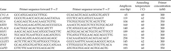
Real-time RT-PCR analysis was performed using Power Sybr Green PCR Master Mix (Applied Biosystems). Reactions were performed in 25 μl, and PCR cycling conditions were 95 °C for 10 min following by 40 cycles of denaturing at 95 °C for 15 s and then annealing at the temperatures given in Table 2 for 1 min. Primer sequences, fragment size, annealing temperature and primer concentrations for each gene are listed in Table 2. Reactions were performed in duplicate for each gene, with an amplification efficiency of >90%.
Information of specific primers used for amplification in the real-time polymerase chain reaction

To determine which housekeeping gene should be used for data normalization, the geNorm program was used (Vandesompele et al., Reference van Wagtendonk-de Leeuw2002). Although the results indicated that cyclophilin-A (CYC-A) was the most stable endogenous control, rather than histone H2A (H2A) and glyceraldehyde-3-phosphate dehydrogenase (GAPDH), the mean expression of these genes was used as endogenous control.
The relative abundance of each gene was calculated using the ΔΔCt method with efficiency correction (Pfaffl, Reference Pfaffl2001). Mean efficiency values for each gene were calculated from the amplification profiles of individual samples using LinRegPCR software (Ramakers et al., Reference Ramakers, Ruijter, Deprez and Moorman2003).
Statistical analysis
The percentage of elongated embryos in tunnels constructed using PBS solution and MQW and the proportion of elongated male and female embryos were evaluated by means of a chi-squared test. Data comparing embryos’ lengths between D11 and D15 did not have normal distribution, even after logarithmic and square root transformations. At this point, they were analysed using the Mann–Whitney test. Gene expression data were analysed by means of a t-test or Mann–Whitney test for those with or without a normal distribution, respectively. All statistical analyses were performed using the Prophet Program, version 5.0; 1997.
Results
A total of 2933 oocytes were used for in vitro embryo production. Blastocyst rates were evaluated on D7 (37%; 1077/2933) and D8 (41%; 1216/2933) post-insemination (pi), the hatching rate was measured on D9 (72%; 873/1216), and on D11, embryos were measured. Then, embryos presenting a diameter ≥ 0.5 mm (39.5%; 345/873) were randomly distributed between two treatments for culture until D15. From the 345 D11 embryos with the minimum size necessary to continue their growth in vitro in the PHD system, 286 (84%) were successfully loaded into the tunnels. Of those, 154 (54%) and 132 (46%) embryos were placed in tunnels made of agarose gel dissolved in MQW and PBS, respectively. The difference in the number of embryos available on D11 and those that were actually loaded into tunnels was due to morphological abnormalities of some embryos (unclear trophoblast and undefined inner cell mass), and the collapse of others when they were removed from the culture drop to be placed into the tunnels.
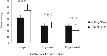
A general overview of embryo behaviour during culture in both PHD systems is shown in Fig. 1. The percentage of embryos elongated at D15 was higher in PBS than in MQW. Nevertheless, the percentages of embryos that grew, but regressed by D15, and those that degenerated were similar for the two types of gel.
Percentage of embryos that elongate, regress and degenerate during culture in a post-hatching development (PHD) system from D11 to D15.

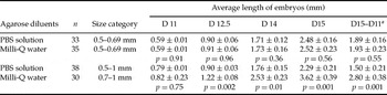
Despite this, embryo development was higher in embryos cultured in MQW than in those cultured in PBS (Table 3). This finding can be observed not only by the size of the embryos on D15 but also by the total length increase during culture, which was calculated by subtracting the final (D15) length from the initial (D11) length for each embryo.
Average length (mm) of elongated bovine embryos (mean ± SEM) on day (D)11, D12.5, D14 and D15 of cultured in a post-hatching development (PHD) system using agarose gel diluted in Milli-Q water or phosphate-buffered saline (PBS) solution

aDifference between the final length of embryos on D15 and the initial length on D11.
To evaluate whether or not the initial embryo size would affect development in the different PHD systems, we separated the embryos according to their size on D11 into two categories (Table 4). When development of embryos from the same size category was compared between two types of gels, we noted that smaller embryos had a similar pattern of development in both treatments. However, the bigger embryos demonstrated higher growth during the whole culture period when cultured in MQW as opposed to PBS.
Average length (mm) of elongated bovine embryos (mean ± SEM) of different size categories, on day (D)11, D12.5, D14 and D15 of cultured in a post-hatching development (PHD) system using agarose gel diluted in Milli-Q water or phosphate-buffered saline (PBS) solution

aSubtraction of the final length of embryos on D15 and the initial length on D11.
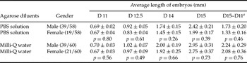
Regarding gender evaluation, there was a higher proportion (p < 0.05) of male than female embryos on D15 of development in both PBS (67% male, n = 39/58 and 33% female, n = 19/58) and MQW treatments (65% male, n = 39/60 and 35% female, n = 21/60). Although more male embryos developed in the PHD system, their morphology, final length and elongation pattern during culture was not affected by gender (Table 5). No differences were observed between the treatments when gender was considered.
Average length (mm) of male and female bovine embryos (mean ± SEM) cultured in a post-hatching development (PHD) system using agarose gel diluted in Milli-Q water or phosphate-buffered saline (PBS) solution on day (D)11, D12.5, D14 and D15 of culture

aSubtraction of the final length of embryos on D15 and the initial length on D11.
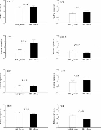
To evaluate the effects of the diluents used for agarose gel on the quality of embryos using molecular profile as an endpoint, the relative expression of eight genes was analysed (Fig. 2). Only the GLUT1 gene showed a tendency (p = 0.08) to be expressed at a lower level in embryos cultured in MQW tunnels when compared with those cultured in PBS tunnels.
Transcript levels of PLAC8, G6PD, GLUT1, GLUT3, HSF1, IFNT, KRT8 and PGK1 genes as determined by real-time polymerase chain reaction in day (D)15 bovine embryos culture in post-hatching system using agarose gel tunnels diluted in Milli-Q water and phosphate-buffered saline (PBS) solution. Data are mean ± SEM. Data were normalized against the mean of CYC-A, GAPDH and H2A genes, with results expressed relative to the control sample using the ΔΔCt method with efficiency correction.

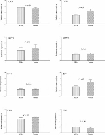
The expression of the same genes between male and female embryos was also compared independently of the treatments. In this analysis, the relative abundance of G6PD (p = 0.05) and KRT8 (p = 0.03) gene transcripts was higher in female than in male embryos on D15 (Fig. 3). The remaining genes showed no difference in expression between genders. Also, no effect of the interaction between diluents and gender was observed in the gene expression of embryos.
Transcript levels of PLAC8, G6PD, GLUT1, GLUT3, HSF1, IFNT, KRT8 and PGK1 genes as determined by polymerase chain reaction in day (D)15 male and female bovine embryos culture in post-hatching system using agarose gel tunnels. Data are mean ± SEM. Data were normalized against the mean of CYC-A, GAPDH and H2A genes, with results expressed relative to the control sample using the ΔΔCt method.

Discussion
In this study, we evaluated the development and gene expression of male and female embryos cultured up to D15 in a PHD system using PBS and MQW tunnels. Our findings demonstrated that tunnel diluents, but not gender, affected embryo development, in which embryo growth in the different tunnels was dependent on the initial embryo size. Additionally, despite the fact that female and male embryos had similar developmental kinetics, they differed in gene expression.
Regarding embryo development, we noted that the overall percentage of embryos that elongated until D15 was higher than that reported by Brandão et al. (Reference Brandão, Hyttel, Lovendahl, Rumpf, Stringfellow and Callesen2004). In addition, the mean length of embryos cultured in tunnels prepared either with PBS or MQW was comparable with the length of IVP embryos, which were transferred on D7 to a synchronized recipient and recovered on D12 (Lazzari et al., Reference Latham2002) and on D14 (Fischer-Brown et al., Reference Ferreira, Machado, Diesel, Carvalho, Rumpf, Melo, Dode and Franco2004). These results suggest that the system we used was suitable to provide growth and elongation of IVP embryos.
Even though more than 40% of the embryos that were loaded in the tunnels on D11 showed continuous growth and were elongated on D15, only 32% of D7 blastocysts were able to reach the minimum size with the morphological normality required to continue PDH culture. A high rate of embryonic loss from D8 to D11 was also reported by Brandão et al. (Reference Brandão, Hyttel, Lovendahl, Rumpf, Stringfellow and Callesen2004). These results suggested that this period of time is critical for the embryo. Indeed, it has been reported that post-hatching embryos may be very susceptible to environmental stress as a result of increased metabolism, protein synthesis and energy demand (Tiffin et al., Reference Tiffin, Rieger, Betteridge, Yadav and King1991; Khurana & Niemann, Reference Khurana and Niemann2000b; Garner, Reference Fukui, Kikuchi, Kondo and Mizushima2008).
The percentage of embryos that grew was higher in the PBS tunnel than in the MQW tunnel; however, the increase in size during development was more pronounced in embryos cultured in MQW gel. Therefore, this gel appeared to give a better indication of embryo quality, as it permitted a greater increase in size than the PBS gel. This assumption is based on other studies, which have reported that morphological characteristics on D14 may be a good indicator of embryo fitness, as these characteristics were associated with pregnancy establishment (Fisher-Brown et al., 2004). In addition, the length of D14 embryos has been positively correlated with IFNT secretion (Block et al., Reference Block, Fischer-Brown, Rodina, Ealy and Hansen2007), suggesting that the more highly developed embryos are better able to prevent initial luteal regression and to maintain pregnancy.
Because Brandão et al. (Reference Brandão, Hyttel, Lovendahl, Rumpf, Stringfellow and Callesen2004) had determined that the size of the embryo on D11 could be used to qualify and to estimate the subsequent development, we investigated whether or not the initial size of the embryo on D11 would affect its growth in the PHD system using different agarose diluents. Therefore, we separated embryos into two categories according to their size. It was observed that the final embryo size and the elongation pattern were similar for both types of gels for smaller embryos. However, larger embryos had a higher developmental rate during the entire culture period in MQW gel. Considering that the elongating stage is an indicator of embryo viability, as stated by Fischer-Brown et al. (Reference Ferreira, Machado, Diesel, Carvalho, Rumpf, Melo, Dode and Franco2004), and has been used as an indicator of embryo quality (Clemente et al., Reference Clemente, de la Fuente, Fair, Al Naib, Gutierrez-Adan, Roche, Rizos and Lonergan2009), we can hypothesise that MQW gel may provide a better indication of those embryos with higher developmental potential.
Aside from testing tunnel gel composition, we focused on the comparison of the morphological characteristics and the kinetics of development of male and female embryos. Gender determination on D15 demonstrated a significant sex ratio shift, in which the majority of the embryos were male. A much more notable imbalance in favour of male embryos on D16 using a PHD system was observed by Brandão et al. (Reference Brandão, Hyttel, Lovendahl, Rumpf, Stringfellow and Callesen2004). Conversely, when IVP embryos were maintained in the recipient uterus from D7 to D16, a tendency of IVP embryos to have more males than females was observed (Bertolini et al., Reference Bertolini, Beam, Shim, Bertolini, Moyer, Famula and Anderson2002). The difference in the sex ratio of IVP embryos has been attributed to several factors, including glucose concentration in the culture medium (Gutiérrez-Adán et al., Reference Gutiérrez-Adán, Oter, Martínez-Madrid, Pintado and De La Fuente2001), which could explain the lower percentage of female embryos on D15 that we observed. Interestingly, we also sexed the embryos that were removed from culture on D9 because they had not hatched, and found that of the discarded embryos, 60% were females (data not shown). Because PHD medium, which contained a high concentration of glucose, was only added to the culture on D9, we might assume that the lesser development of females was not due to the presence of a high glucose concentration. It is possible that a greater female sensitivity to the culture environment may be due to the process of X chromosome inactivation, which should occur before the embryos were placed in the PHD system, as stated by others (Latham, Reference Khurana and Niemann1996; Gutiérrez-Adán et al., Reference Gutiérrez-Adán, Oter, Martínez-Madrid, Pintado and De La Fuente2001; Okamoto et al., Reference Narayanan, Xiong and Liu2004; Ferreira et al., Reference Ellis, Bondiolli and Williams2010). In addition, it is very likely that the difference in the sex ratio observed on D15 was already present on D11 when embryos were placed in the agarose tunnels.
Although a lower percentage of female embryos reached the size required to be placed in the tunnels, those that were loaded showed similar size and a similar pattern of development to that of male embryos. Because the period of greatest embryonic loss is between D7 and D11 and females are more susceptible to culture environment, those that survived and reached D11 had most likely gone through a robust selection process and were similar in quality to male embryos.
Gene expression has been widely used to evaluate embryo quality (Rizos et al., Reference Rizos, Lonergan, Boland, Arroyo-Gárcia, Pintado, Fuente and Gutiérrez-Adán2002; Gutiérrez-Adán et al., Reference Gutiérrez-Adán, Lonergan, Rizos, Ward, Boland, Pintado and De La Fuente2004; Lonergan et al., Reference Lonergan, Rizos, Gutiérrez-Adán, Moreira, Pintado, De La Fuente and Boland2006). Therefore, we also used this approach to examine the suitability of the utilized PHD system and to evaluate gender differences. The significance of the genes we chose resides in their close association with placenta development, heat stress, maternal recognition of pregnancy and glucose metabolism.
When comparing agarose gels treatments, only the transcript levels of the GLUT1 gene had a tendency (p = 0.08) to be more abundant in the elongated D15 embryos cultured in PBS gel. Different groups have studied gene expression patterns of glucose transporters during the preimplantation period in bovines, but the results were variable when in vivo and in vitro embryos were compared (Lazzari et al., Reference Latham2002; Harvey et al., Reference Gutiérrez-Adán, Rizos, Fair, Moreira, Pintado, Fuente, Boland and Lonergan2004; Balasubramanian et al., Reference Balasubramanian, Son, Mohana Kumar, Ock, Yoo, Im, Choe and Rho2007). In the present study, GLUT1, but not GLUT3, had a tendency to be more highly expressed in PBS gel. This differential expression suggests that diluents may affect glucose uptake and embryo metabolism, but the reason for this is not clear. It is important to highlight the fact that GLUT1 has a role in intercellular, rather than apical, transport of glucose, meaning that its expression does not necessarily imply a dominant role in embryonic uptake from the environment, as it does with GLUT3 (Augustin et al., Reference Augustin, Pocar, Navarrete-Santos, Wrenzycki, Gandolfi, Niemann and Fischer2001).
There is a general understanding that male IVP embryos develop faster than female embryos, which has been attributed to higher glucose uptake and metabolism (Gutiérrez-Adán et al., Reference Gutiérrez-Adán, Oter, Martínez-Madrid, Pintado and De La Fuente2001; Rheingantz et al., Reference Rheingantz, Pegoraro, Dellagostin, Pimentel, Bernardi and Deschamps2004; Morton et al., Reference Maddox-Hyttel, Alexopoulos, Vajta, Lewis, Rogers, Cann, Callesen, Tveden-Nyborg and Trounson2007). In the present study, we were not able to find a difference in the expression of glucose transport genes (GLUT1 and GLUT3) between male and females embryos. Bertolini et al. (Reference Bertolini, Beam, Shim, Bertolini, Moyer, Famula and Anderson2002) found that D16 male embryos expressed more transcripts for GLUT1 and GLUT3 than did female embryos. However, they also demonstrated that expression of those genes was correlated with embryo development, as evaluated by physical length. These observations could explain our results, as D15 male and female embryos had similar development rates and lengths.
Even though glucose transport and uptake are part of the complex metabolism of glucose, there are other factors involved that can differ according to gender, such as X chromosome-linked enzymes (Tiffin et al., Reference Tiffin, Rieger, Betteridge, Yadav and King1991; De La Fuente et al., Reference de Oliveira, Lopes and Rodrigues1999). G6PD and PGK1 are some of the enzymes related to energetic metabolism that are linked to the X chromosome. In the present study, a higher expression of the G6PD gene was detected in D15 female embryos than in male embryos. Results of several experiments demonstrated that female in vitro embryos on D7 presented a greater expression of G6PD than did male embryos (De La Fuente et al., Reference de Oliveira, Lopes and Rodrigues1999; Gutierrez-Adán et al., 2000). Hence, it could be speculated that an in vitro culture environment may somehow affect the X-inactivation process by modifying the pattern of G6PD gene expression (Lonergan et al., Reference Lonergan and Fair2003; Balasubramanian et al., Reference Balasubramanian, Son, Mohana Kumar, Ock, Yoo, Im, Choe and Rho2007; Morton et al., Reference Maddox-Hyttel, Alexopoulos, Vajta, Lewis, Rogers, Cann, Callesen, Tveden-Nyborg and Trounson2007). However, considering the importance of that process for female embryo survival, we believe that if any alteration had occurred, the female embryos would not be able to develop until D15 and instead would halt their development at earlier stages. In addition, no changes were observed in the PGK1 gene, which is also an X-linked gene. Therefore, we can hypothesise that the increase in the expression of the G6PD gene, as observed here, may be more highly associated with higher viability, in response to adverse conditions, than to problems in the X chromosome inactivation.
This hypothesis is supported by the greater expression, in D15 female embryos, of the KRT8 gene, which is involved in placentation and subsequent implantation, than that in male embryos. The KRT8 protein is critical for post-hatching development of bovine embryos (Maddox-Hyttel et al., Reference Machado, Carvalho, Siqueira Filho, Caixeta, Franco, Rumpf and Dode2003) and its deficiency has been associated with embryonic death in mice (Jaquemar et al., Reference Holm, Booth, Schimidt, Greve and Callesen2003). Lower expression of this gene has been reported in nuclear transfer (NT) embryos when compared with IVF (Pfister-Genskow et al., Reference Pfister-Genskow, Myers, Childs, Lacson, Patterson, Betthauser, Goueleke, Koppang, Lange, Fisher, Watt, Forsberg, Zheng, Leno, Schultz, Liu, Chetia, Yang, Hoeschele and Eilertsen2005).
In conclusion, we noted that agarose diluent used for tunnel construction affects embryo development in the PHD system and that the use of MQW was most indicative of the ability to evaluate embryo quality. Furthermore, it is important to highlight that although fewer female embryos developed to D11, those that survived this critical phase and were submitted to the PHD system had a similar morphology and elongation pattern to that seen in male embryos. Also, the differences we found in the expression of some genes evaluated were more strongly related to gender than to embryo quality. Therefore, embryo development in the PHD system is not influenced by gender and can be used to compare D15 embryos.
Acknowledgements
G. M. Machado received a scholarship from CAPES–Brazil. This research was supported by Embrapa (Grant no. 003.0709040000) and CNPq (Grant no. 475412/2007–9), Brazil. The authors thank the Laboratory of Ovarian Molecular Physiology, UNESP–Botucatu, São Paulo, Brazil, for assistance in the gene expression analysis of bovine embryos.