Introduction
Laryngeal papillomatosis is a benign disease caused by human papillomavirus types 6 and 11,Reference Hughes1,Reference Trzcinska, Zhang, Gitman and Westra2 and malignant transformation can occur in 3‒7 per cent of cases.Reference Derkay and Wiatrak3 The glottis is the most common sub-anatomical unit of laryngeal papillomavirus infection.Reference Preuss, Klussmann, Jungehulsing, Eckel, Guntinas-Lichius and Damm4 Complete surgical resection of lesions is the primary treatment method at present. The methods used include surgical power tool resection, laser resection and laser gasification of lesions. Adjuvant treatment includes antiviral therapy, chemotherapeutic drugs and photodynamic therapy.Reference Derkay and Wiatrak3,Reference Armstrong, Derkay and Reeves5 Although surgery can achieve a good therapeutic effect, the recurrence rate of laryngeal papillomatosis is very high, and there is no radical cure at present.
The key to surgical treatment is complete removal of diseased tissue during an operation. However, identifying all papilloma lesions during the process is usually challenging. Some lesions, especially in cases of multiple laryngeal papillomatosis, are often undiagnosable, and may even be missed during pre-operative endoscopy and intra-operative microscopy because of their small volumes. Therefore, we studied the effectiveness of haemoglobin absorption spectral imaging (vascular enhanced imaging) in the intra-operative evaluation of patients with laryngeal papillomatosis and analysed whether this method is helpful in identifying additional papilloma lesions during surgery.
At present, narrow-band imaging technology has been widely used and affirmed in the early diagnosis and treatment of malignant laryngeal tumours,Reference Ni, He, Xu, Lü, Gao and Yuan6–Reference Sun, Han, Li, Zhang and Du8 and there have been a few applications in the treatment of laryngeal papilloma.Reference Loizou, Laurell, Lindquist and Olofsson9,Reference Dippold, Becker, Nusseck, Richter and Echternach10 Nevertheless, there are no reports on treating laryngeal papillomatosis using haemoglobin absorption spectral imaging. Based on the similar imaging principles of haemoglobin absorption spectral imaging and narrow-band imaging, we conducted a series of statistical analyses to determine whether the haemoglobin absorption spectral imaging mode is helpful in accurately locating papilloma lesions, with preliminary exploration of the feasibility of haemoglobin absorption spectral imaging technology in laryngeal papilloma surgery.
Materials and methods
Seventeen patients (12 males (70.59 per cent) and 5 females (29.41 per cent)) with laryngeal papilloma who underwent surgical treatment in our department from September 2019 to September 2021 were selected. The patients’ ages ranged from 21 to 75 years (mean, 41.18 years). A range of one to six operations had been performed previously (average of 2.3 procedures per patient).
None of the patients had a previous history of malignant tumours or tracheostomy. All patients were examined before the procedure using a fibre-laryngoscope and/or fibre-bronchoscope, with preliminary exclusion of tracheal lesions. All lesions were confirmed as papilloma based on intra-operative or post-operative pathology findings.
All patients underwent carbon dioxide (CO2) laser surgery under a microscope, and lesion sites were viewed in white light mode and vascular enhanced imaging (haemoglobin absorption spectral imaging) mode during the operation.
The instruments used in the procedures were the German Atmos® i View haemoglobin absorption spectral imaging operating microscope, a Deka (Calenzano, Italy) second-generation CO2 laser, an anti-laser supporting laryngoscope, anterior commissuroscope (standby) and laryngeal microsurgery instruments.
Each patient, in the supine position, was intubated through the mouth for intravenous compound anaesthesia. Their upper and lower teeth were protected with tooth pads, the laryngeal cavity was fully exposed with a supporting laryngoscope, and the microscope and CO2 laser were connected.
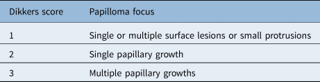
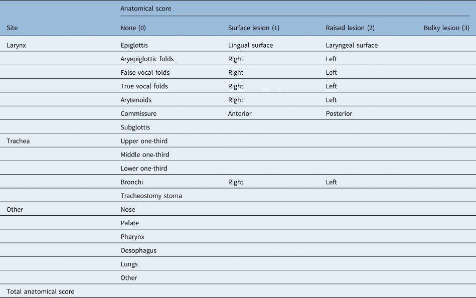
The laryngeal lesions were carefully observed in white light mode and vascular enhanced imaging mode (haemoglobin absorption spectral imaging), and evaluated by two experienced throat, head and neck doctors. First, the lesions were scored using the Dikkers scoring system (Table 1). Then, each part of the throat (epiglottis, aryepiglottic folds, false vocal folds, true vocal folds, arytenoid cartilages, anterior commissure, posterior commissure and subglottis) and other parts were evaluated (Table 2) for the following: (1) surface lesions; (2) raised lesions; and (3) bulky lesions. At the end of the white light and haemoglobin absorption spectral imaging examination, the lesion scores observed in the two modes were evaluated using the Derkay anatomy scale. The two modes were compared for their accuracy in detecting laryngeal papillomatosis.
Dikkers scoring criteria

Derkay scoring criteria for each anatomical site

The CO2 laser power was then adjusted to 2 W or 3 W. The 1.3 mm line mode was used (with dwell time adjusted to 0.2 ms) or no scan mode was employed. The tumour was completely excised with the laser or with laser vaporisation along the base of the lesion.
After the operation, one patient was transferred to the intensive care unit for observation for 1 night. After waking up, the other patients were extubated and returned to the general ward, and were treated with compression atomisation inhalation and antibiotics for 3–5 days. Patients with throat reflux were treated with proton-pump inhibitors for 8–12 weeks to avoid delayed wound healing or local hyperplasia caused by reflux.
Statistical analysis
A paired t-test was used to compare the score results of each case evaluated in white light mode and haemoglobin absorption spectral imaging mode. Analysis of variance (ANOVA) was used to compare whether the additional scores for the haemoglobin absorption spectral imaging mode were more meaningful than the white light mode scores according to different Dikkers scores. A p-value of less than 0.05 reflected statistical significance. The data analysis applications used were IBM SPSS® Statistics software version 26.0 and Microsoft Excel® spreadsheet software version 2019.
Results
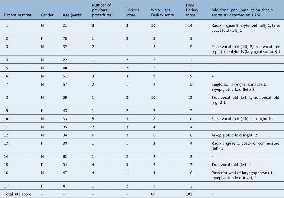
Microscopically, there was a significant difference in the Derkay scores between the white light mode and haemoglobin absorption spectral imaging mode (paired t-test, p = 0.002001). The haemoglobin absorption spectral imaging mode showed increased pathological tissues in 9 out of 17 patients (52.94 per cent). A total of 18 additional papilloma lesions were detected in all patients (Table 3) (2 in radix linguae (11.11 per cent), 3 in the false vocal fold (16.67 per cent), 4 in the true vocal fold (22.22 per cent), 1 in the arytenoid (5.56 per cent), 2 in the epiglottis (11.11 per cent), 3 in the aryepiglottic fold (16.67 per cent), 1 in the posterior commissure (5.56 per cent), 1 in the posterior wall of the laryngopharynx (5.56 per cent) and 1 in the subglottis (5.56 per cent)).
Summary of patient information

HASI = haemoglobin absorption spectral imaging
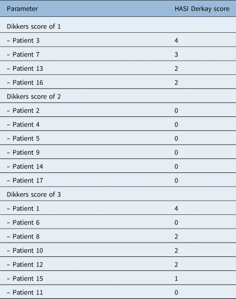
There was a statistically significant difference between the additional haemoglobin absorption spectral imaging mode scores corresponding to the different Dikkers scores in the three groups (one-way ANOVA, p = 0.002780). There was a statistically significant difference between the haemoglobin absorption spectral imaging mode scores corresponding to Dikkers scores 1 and 2 (one-way ANOVA, p = 0.000087) and between the scores corresponding to Dikkers scores 2 and 3 (one-way ANOVA, p = 0.019332). However, there was no statistically significant difference between the haemoglobin absorption spectral imaging mode scores corresponding to Dikkers scores 1 and 3 (one-way ANOVA, p = 0.172161).
The Dikkers score was 1 for patients with single or multiple surface lesions or small protrusions; four out of four patients (100 per cent) with this score showed additional papilloma lesions in the haemoglobin absorption spectral imaging mode. In patients with a Dikkers score of 2 (single papillary growth), no additional papilloma lesions were detected in the haemoglobin absorption spectral imaging mode. In patients with a Dikkers score of 3 (multiple papillary growths), five out of seven patients (71.43 per cent) showed additional papilloma lesions in the haemoglobin absorption spectral imaging mode (Table 4).
Additional HASI Derkay scores according to Dikkers scoring

HASI = haemoglobin absorption spectral imaging
Discussion
This study focused on the application of haemoglobin absorption spectral imaging in laryngeal papilloma surgery. Haemoglobin absorption spectral imaging is similar to narrow-band imaging; it enhances light with a spectrum of 450–570 nm (produced by light-emitting diodes) through the haemoglobin absorption spectral imaging system. The light absorption for superficial mucosal blood vessels occurs at 450 nm, and that for submucosal blood vessels occurs at 570 nm. Using the contrast of reflected and non-reflected light to display high-resolution images, we can view the vascular patterns unseen under white light endoscopy, and help identify the superficial capillaries and neo-angiogenesis in abnormal mucosa.
In many studies, narrow-band imaging has been reported to have high sensitivity, specificity, and negative and positive predictive values in detecting head and neck pre-cancerous lesions.Reference Ni, He, Xu, Gao, Lu and Yuan11–Reference Nguyen, Bashirzadeh, Hodge, Agnew, Farah and Duhig13 Domestic and foreign scholars have also studied the application of narrow-band imaging in the diagnosis and treatment of laryngeal papilloma. They believe that narrow-band imaging can improve the detection rate of laryngeal papilloma.Reference Zhang, He, Huang, Jiang and Wei14–Reference Imaizumi, Okano, Tada and Omori18 However, haemoglobin absorption spectral imaging based on similar imaging principles has not been reported. Therefore, we decided to systematically summarise and compare the microscopical haemoglobin absorption spectral imaging mode and the white light mode in evaluating papilloma lesions.
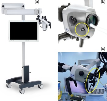
The haemoglobin absorption spectral imaging system was attached to the Atmos microscope (with high-definition camera) (Figure 1). The normal state is the common white light mode. The haemoglobin absorption spectral imaging mode can be found by rotating the filter knob on the side of the microscope barrel. During the operation, the knob can be switched between the white light mode and the haemoglobin absorption spectral imaging mode arbitrarily, to distinguish diseased tissue from normal tissue. It is possible to combine the respective advantages of the two modes to improve operational efficiency.
(a) Atmos i View haemoglobin absorption spectral imaging operating microscope. (b) The mode switch (circled) to change between white light and haemoglobin absorption spectral imaging modes. (c) Microscope coupled carbon dioxide lasers (circled).

As there is no diagnostic standard for the haemoglobin absorption spectral imaging mode, we observed papilloma lesions according to the throat narrow-band imaging classification proposed by Ni.Reference Ni19,Reference Ni20 In the haemoglobin absorption spectral imaging mode, the lesions were mainly intrapapillary capillary loop small spot type IV, type Vb resembles a serpentine, earthworm, or tadpole shape, and some single papilloma lesions were type III without vasodilation.
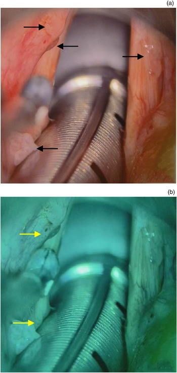
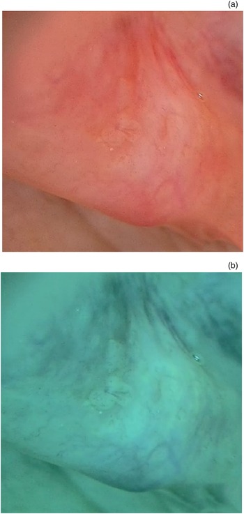
The findings indicated statistical differences between the two modes in terms of different Dikkers scores. In cases with a Dikkers score of 2, papilloma were single lesions. It is not difficult to find the lesions using an ordinary laryngoscope or under a microscope using a white light mode, and there are generally no missed diagnoses. For cases with a Dikkers score of 1, given the possibility of multiple surface lesions, some small lesions are easily missed in white light mode, resulting in omission; this is thought to be one of the causes of multiple recurrences in some cases. The cases with a Dikkers score of 3 had multiple papillary tumours. The statistical results showed a high incidence of missed diagnoses in: the posterior and lateral edges of the arytenoid cartilage, the epiglottic nodule near the epiglottic laryngeal surface, the laryngeal chamber between the upper surface of the vocal fold, and the lower edge of the ventricular band. The most common reasons for missing such cases in white light mode during the operation were: the small volume of the lesion, the concealed location, and the colour being similar to that of surrounding mucosa. However, the detection rate could be improved with the haemoglobin absorption spectral imaging mode (Figure 2 and Figure 3).
(a) Papillomas of the left false vocal fold, left arytenoid and right true vocal fold can be seen in white light mode (black arrows). (b) Additional left false vocal fold, posterior commissure papillomas were detected on haemoglobin absorption spectral imaging (yellow arrows).

(a) The left arytenoid lesion in white light mode is easily missed. (b) Small focal surface spots are visible in haemoglobin absorption spectral imaging mode.

The cases with a Dikkers score of 1 had a 100 per cent missed diagnosis rate in the white light mode, hence we believe such cases should be given special attention in surgery. The white light and haemoglobin absorption spectral imaging modes should be combined during the operation, and various laryngeal sub-anatomical units should be repeatedly examined to avoid missing small lesions.
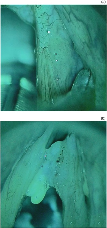
In cases of single papillary growth (Dikkers score of 2), the haemoglobin absorption spectral imaging mode seems to be of little value in terms of improving the detection rate. However, during actual operations, the haemoglobin absorption spectral imaging mode was found to: more clearly define the tumour boundary, help improve the accuracy of the surgical process, and reduce damage to the epithelium and superficial lamina propria of the vocal fold. It is also conducive to post-operative voice recovery (Figure 4). Ochsner and KleinReference Ochsner and Klein17 found that narrow-band imaging improved the visualisation of diseased tissues in 90.0 per cent of patients, showed additional diseased tissue areas in 46.7 per cent and more clearly defined the papilloma boundary in 76.7 per cent; it was also concluded that the use of narrow-band imaging endoscopy before surgery can help reduce the number of operations required. Imaizumi et al.Reference Imaizumi, Okano, Tada and Omori18 demonstrated that using narrow-band imaging to identify lesion boundaries aids effective removal of papillomas using a CO2 laser or surgical power tool system. Our statistical results are consistent with the conclusions of the above scholars regarding the role of narrow-band imaging in the diagnosis and treatment of papilloma.
(a) The tumour boundary on the upper surface of the right vocal fold is clear. (b) The tumour boundary on the right vocal fold is clear.

Narrow-band imaging endoscopy is primarily used in pre-operative examinations and post-operative follow-up appointments. After intra-operative anaesthesia, given the change in the patient's body position, the use of a supporting laryngoscope, the application of muscle relaxants and other factors, there can be a subjective position-related deviation between the position of lesions seen during the operation and the results of the pre-operative examination. This will affect the implementation of the procedure to varying degrees, and narrow-band imaging endoscopy cannot be coupled with a laser. In CO2 laser operations, if the narrow-band imaging mode is switched frequently, the operation progress is bound to be affected. The microscope with haemoglobin absorption spectral imaging mode can avoid the above shortcomings so that the area of focus can be observed in real time and dynamically during the operation. It has the advantage of not affecting the operation of coupled CO2 lasers, and no extra assistants are required.
• The recurrence rate of laryngeal papillomatosis is very high; complete removal of diseased tissue peri-operatively is crucial to surgical success
• Haemoglobin absorption spectral imaging is similar to narrow-band imaging, enabling visualisation of vascular patterns unseen under white light endoscopy
• The haemoglobin absorption spectral imaging mode on an operating microscope does not affect the operation of coupled carbon dioxide lasers, and no extra assistants are required
• Haemoglobin absorption spectral imaging mode as an additional tool for intra-operative microscopy can improve the laryngeal papilloma detection rate
Nevertheless, we should realise it is unrealistic to expect to cure laryngeal papilloma with just one operation. During surgery, normal airway mucosa should be protected as much as possible to reduce the impact on patients’ vocal and respiratory functions. The main purpose of the haemoglobin absorption spectral imaging mode is to improve the detection rate and avoid unnecessary re-operation associated with missing lesions during surgery. The edge of the lesion should be clearly identified to achieve more accurate treatment.
Although haemoglobin absorption spectral imaging is easy to use (one knob enables the switch between white light and haemoglobin absorption spectral imaging modes), we also realise that the haemoglobin absorption spectral imaging mode has some limitations. First, the light wave used in the haemoglobin absorption spectral imaging mode can be captured by haemoglobin. Therefore, when there is inflammatory congestion and active bleeding in the mucosa, the diseased tissue cannot be accurately evaluated,Reference Yue, Zhang, Wang, Du and Lin21 which seriously affects intra-operative judgment. Gentle intubation and careful operation are crucial, requiring close co-operation with experienced anaesthesiologists. Second, given the small sample size and short study time, objective evaluation is lacking. Therefore, it is necessary to conduct a long-term study on the treatment and post-operative follow up of patients with laryngeal papilloma, to confirm the real benefit of haemoglobin absorption spectral imaging mode in the surgical treatment of laryngeal papilloma. Nevertheless, our study shows that haemoglobin absorption spectral imaging technology remains of great value in treating patients with laryngeal papilloma.
Conclusion
The most critical aspect of laryngeal papilloma treatment is identifying all diseased tissues and removing them completely. Based on our study, use of the haemoglobin absorption spectral imaging mode as an additional tool for intra-operative microscopy can improve the detection rate of these diseased tissues.
Competing interests
None declared