INTRODUCTION
Photorhabdus are motile Gram-negative bacteria of the family Enterobacteriacae that form a mutualistic association with soil nematodes of the genus Heterorhabditis. Together this complex can infect a wide range of insects. The bacteria are normally found colonizing the gut of the free-living, soil-dwelling infective juvenile (IJ) stage of the nematode. The IJs actively seek and physically penetrate a potential insect host before migrating to the insect haemocoel, the body cavity that also constitutes the open circulatory system of the insect (Klowden, Reference Klowden and Klowden2007; Nation, Reference Nation and Nation2008). The symbiotic bacteria are then regurgitated directly into the haemocoel (Ciche and Ensign, Reference Ciche and Ensign2003), where they multiply rapidly, converting the insect biomass into a nutrient ‘soup’ that supports nematode growth and development. Photorhabdus bacteria colonize specific tissues within the insect such as the midgut and fat body causing toxaemia and septicaemia in the host by the secretion of several toxins and virulence factors that kill the insect within 48 h (Silva et al. Reference Silva, Waterfield, Daborn, Dean, Chilver, Au, Sharma, Potter, Reynolds and ffrench-Constant2002). After several rounds of both bacterial and nematode replication, a new generation of nematodes selectively re-packages. Photorhabdus and the complex emerges from the carcass in search of a new insect host. Re-association between the IJ and its specific related strain of Photorhabdus is specific; therefore a bacterial strain representative of one species will not re-associate and re-colonize nematodes of another species, although nematode growth and development may be supported by Photorhabdus from another species (Watson et al. Reference Watson, Joyce, Spencer and Clarke2005; Joyce et al. Reference Joyce, Watson and Clarke2006).
Insects activate their innate immune system through pattern recognition receptors (Yu et al. Reference Yu, Zhu, Ma, Fabrick and Kanost2002), which leads to the expression of highly efficient immune mechanisms involving both humoral and cellular responses against microbial infections (Kanost et al. Reference Kanost, Jiang and Yu2004; Lemaitre and Hoffmann, Reference Lemaitre and Hoffmann2007). Humoral defences include the transcriptional activation of several genes that results in the production of peptides and effector molecules with both recognition and antimicrobial properties (Ferrandon et al. Reference Ferrandon, Imler, Hetru and Hoffmann2007). Cellular responses in insects are mediated by the activity of circulating blood cells, referred to as haemocytes, which participate in multiple immune functions including phagocytosis, the formation of multicellular haemocyte aggregates and nodules, encapsulation of invading bacteria, and the production of reactive nitrogen and oxygen radicals (Jiravanichpaisal et al. Reference Jiravanichpaisal, Lee and Söderhall2006). An important player of both arms of the insect immune system is the activation of phenoloxidase (PO) through a complex proteolytic cascade that is initiated upon recognition of non-self microorganisms leading to the regulation of haemolymph coagulation and melanization, wound healing, and the release of microbicidal reactive intermediates (Cerenius et al. Reference Cerenius, Lee and Söderhäll2008).
Bacterial pathogens have developed 2 main strategies to escape the systemic immune response: they avoid detection because they lack (or hide) immune elicitors on their cell surfaces or they suppress the immune response (Vallet-Gely et al. Reference Vallet-Gely, Lemaitre and Boccard2008). It has previously been shown that Photorhabdus bacteria are able to evade both the antimicrobial peptides and the professional phagocytes that also patrol the haemocoel. The bacteria can interfere with haemocyte phagocytosis (ffrench-Constant et al. Reference ffrench-Constant, Waterfield, Daborn, Joyce, Bennett, Au, Dowling, Boundy, Reynolds and Clarke2003), block the production of highly reactive tyrosine metabolites with bacteriostatic actions (Eleftherianos et al. Reference Eleftherianos, Boundy, Joyce, Aslam, Marshall, Cox, Simpson, Clarke, ffrench-Constant and Reynolds2007 a, Reference Eleftherianos, Waterfield, Bone, Boundy, ffrench-Constant and Reynolds2009) and also destroy haemocytes using specific toxins (Daborn et al. Reference Daborn, Waterfield, Silva, Au, Sharma and ffrench-Constant2002). However, in all these studies insects were injected directly with bacteria alone, which although it simplifies interpretation of the virulence role of the bacteria, does not provide information about that of the corresponding Heterorhabditis nematodes.
Here, we use the Photorhabdus-Heterorhabditis-Manduca tripartite model to investigate insect immune responses against nematodes carrying or lacking their symbiotic bacteria and bacteria alone. The goal was to examine whether the contributions to virulence of the partners in symbiosis are quantitatively equal, and whether heterorhabditid nematodes have their own distinct effects on host immunity compared to those of their associated bacteria.
MATERIALS AND METHODS
Insects and pathogens
Larvae of the Tobacco Hornworm, Manduca sexta (L.) (Lepidoptera: Sphingidae), were maintained individually on a wheat germ-based artificial diet at 25°C and a photo-period of 17 h light:7 h dark (Reynolds et al. Reference Reynolds, Nottingham and Stephens1985). Newly moulted (day 0) fifth-stage larvae were used for all experiments. Insects were naturally infected by placing them on wet filter paper onto which a suspension of nematodes had been pipetted. The nematodes had either previously been allowed to re-associate with their own symbiotic bacteria, (Heterorhabditis bacteriophora carrying Photorhabdus luminescens subsp. laumondii strain TT01, Heterorhabditis downesi carrying P. temperata strain K122, Heterorhabditis gerrardi carrying P. asymbiotica strain ATCC43949), or were axenic (i.e. the nematodes were free of live bacteria – see below). Other insects were injected with Photorhabdus bacteria alone. One hundred 6-day-old nematodes were suspended in 50 μl of sterile phosphate-buffered saline (PBS; 0·15 m sodium chloride, 10 mm sodium phosphate buffer, pH 7·4) and the suspension was pipetted onto wet filter paper, which supported 2 Manduca caterpillars.
Bacterial cultures were prepared as previously described (Eleftherianos et al. Reference Eleftherianos, Marokhazi, Millichap, Hodgkinson, Sriboonlert, ffrench-Constant and Reynolds2006 a, Reference Eleftherianos, Millichap, ffrench-Constant and Reynoldsb). For insect injection, 50 μl of a PBS suspension containing an estimated 1×10³ washed Photorhabdus cells were injected directly into the haemocoel of Manduca larvae using a 100 μl disposable syringe with a 30-gauge needle. The DH5α strain of E. coli was used as a control in all experiments (50 μl of PBS containing approximately 3×10⁷ washed DH5α cells). PBS-injected insects served as negative controls. After treatment, experimental larvae were held at 28°C for 96 h.
Axenicized IJ nematodes
Nematodes devoid of bacteria were prepared as described by Joyce and Clarke (Reference Joyce and Clarke2003) . Briefly, Heterorhabditis downesei IJ nematodes were surface sterilized and inoculated onto Lipid Agar plates that were pre-seeded with either their natural symbiont P. temperata strain K122 or their non-natural symbiont P. luminescens strain TT01. Similarly, Heterorhabditis bacteriophora IJ nematodes were surface sterilized and inoculated onto Lipid Agar plates that were pre-seeded with either their natural symbiont P. luminescens strain TT01 or their non-natural symbiont P. temperata strain K122. Plates were incubated in the dark at 28°C and after 3 weeks IJ stages were collected from the plates. The IJs recovered from all 4 source plates were surface sterilized. Ten individual nematodes from each source were chosen randomly and individually crushed using a motorized mortar and pestle in a 1·5 ml Eppendorf containing 100 μl of LB. The contents were then transferred to LB agar and the number of colony-forming units (CFU) was counted. These counts confirmed that nematode species grown using Photorhabdus from another species of nematode did not re-package any bacteria either in the nematode gut or in the intracuticular spaces. Those nematodes grown on their own natural mutualistic Photorhabdus therefore contained only their own species of Photorhabdus. The surface sterilized nematodes were diluted to 100 IJs per ml in sterile PBS and were subsequently used for insect infections. Control infections using Galleria mellonella, a host that requires a Lethal Dose 50% (LD50) of only 1–3 colony-forming units (CFU) of Photorhabdus, irrespective of its species, were used as an internal control to test for the presence of Photorhabdus bacteria in the axenized and re-complexed nematodes (results not shown).
RNA extraction and RT-PCR
At 18 h after infection, insects were chilled on ice for 15 min, surface sterilized with 70% ethanol and then dissected to collect the fat body tissue (approximately 100 mg), as previously described (Eleftherianos et al. Reference Eleftherianos, Marokhazi, Millichap, Hodgkinson, Sriboonlert, ffrench-Constant and Reynolds2006 a, Reference Eleftherianos, Millichap, ffrench-Constant and Reynoldsb). To isolate total RNA, extracted tissues were homogenized in TRI reagent (Sigma, UK). Single-step reverse transcription (RT)-PCR was performed with the ‘OneStep’ RT-PCR kit (Qiagen, UK). Each reaction was carried out in a 50 μl volume containing 0·6 μm of forward and reverse gene primers and 2 μg of RNA template (Eleftherianos et al. Reference Eleftherianos, Marokhazi, Millichap, Hodgkinson, Sriboonlert, ffrench-Constant and Reynolds2006 a, Reference Eleftherianos, Millichap, ffrench-Constant and Reynoldsb). Primers were gene-specific and their sequences have been given before (Eleftherianos et al. Reference Eleftherianos, Marokhazi, Millichap, Hodgkinson, Sriboonlert, ffrench-Constant and Reynolds2006 a, Reference Eleftherianos, Boundy, Joyce, Aslam, Marshall, Cox, Simpson, Clarke, ffrench-Constant and Reynolds2007 a). Amplifications were performed on a PTC-100 thermal controller (MJ Research, USA) under cycling conditions as previously stated (Eleftherianos et al. Reference Eleftherianos, Marokhazi, Millichap, Hodgkinson, Sriboonlert, ffrench-Constant and Reynolds2006 a, Reference Eleftherianos, Millichap, ffrench-Constant and Reynolds2006 b). RT-PCR control reactions for ribosomal protein S3 (rpS3) (Jiang et al. Reference Jiang, Wang and Kanost1996) were performed as before (Eleftherianos et al. Reference Eleftherianos, Marokhazi, Millichap, Hodgkinson, Sriboonlert, ffrench-Constant and Reynolds2006 a, Reference Eleftherianos, Millichap, ffrench-Constant and Reynoldsb). The control experimental treatment was fat body tissue from insects injected with PBS. An RT-PCR control without template was also included. In both of these controls, the levels of gene transcripts of the immune-related genes were too low to detect (not shown). Gene transcription levels were assessed in duplicate for each treatment and the reactions were repeated 3 times.
Numbers of haemocytes and bacteria
Day 0 fifth-stage Manduca larvae were infected as previously described and 18 h later were chilled on ice, cut at the midpoint of the dorsal horn and bled into pre-chilled sterile polypropylene tubes. A standard volume of haemolymph (500 μl) was extracted from each caterpillar. Aliquots of haemolymph (50 μl) were immediately added to pre-chilled Grace's Insect Medium (GIM – Sigma, UK) (450 μl). Haemocyte numbers were counted at 100× using a Neubauer haemocytometer.
To determine the number of surviving bacteria, serial dilutions of the haemolymph were plated onto Petri dishes containing 2·5% Luria-Bertani (LB) and 1·5% agar (Difco Laboratories, USA) and the number of E. coli CFU was recorded 24 h later. The average number of 2 counts per insect of 9 insects per treatment was calculated and the experiment was replicated 3 times.
Haemocyte viability
Manduca larvae were chilled at 18 h after infection, surface sterilized and bled into pre-chilled GIM and cell density was adjusted accordingly to 1×10⁶/ml using a haemocytometer. Haemocyte suspensions (100 μl) were seeded onto each cover-slip (10 mm in diameter) and left for 30 min at room temperature to allow haemocytes to attach. The monolayers were washed 3 times with GIM then incubated in single wells of a 24-well plate (Nunc, UK) in 400 μl of fresh GIM and kept at room temperature. To assess haemocyte viability, monolayers were removed from the wells, washed 3 times with GIM, stained with 0·2% (w/v) Trypan blue (Sigma, UK) in PBS for 10 min at room temperature, washed 3 times with PBS and then fixed with 4% paraformaldehyde in PBS. Trypan blue-stained and total cell counts were visualized and recorded with a light microscope at 200×magnification. The mean of 2 monolayers per insect of 9 insects per treatment for 3 independent experiments was estimated.
Phenoloxidase activity
Insects were infected and bled as before. Phenoloxidase (PO) activity in the haemolymph of day 0 fifth-stage Manduca larvae was quantified by using a microplate enzyme assay as described previously (Eleftherianos et al. Reference Eleftherianos, Millichap, ffrench-Constant and Reynolds2006 b). Briefly, a reaction mixture containing 115 μl of 50 mm PBS buffer (pH 6·5), 10 μl of diluted haemolymph plasma, and 2 μl of E. coli LPS (5 mg/ml) (Sigma, UK) was left for 1 h on a shaker at slow speed (1 rpm) and at room temperature to allow the activation of prophenoloxidase (PPO) to its active form, and the reaction started by adding 25 μl of 20 mm 4-methyl catechol (Sigma, UK). The change in absorbance was read at 490 nm for 1 h at room temperature with a reading taken every 1 min. Eight insects were used per treatment and the experiment was repeated 3 times.
Haemocyte phagocytosis
E. coli bacteria were stained using the ‘BacLight’ green bacterial stain kit (Molecular Probes, UK) as previously described (Eleftherianos et al. Reference Eleftherianos, Gökçen, Felföldi, Millichap, Trenczek, ffrench-Constant and Reynolds2007 b). Insects were infected as before and 2 h later injected with 1×10³ green fluorescent E. coli cells. Haemolymph samples were extracted after 18 h. Haemocyte monolayers were prepared as previously described (Eleftherianos et al. Reference Eleftherianos, Gökçen, Felföldi, Millichap, Trenczek, ffrench-Constant and Reynolds2007 b). Monolayers were incubated overnight at 4°C in hybridoma supernatant containing the monoclonal antibody MS77 (Willott et al. Reference Willott, Trenczek, Thrower and Kanost1994), without dilution. Cells were then incubated with secondary antibody solution (TRITC-conjugated goat anti-mouse IgG) (Sigma, UK) at 1:100 (v/v) dilution in 0·3% bovine serum albumin (BSA) in PBS. Cover-slips were finally mounted at room temperature in 5 μl of Mowiol-4-88 solution (Calbiochem, UK). Control slides were incubated with the secondary antibody only. The phagocytic competence of haemocytes from infected day 0 fifth-stage Manduca larvae and the numbers of ingested E. coli cells per haemocyte were examined using a Zeiss LSM-510 laser scanning confocal microscope. Nine insects were used for each treatment and the experiment was replicated 3 times.
Nodule formation
Day 0 fifth-stage Manduca larvae were infected with Heterorhabditis with or without Photorhabdus and with bacteria alone, and nodule formation was assessed after 18 h as described before (Eleftherianos et al. Reference Eleftherianos, Gökçen, Felföldi, Millichap, Trenczek, ffrench-Constant and Reynolds2007 b). Melanized nodules within the haemocoel were observed using a stereomicroscope and nodule numbers were manually counted with the help of a tally counter. Nine insects per treatment were examined and the experiment was repeated 3 times.
Haemocyte aggregation
Insects were infected as before and 18 h later were bled (100 μl haemolymph) into 1·5 ml polypropylene tubes containing Manduca buffered saline solution (MBS; 4 mm NaCl, 40 mm KCl, 18 mm MgCl2·6H20, 1·5 mm Na2HPO4, 1·5 mm NaH2PO4, 192·8 mm sucrose, pH 4·5). Haemocytes were centrifuged at 200 g for 10 min at 4°C and the cell pellets were re-suspended in 200 μl of cold GIM. Cell density of the suspensions was adjusted to 1×10⁶ cells/ml using a haemocytometer, samples were transferred to individual wells of a 96-well microtitre plate and the micro-aggregates were counted using a Nikon TMS-F inverted microscope. Microaggregates of 5 or more cells were counted as incipient nodules. The mean of 9 larvae per treatment was calculated and the assay was replicated 3 times.
Statistical analysis
All values were expressed as means±standard deviation. Statistics were performed using the commercial computer software package GraphPad Prism version 5·01 for Windows (GraphPad Software, San Diego California, USA). For data analyses, means were compared using a one-way analysis of variance (ANOVA) with a Tukey post-hoc test for multiple comparisons. The effect of infection with E. coli on the ability of these bacteria to survive in Manduca haemolymph, compared to pre-treatment with PBS, was determined using the unpaired two-tailed Student's t-test. Comparison between survival curves (fraction death) was conducted using a Log-rank (Mantel-Cox) test. P values below 0·05 were considered statistically significant.
RESULTS
Insect survival decreases upon infection with axenic Heterorhabditis
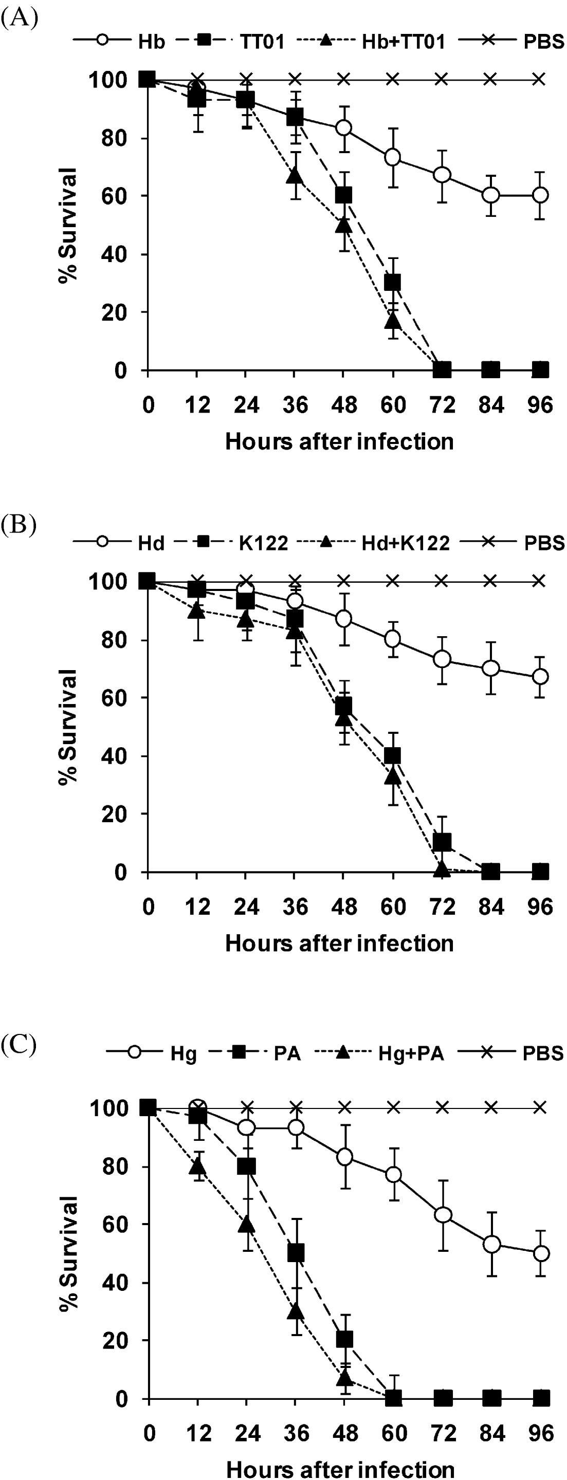
We compared survival between insects injected with a standard dose of Photorhabdus, bacteria alone, and insects naturally infected with either axenic Heterorhabditis or nematodes carrying their own symbiotic bacteria (Fig. 1). In each case, there were large differences in percentage survival between Heterorhabditis-infected larvae and those challenged with bacteria alone or the nematode-bacterium complex from 36 h or 48 h and up to 96 h after infection. In particular, we found significant differences in survival curves between axenic and non-axenic nematode treatments (for example comparison between the Hb and Hb+TT01 survival curves: Mantel-Cox test, χ2=8·77, P<0·05). However, there were no differences in survival curves between the Photorhabdus (TT01, K122 or ATCC43949) and the Heterorhabditis+Photorhabdus (Hb+TT01, Hd+K122 or Hg+ATCC43949) treatments (Mantel-Cox test, P>0·05). Negative controls including injection of PBS or a suspension of E. coli in PBS caused no mortality (results not shown).
Insect survival following challenge with axenic nematodes, nematodes carrying symbiotic bacteria and pathogenic bacteria alone. Day 0 fifth-stage Manduca larvae were infected with 100 six-day-old axenic Heterorhabditis bacteriophora (Hb), Heterorhabditis downesi (Hd) or Heterorhabditis gerrardi (Hg) nematodes, 1×10³ cells of Photorhabdus luminescens strain TT01, Photorhabdus temperata strain K122, or Photorhabdus asymbiotica (PA) strain ATC43949, or 100 nematodes carrying their cognate symbiotic bacteria (Hb+TT01, Hd+K122 and Hg+ATCC43949). Insect mortality was then monitored for 96 h. Note that nematodes lacking bacteria symbionts decrease insect survival, but they are less able to kill insect larvae compared to pathogenic bacteria and Heterorhabditis carrying Photorhabdus. Bars show means±standard deviations (s.d.) (n=3 replicated per point, 10 insects were used per replicate). PBS injection was used as a negative control.

Immune gene mRNA levels are lower in insects infected with axenic nematodes than with bacteria-associated nematodes
In order to determine the mRNA transcription of 3-pattern recognition protein genes (haemolin, immulectin-2 and peptidoglycan recognition protein-1A), the precursor of the phenoloxidase enzyme (prophenoloxidase-2) and 1 antibacterial effector gene (cecropin), we used semi-quantitative RT-PCR on RNA extracted from fat body of day 0 fifth-stage Manduca larvae infected with axenic nematodes, nematodes carrying symbiotic Photorhabdus or bacteria alone (Fig. 2). We found that 18 h after challenge with axenic nematodes immune genes were either transcribed at levels too low to detect in our experiments, or were transcribed at lower levels (immulectin-2) compared to the other treatments. Control insects injected with PBS showed undetectable levels of all 5 transcripts (results not shown). Transcription of the ribosomal protein S3 gene (rpS3) was used as a loading control.
Immune gene transcription in day 0 fifth-stage Manduca larvae. Genes tested were: hemolin (HEM), immulectin-2 (IML-2), peptidoglycan recognition protein-1A (PGRP-1A), prophenoloxidase-2 (PPO-2) and cecropin (CEC). Insects were infected with axenic Heterorhabditis (N), Photorhabdus alone (B) or Heterorhabditis carrying symbiotic Photorhabdus (N+B). Abbreviations as in Fig. 1. Semi-quantitative RT-PCR was used to assess levels of mRNA in fat body tissue 18 h following the experimental infection. Note that transcription of immune genes decreases in insects infected with axenic nematodes. Sizes of the PCR products are indicated. PCR amplifications used 35 cycles for each combination of primers and templates. A loading control (Manduca sexta ribosomal protein S3 – rpS3) was used to ensure equal loading in each lane. Each RT-PCR experiment was performed in duplicate.

Infection with axenic nematodes suppresses Manduca PO activity, nodulation and haemocyte aggregation
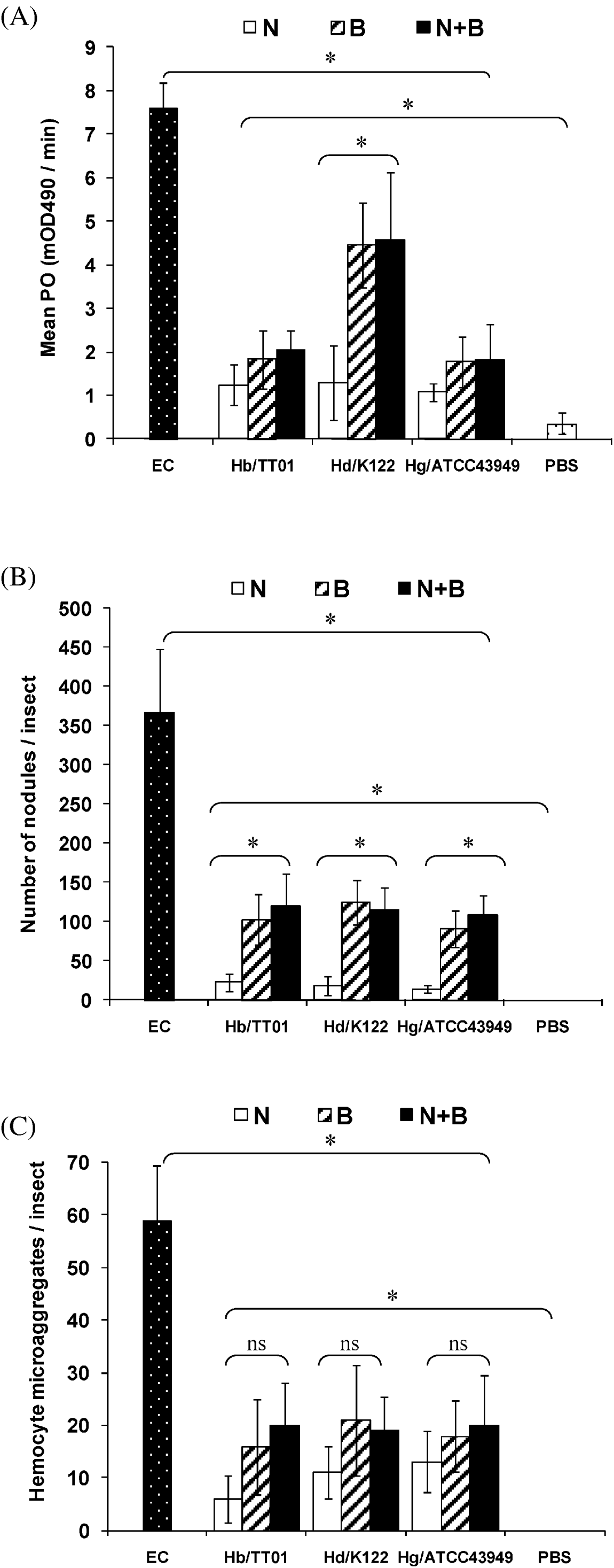
We also measured PO activity within haemolymph extracted from 18 h infected Manduca (Fig. 3A). PO levels were significantly higher in cell-free haemolymph plasma from insects infected by axenic Heterorhabditis, the Heterorhabditis-Photorhabdus complex or the pathogenic bacteria alone compared to the PBS control (one-way ANOVA, d.f.=9,70, F=33·650; P<0·0001; Tukey post-hoc test, P<0·05). In all cases, PO values were lower than for the E. coli treatment (one-way ANOVA, d.f.=9,70, F=56·487; P<0·0001; Tukey post-hoc test, P<0·05). It should be noted, however, that the number of E. coli cells initially injected was considerably greater than for Photorhabdus, so that the difference between these bacteria is not necessarily functionally significant, but the E. coli treatment nevertheless serves as a positive control. This result indicates that PO activity is less than maximal in insects infected with nematodes alone. The reason for this is not known, but indicates either that nematodes are not recognized by the PO-activating system, or that they produce inhibitor(s) of PPO activation or of activated PO. There were no differences between different strains of Heterorhabditis (one-way ANOVA, P>0·05), implying that all are recognized to the same extent.
(A) Phenoloxidase (PO) activity, (B) number of melanotic nodules and (C) number of haemocyte micro-aggregates in day 0 fifth-stage Manduca larvae 18 h after infection with axenic nematodes alone (N), bacteria alone (B) or nematodes with symbiotic bacteria (N+B). Abbreviations as in Fig. 1. Injections with E. coli (3×10⁷ washed DH5α cells) and PBS were used as positive and negative controls, respectively. Note the reduced PO activity, nodulation and haemocyte aggregation in insects infected with axenic nematodes compared to the other treatments. Bars show means±s.d. (n=3 replicates). Significant differences are indicated with an asterisk (ANOVA, *P<0·0001); ns denotes non-significant differences between treatments.

PO activity was also submaximal (i.e. less than following E. coli) in insects infected with all 3 Photorhabdus sp. (one-way ANOVA, d.f.=3,28, F=58·641; P<0·0001; Tukey post-hoc test, P<0·05), despite the fact that all the bacterial treatments would have led to proliferation of bacteria within the insect's haemocoel with ultimately lethal effects. This may reflect the fact that Photorhabdus is known to produce inhibitors of activated PO (Eleftherianos et al. Reference Eleftherianos, Boundy, Joyce, Aslam, Marshall, Cox, Simpson, Clarke, ffrench-Constant and Reynolds2007 a, Reference Eleftherianos, Waterfield, Bone, Boundy, ffrench-Constant and Reynolds2009).
Interestingly, PO values in haemolymph from larvae naturally infected with axenic H. downesi nematodes were significantly lower than PO values from insects infected with the same nematodes carrying P. temperata strain K122 bacteria or P. temperata-injected individuals (one-way ANOVA, d.f.=2,21, F=36·170; P<0·0001; Tukey post-hoc test, P<0·05), whereas this was not the case for the other 2 Photorhabdus species. This may reflect a lower PO-inhibitory ability of this Photorhabdus species, or a different rate of proliferation of the bacterium within the host. No significant differences in haemolymph PO activity were noted among insects infected with the other pathogens (one-way ANOVA, P>0·05).
At 18 h post-infection we also estimated the extent of nodulation within dissected caterpillars (Fig. 3B). Again, we found that both nematodes and symbiotic bacteria of all 3 species pairs elicited higher numbers of nodules than the PBS control treatment, implying that both are recognized by the nodule forming system. As before, we found that a significantly smaller number of melanotic nodules was formed in tissues of Manduca infected with Heterorhabditis associated with Photorhabdus, axenic Heterorhabditis, or injected only with symbiotic bacteria, than in tissues of larvae injected with non-pathogenic E. coli (one-way ANOVA, d.f.=9,80, F=74·503; P<0·0001; Tukey post-hoc test, P<0·05). Again, this probably reflects the larger number of E. coli cells that had been injected, but also clearly indicates that the nodulation response is submaximal following infection by all 3 Heterorhabditis-Photorhabdus species pairs. Notably, for all 3 Photorhabdus species, the number of dark nodules was significantly less in tissues of insects infected with axenic nematodes compared to nematodes with bacteria, or bacteria alone (one-way ANOVA, d.f.=8,72, F=30·815; P<0·0001; Tukey post-hoc test, P<0·05). This indicates either that nematodes are much less well recognized than bacteria, or despite being recognized are much less efficient in provoking a nodulation response. In evaluating this difference, it should be borne in mind that the Photorhabdus bacteria would have multiplied in number within the host during the 18 h since infection, while the nematodes would not. There was no difference in the extent of nodulation in insects infected with P. temperata K122, compared to the other two Photorhabdus species (one-way ANOVA, P>0·05), which implies that all 3 bacteria are equally well recognized, and suggests that the previously observed strain-related difference in PO activity may be due to a lesser ability of K122 to inhibit PO activation or activity.
Next, we examined the in vivo ability of washed haemocytes to form aggregates around naturally infected Heterorhabditis nematodes or injected Photorhabdus bacterial cells (Fig. 3C). This is commonly taken to be a measure of nodule formation that is distinct from melanization (Miller and Stanley, Reference Miller, Stanley and Weisner1998). Again, we noticed that the number of haemocyte micro-aggregates in insects infected with nematodes, with or without symbiotic bacteria, or Photorhabdus alone, was significantly greater than for the PBS controls (one-way ANOVA, d.f.=9,80, F=8·399; P<0·0001; Tukey post-hoc test, P<0·05). Although the number of micro-aggregates following infection with axenic Heterorhabditis was somewhat lower than the other treatments, which involved Photorhabdus bacteria with or without nematodes, these values did not differ significantly (one-way ANOVA, P>0·05). These results clearly show that both nematodes and their symbiotic bacteria are recognized by the system that initiates haemocyte aggregation. The number of micro-aggregates was, however, lower in all cases than following injection of E. coli (one-way ANOVA, d.f.=9,80, F=29·393; P<0·0001; Tukey post-hoc test, P<0·05). As previously noted, this result is not necessarily significant in itself, since the number of E. coli bacteria injected was much greater than the number of either nematodes or Photorhabdus, but does indicate that haemocyte aggregation is submaximal for all 3 Heterorhabditis-Photorhabdus species pairs.
The Heterorhabditis-Photorhabdus complex reduces haemocyte viability and phagocytosis in Manduca
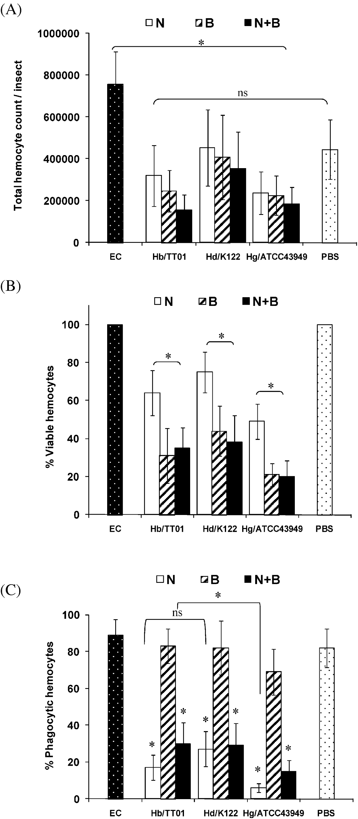
We recorded the total numbers of circulating haemocytes recovered from Manduca fifth-stage larvae infected with axenic nematodes, nematodes carrying bacterial symbionts, or Photorhabdus alone (Fig. 4A); for all 3 species pairs, we found no significant differences between all these treatments at 18 h after the infection (one-way ANOVA, P>0·05). Although total haemocyte counts in insects challenged with axenic Heterorhabditis, non-axenic nematodes, or Photorhabdus were not different from the PBS control (one-way ANOVA, P>0·05), they were significantly lower than in those insects subjected to E. coli injection (one-way ANOVA, d.f.=9,80, F=14·812; P<0·0001; Tukey post-hoc test, P<0·05). We previously noted (Au et al. Reference Au, Dean, Reynolds and ffrench-Constant2004) that in Manduca, total haemocyte numbers increase after injection of E. coli, although the time-course was different to that seen here. Au et al. (Reference Au, Dean, Reynolds and ffrench-Constant2004) also observed that injections of P. temperata K122 and P. luminescens W14 were followed by falls in the total number of haemocytes. It is not clear why this did not happen here.
(A) Total numbers of haemocytes, (B) viable haemocytes and (C) percentage of phagocytic haemocytes recovered from Manduca larvae 18 h post-infection with axenic nematodes alone (N), bacteria alone (B) or nematodes with symbiotic bacteria (N+B). Abbreviations as in Fig. 1. As before, injections with E. coli and PBS were the positive and negative controls, respectively. Note the increase in free haemocytes after E. coli injection and the decrease following infection with Photorhabdus. Also, note the decrease in the number of viable haemocytes following infection with Photorhabdus alone or with nematodes carrying pathogenic bacteria, and the decrease in numbers of phagocytic haemocytes in insects infected with axenic nematodes or nematodes carrying Photorhabdus. Bars show means±s.d. (n=3 replicates). The asterisks show a value that is significantly different from the other treatments (ANOVA, *P<0·0001) and ns denotes non-significant differences between treatments.

In the same experiment, we counted numbers of viable haemocytes (i.e. those able to adhere to a glass surface and form a monolayer) (Fig. 4B); the results revealed that Manduca larvae naturally infected with any of the 3 species of Heterorhabditis carrying their own species of Photorhabdus, or injected with Photorhabdus alone, yielded significantly fewer viable haemocytes than did larvae infected only with axenic nematodes (one-way ANOVA, d.f.=8,72, F=23·909; P<0·001; Tukey post-hoc test, P<0·05). This is consistent with the previous finding that Photorhabdus bacteria produce toxins that have adverse effects on haemocyte viability (Au et al. Reference Au, Dean, Reynolds and ffrench-Constant2004; Vlisidou et al. Reference Vlisidou, Dowling, Evans, Waterfield, ffrench-Constant and Wood2009).
We also estimated the number of phagocytic haemocytes in treated insects. To do this, we pre-treated insects with the various pathogens, and subsequently injected them with a standard dose of fluorescent-labelled E. coli and assessed the number of labelled bacteria that could later be seen inside haemocytes. Strikingly, for all 3 Heterorhabditis-Photorhabdus species pairs, we found that the number of haemocytes containing fluorescent bacterial cells (Fig. 4C) was significantly lower in insects that had been pre-infected with axenic Heterorhabditis, or with nematodes carrying Photorhabdus, compared to the number of phagocytic haemocytes in larvae previously injected with Photorhabdus alone (one-way ANOVA, d.f.=8,72, F=78·972; P<0·0001; Tukey post-hoc test, P<0·001). The number of phagocytic haemocytes did not differ between those insects that had been pre-treated with any of the 3 species of Photorhabdus alone, and the 2 controls that had been pre-injected with either E. coli or PBS (one-way ANOVA, P>0·05). This clearly indicates that all 3 heterorhabditids, but not their associated Photorhabdus spp., are able to depress the phagocytic capability of the insect host. The number of phagocytic cells was also significantly lower in insects pre-infected by axenic H. gerrardi nematodes compared to the other 2 axenic nematode treatments (one-way ANOVA, d.f.=2,24, F=19·808; P<0·0001; Tukey post-hoc test, P<0·5). This may indicate that this species of Heterorhabditis is particularly potent in suppressing phagocytosis.
Entomopathogenic nematodes and symbiotic bacteria affect phagocytosis and clearance of E. coli in vivo
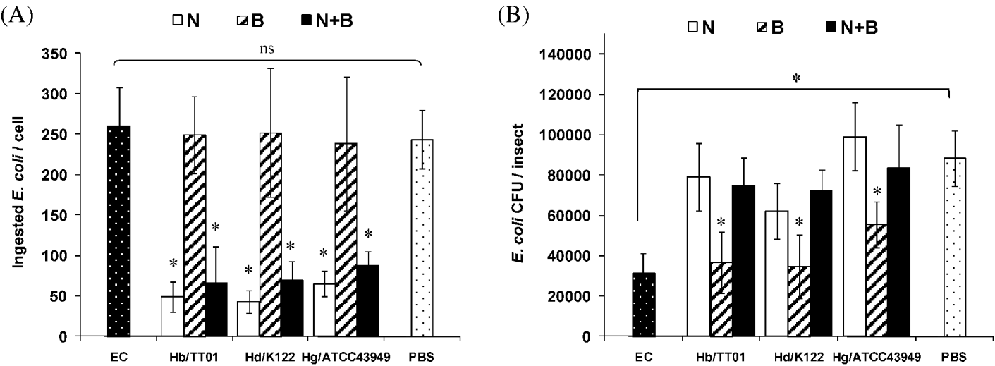
We looked at the ability of infected insects to remove bacteria from the circulation. To do this we first counted numbers of fluorescence-labelled E.coli cells in phagocytic haemocytes extracted from Manduca that had been previously infected with the pathogens (Fig. 5A). We found that the number of E. coli cells contained in haemocytes from larvae that had been pre-infected with nematodes carrying or lacking symbiotic bacteria was significantly lower than the number of bacterial cells in haemocytes from insects pre-injected with pathogenic Photorhabdus alone (one-way ANOVA, d.f.=8,72, F=36·151; P<0·0001; Tukey post-hoc test, P<0·05). No statistically significant differences were observed between numbers of ingested labelled E. coli in haemocytes from insects that had been pre-injected with any of the Photorhabdus strains alone, with E. coli, or with PBS (one-way ANOVA, P>0·05). These results indicate that all 3 nematode species inhibit the extent of phagocytosis by host haemocytes, whether or not their symbiotic Photorhabdus bacteria are present, and that Photorhabdus alone does not have this effect. This is consistent with the result of the previous experiment showing that nematodes (but not bacteria) reduce the number of phagocytic cells.
(A) Numbers of E. coli ingested cells and (B) numbers of recoverable E. coli bacteria (CFU, colony forming units) from day 0 fifth-stage Manduca first infected with axenic nematodes (N), bacteria alone (B) or nematodes with symbiotic bacteria (N+B) and 2 h later challenged with E. coli-labelled bacteria. Larvae were bled 18 h following the first infection. Abbreviations as in Fig. 1. Injections with E. coli alone and PBS served as controls. Note the decrease in the number of engulfed E. coli cells and the increase in the number of recoverable E. coli bacteria in insects infected with axenic nematodes or nematodes carrying Photorhabdus compared to the treatment involving injection of pathogenic bacteria alone. Bars show means±s.d. (n=3 replicates, 20 haemocytes per insect were examined). The asterisk shows a value that is significantly different from the other treatments (ANOVA, *P<0·0001) and ns denotes non-significant differences between treatments.

Finally, we determined the influence of infection with nematodes and symbiotic bacteria on the ability of harmless bacteria to survive in Manduca hemolymph. To do this, we pre-infected insects with nematodes, symbiotic bacteria, or both, and 2 h later injected a large number of E. coli cells. We then estimated the number of viable cells recoverable from the haemolymph of insects as colony forming units (CFU) (Fig. 5B). As we previously found (Eleftherianos et al. Reference Eleftherianos, Marokhazi, Millichap, Hodgkinson, Sriboonlert, ffrench-Constant and Reynolds2006 a), we noticed that pre-treatment of the insects with E. coli caused a significant decrease in the survival of bacteria that were injected later, compared to pre-treatment with PBS (Unpaired t-test, d.f.=16, t=9·976; P<0·0001). This is evidence for ‘priming’ of the insect's innate immune system.
When we treated the insects with components of the Heterorhabditis-Photorhabdus system either together or alone, however, we found that significantly greater numbers of live E. coli cells (Fig. 5B) were recoverable from haemolymph samples when the pre-treatment had involved natural infection by Heterorhabditis, with or without their bacterial symbionts, than from insects injected with Photorhabdus only (one-way ANOVA, d.f.=8,72, F=17·452; P<0·0001; Tukey post-hoc test, P<0·5). Further, there were no significant differences between numbers of E. coli CFU recovered from insects pre-treated with E. coli, or with any of the pathogenic Photorhabdus strains alone (one-way ANOVA, P>0·05). Notably, all of these bacteria-only treatments led to a smaller number of CFU being recovered than in the case of the control pre-treatment with PBS alone (one-way ANOVA, d.f.=4,40, F=28·339; P<0·0001; Tukey post-hoc test, P<0·5). These results indicate that both Photorhabdus and E. coli can ‘prime’ the Manduca immune system (as previously noted by Eleftherianos et al. Reference Eleftherianos, Marokhazi, Millichap, Hodgkinson, Sriboonlert, ffrench-Constant and Reynolds2006 a), but that none of the 3 species of Heterohabditis nematode elicits priming in this way. Further, the simultaneous presence of nematodes evidently prevents priming by Photorhabdus; it might be conjectured that this is because the symbiotic bacteria are not exposed to recognition by the insect when in association with nematodes, as they would be if they were simply injected; however, this is not in accord with our other observations on host PO activity, which indicate that Photorhabdus can exert specific effects on host immunity whether it is associated with nematodes or not. A possible reconciliation is that in the PO experiments, a period of 18 h elapsed between pre-infection and the biological assay, whereas in the case of the observations on E. coli survival, there was only 2 h between pre-infection with the Heterorhabditis-Photorhabdus pair and the later injection of E. coli.
DISCUSSION
Symbioses between entomopathogenic nematodes of the genus Heterorhabditis and bacteria of the genus Photorhabdus are mutualistic associations in which both partners derive fitness benefits from each other in terms of their ability to cause disease in a third organism, generally an insect (Waterfield et al. Reference Waterfield, Ciche and Clarke2009). This relationship has been extensively studied in terms of the pathogenic potential of the bacteria alone (Vallet-Gely et al. Reference Vallet-Gely, Lemaitre and Boccard2008), but the contribution of the nematode to pathogenesis is much less well understood. The goal of the present study was to address the question of the impact of Heterorhabditis on the outcome of insect infection, and to clarify the relative roles of the nematode and its symbiotic bacterium against the humoral and cellular immune responses of the host.
As in previous studies (Ciche and Ensign, Reference Ciche and Ensign2003; Eleftherianos et al. Reference Eleftherianos, Millichap, ffrench-Constant and Reynolds2006 b), we found that a relatively small number of bacterial cells is sufficient to kill the host within a few days. Importantly, we also found that administering Photorhabdus together with their associated Heterorhabditis nematodes did not increase the speed or extent of mortality compared to bacteria alone. Thus, at least under the experimental infection conditions used, Photorhabdus bacteria clearly play a quantitatively predominant role in killing the host.
However, our results also showed that although natural infections using axenic (germ-free) nematodes alone were significantly less pathogenic than when bacteria were present, nematodes without bacteria were nevertheless able to cause significant mortality. Thus, the symbiotic nematode partner also contributes directly to virulence. Previous workers have also found that axenic Heterorhabditis can efficiently infect insects, but cause much less (or even no) insect mortality compared to symbiont-bearing infective juveniles, and are subsequently unable to reproduce in the host (Han and Ehlers, Reference Han and Ehlers2000; Hallem et al. Reference Hallem, Rengarajan, Ciche and Sternberg2007). On the other hand, axenic entomopathogenic nematodes of another genus (Steinernema) are known to produce toxins that are lethal to the host, even without their associated symbiotic bacteria (Brivio et al. Reference Brivio, Mastore and Pagani2005). All this suggested to us that heterorhabditid nematodes might produce factors that affect the host, even if they are not very effective (or even insufficient) in causing host mortality. We therefore studied the ways in which such factors might influence host immunity, and thus determine the outcome of the infection.
In the experiments reported here, the mRNA levels of certain pattern recognition proteins (Hemolin and PGRP-1A) in response to injected bacteria alone were much the same as when a similar number of bacteria were delivered through a natural nematode infection. In addition, we found that mRNA levels of a representative antimicrobial peptide (AMP) (Cecropin) were very low (undetectable under our conditions) in fat body of insects infected only with axenic nematodes. These results together imply that (i) nematodes are not recognized by the insect, or are much less efficiently recognized, than their symbiotic bacteria and (ii) nematodes do not markedly influence the host's recognition of, and response to, pathogenic bacteria.
The observed failure of heterorhabditid nematodes without symbiotic bacteria to provoke a host immune response agrees with previous findings. It has previously been shown that there is no defensin transcription in the fat body of Armigeres subalbatus in response to Brugia malayi (Lowenberger et al. Reference Lowenberger, Ferdig, Bulet, Khalili, Hoffmann and Christensen1996). More recently, axenic Heterorhabditis failed to induce significant expression of any of 7 AMP genes in Drosophila larvae (Hallem et al. Reference Hallem, Rengarajan, Ciche and Sternberg2007). Nevertheless, it is not yet clear whether the nematodes are able to interfere with host antibacterial activity (Richman and Kafatos, Reference Richman and Kafatos1995). In the case of Manduca, the specific roles of Cecropins and other AMPs in targeting and/or disrupting the anti-parasite response remain thus far unknown, and will be the focus of future experiments.
In the present study we further show that natural infection with axenic Heterorhabditis nematodes fails to provoke transcription of PPO-2 in Manduca fat body. Because activation of the PO cascade is one of the earliest processes that can effectively prevent the growth and diffusion of potential pathogens (Zhao et al. Reference Zhao, Li, Wang and Jiang2007), inhibition of active PO may be conjectured to be a major contribution to successful parasitization. Brivio et al. (Reference Brivio, Pagani and Restelli2002) observed a drastic PPO system inactivation in Galleria mellonella larvae following short-term infection with the entomopathogenic nematode Steinernema feltiae (Brivio et al. Reference Brivio, Pagani and Restelli2002); it was subsequently suggested (Brivio et al. Reference Brivio, Mastore and Moro2004) that to ensure successful infection, PO depression must be achieved by the nematode before its symbiotic bacteria (Xenorhabdus) are released. Our observation of low levels of PO in Manduca infected by heterorhabditid nematodes, compared to levels in those given Photorhabdus K122 bacteria, is consistent with this idea. As explained in the results section, however, other strains of Photorhabdus also produce their own PO inhibitors, which would probably obscure the PO-inhibiting effects of the corresponding nematodes.
Besides humoral responses, insect hosts are also able to mount cellular immune reactions against parasitic nematodes and their symbiotic bacteria (Strand, Reference Strand2008). It would therefore be expected that successful parasites and pathogens would possess adaptations to abrogate these defences. Here we show that Photorhabdus and Heterorhabditis (injected separately or together) reduce both total haemocyte numbers, and counts of viable haemocytes, and also suppress haemocyte aggregation. Interestingly, axenic Heterorhabditis do not cause such effects. This implies that the principal agent of these anti-haemocytic effects is the bacterial symbiont. By contrast, both axenic Heterorhabditis and nematodes carrying symbiotic Photorhabdus, but not bacteria alone, adversely affect phagocytosis (measured as the number of phagocytically active haemocytes, and the number of ingested bacterial cells) thus interfering with the ability of Manduca haemocytes to clear non-pathogenic E. coli from the haemolymph. Similarities of the cellular defences between bacterial and nematode infections may suggest that Photorhabdus bacteria, released from Heterorhabditis, could be responsible for the observed response. It is well documented that Photorhabdus secretes certain toxins that actively kill haemocytes or have an adverse effect on haemocyte function (Waterfield et al. Reference Waterfield, Ciche and Clarke2009). However, immuno-evasion strategies employed by insect nematodes are often attributable to the mimetic properties of the parasite body surface rather than to the cells being damaged by the worm. For example, G. mellonella immunocompetent haemocytes are unable to recognize S. feltiae parasites as non-self (Brivio et al. Reference Brivio, Mastore and Moro2004). Nevertheless, the nematobacterial complex S. carpocapsae-X. nematophilus produces unidentified molecules having toxic activity in vitro towards insect haemocytes (Ribeiro et al. Reference Ribeiro, Duvic, Oliveira, Givauda, Palha, Simoes and Brehélin1999). Previously, it has been reported that H. bacteriophora is able to escape encapsulation in Popillia japonica larvae by an unknown mechanism (Wang et al. Reference Wang, Gaugler and Cui1994, Reference Wang, Campbell and Gaugler1995). It remains now to be clarified whether Heterorhabditis produces anti-phagocytic factors that have direct and/or indirect pathological effects on Manduca haemocytes.
In conclusion, this study emphasizes the complexity found in similar interactions between mutualistic organisms. By studying systems based on entomopathogenic nematodes, we can investigate a diverse array of functions in insect innate immunity, as the insect defends against not only the nematode vector, but also against the bacterial symbiont. The present work could also give important insight into recognition mechanisms required to trigger a rapid and efficient anti-parasite immune response and use this information to identify specific molecular components involved in vector competence and disease transmission. From the practical point of view, since entomopathogenic nematodes represent an alternative to chemicals for insect pest control, it is crucial to elucidate the basis of infectivity of the nematode-bacterium complex and the interaction with the host immune system. Although the effects of Photorhabdus infection on insect immune responses have been extensively studied before (Waterfield et al. Reference Waterfield, Ciche and Clarke2009), the role of Heterorhabditis in pathogenesis and its impact on host immune function are not fully established. The present study shows that the role of the nematode is quantitatively less important; however, this does not imply that the contributions of the two partners to the entire symbiotic life cycle are unequal. Important benefits may also be conferred by the nematode on its bacterial partner through enhancements in the efficiency of transmission and survival between infections (Goodrich-Blair and Clarke, Reference Goodrich-Blair and Clarke2007). Thus the symbiosis appears to be a division of labour, rather than an even-handed collaboration.
ACKNOWLEDGEMENTS
We thank Sandra Barnes for rearing the insects. This project was supported by a grant to S.E.R., R.ff.-C. and D.J.C. from the Exploiting Genomics Initiative of the UK Biotechnology and Biological Sciences Research Council (BBSRC).