Introduction
The genus Akonesis Opitz (Coleoptera: Cleridae: Peloniinae) was established by Opitz (Reference Opitz2014b). The two known species included in that publication, and A. ridens (Gorham) included herein, were previously classified in Cregya LeConte. The placement of these species in Akonesis follows the definition of the genus, which is characterised primarily by having 1-2-2 tibial spur formula. Placement of Akonesis in Peloniinae follows the definition of the subfamily as defined in Opitz (Reference Opitz2010: 97) and expanded in Opitz (Reference Opitz2016: 261, Reference Opitz2017: 32).
The value of elytral colour as a parameter for prediction of species status is inconsistent among checkered beetles. Elytral infraspecific variation in colour patterns abounds in genera such as Trichodes Herbst (Foster Reference Foster1976), Perilypus Spinola (Ekis Reference Ekis1977), Eurymetopum Blanchard (Solervicens Reference Solervicens1986), and Agnatis Opitz (Opitz Reference Opitz2014a). Conversely, slight differences in elytral colour patterns are species specific in Aulicus Spinola (Barr and Foster Reference Barr and Foster1979), Madoniella Pic (Opitz Reference Opitz2011), Enoclerus Gahan (Rifkind Reference Rifkind2017), and Plocamocera Spinola (Opitz Reference Opitz2004). Recently acquired information about aedeagal morphology confirms that species of Akonesis fall into the last-mentioned group of genera. The purpose of this treatise is to improve our understanding of the species composition in the subfamily Peloniinae.
Materials and methods
Methods involving dissections, measurements, and morphological terminology follow those described in Opitz (Reference Opitz2010: 35). Brown (Reference Brown1956) was used to form scientific names. All measurements were made at 250×. Line drawings were made with an M5 Wild stereoscopic microscope with camera a lucida attachment (Leica, Wetzlar, Germany). Habitus photographs were taken with a Leica Z 16 APO microscope equipped with a JVC KY-F75U-CCD camera and controlled by Syncroscopy Auto Montage software (Cambridge, United Kingdom). The scanning electron micrographs were produced with a Scanning Electron Microscope-S-3500N (Hitachi Science Systems, Tokyo, Japan). To facilitate the identity of type specimens, I transcribed their locality information verbatim.
Although morphological criteria are used to determine species status, I adhere to the biological species concepts as discussed by Standfuss (Reference Standfuss1896), Dobzhansky (Reference Dobzhansky1937), and Mayr (Reference Mayr1963). I have found that broad experience in morphology and morphological divergence among population samples of a group provides a reasonable criterion for predictions of reproductive isolation. In this study, determination of species status is based on the shape of the capitulum, configuration of the elytral markings. The rationale of Hennig (Reference Hennig1966) is followed for speculations of supra-specific relationships, although I am in agreement with Tuomikoski (Reference Tuomikoski1967), who advocates the use of “apotypic” and “plesiotypic” instead of “apomorphic” and “plesiomorphic” because phylogenetic work may not be restricted to morphological criteria.
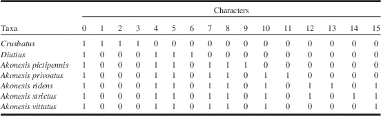
A total of 16 character states were organised into a matrix (Table 1), which was then analysed with NONA (Goloboff Reference Goloboff2003) in combination with WINCLADA version 100.80 (Nixon Reference Nixon2002), to find to most parsimonious phylogenetic tree. These programmes produced one tree produced via a heuristic analysis (maximum trees (held)=100, number of replications 1 (mult)=100, and multiple TBR=TBR (mult max) were used). The tree indices involve: length=15, and confidence indices of confidence index=100 and retention index=100. Character states given the value of “0” are assessed as plesiotypic whereas those judged a value of “1” are assessed as apotypic. Mesquite v3.2 build 801 (Maddison and Maddison Reference Maddison and Maddison2017) was used to prepare a nexus formatted data matrix. This was used in PAUP٭ v4.0a build 157 (Swofford Reference Swofford2002) to conduct 1000 branch and bound bootstrap replicates under the parsimony criterion. The genera Crusbatus Opitz and Diutius Opitz, and my general knowledge of other Peloniinae genera, were used as outgroups to assist in character state assessments and to predict the evolutionary states of characteristics. Moreover, I also relied on the methods of character-state analysis employed by Ekis (Reference Ekis1977), Watrous and Wheeler (Reference Watrous and Wheeler1981), and Nixon and Carpenter (Reference Nixon and Carpenter1993).
Character matrix of 16 adult morphological characters of Crusbatus (outgroup), Diutius (outgroup), and Akonesis.

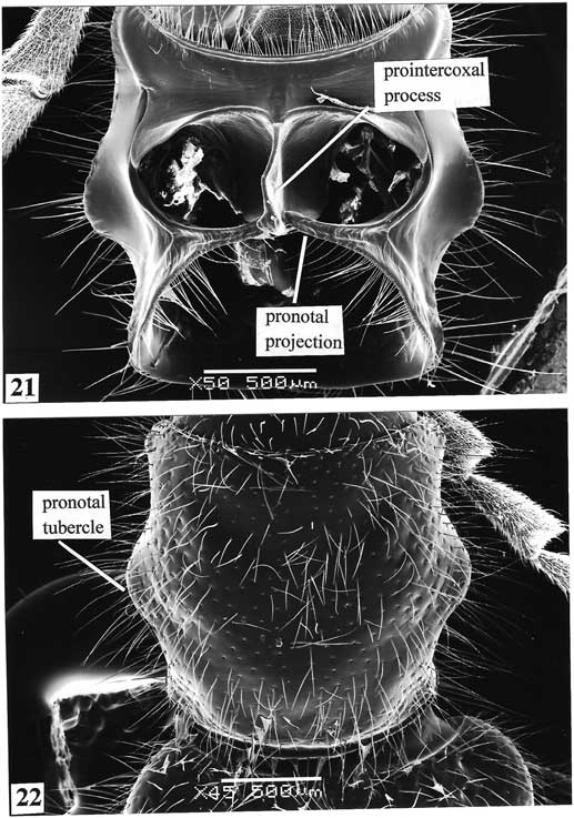
Character 0 – Pronotal projection: (1) short, less than 1/2 distance to prointercoxal process; (2) long, more than 1/2 distance to prointercoxal process (Fig. 21)
Character 1 – Sixth antennomere: (0) not enlarged; (1) enlarged
Character 2 – Pronotum: (0) quadrate; (1) oblong
Character 3 – Protibia: (0) not spinous; (1) spinous
Character 4 – Phallobase: (0) with struts; (1) without struts
Character 5 – Capitular colour: (0) without marginal black streak; (1) with marginal black streak
Character 6 – Phallic plates: (0) not serrate; (1) serrate
Character 7 – Pronotal tubercle: (0) not well developed; (1) well developed (Fig. 22)
Character 8 – MP3 vein: (0) present; (1) absent
Character 9 – Sixth ventrite: (0) not modified; (1) modified
Character 10 – Configuration of elytral marking: (0) not vittate; (1) vittate
Character 11 – Capitular size: (0) expanded; (1) not expanded (Fig. 3)
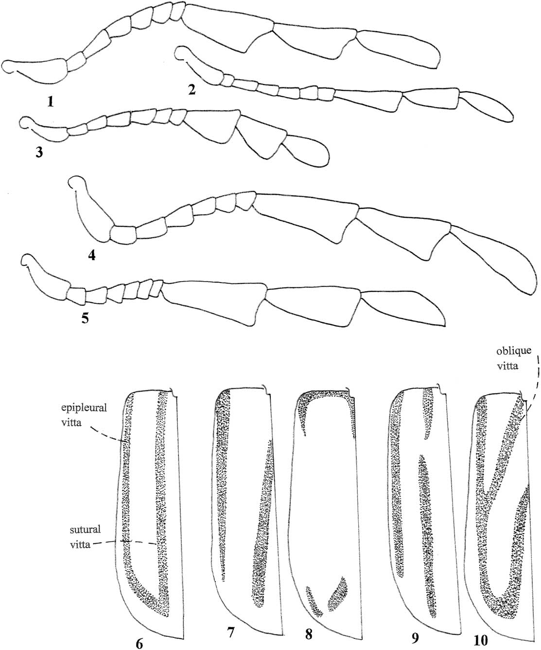
Antenna and elytra. 1. Antenna, Akonesis ridens, female. 2. Antenna, Akonesis pictipennis, male. 3. Antenna, Akonesis privoatus, female. 4. Antenna, Akonesis strictus, male. 5. Antenna, Akonesis vittatus, male. 6. Elytra, Akonesis strictus. 7. Elytra, Akonesis vittatus. 8. Elytra, Akonesis pictipennis. 9. Elytra, Akonesis privoatus. 10. Elytra, Akonesis ridens.
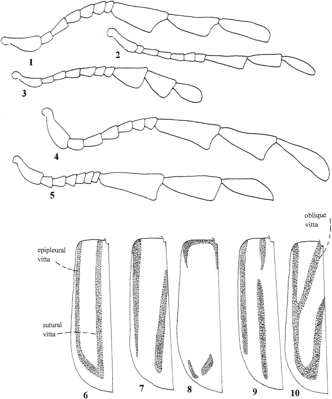
Character 12 – Sutural and epipleural elytral vitta: (0) not confluent (Fig. 7); (1) confluent (Figs. 6, 10)
Character 13 – Oblique elytral vitta: (0) absent; (1) present (Fig. 10)
Character 14 – Elytral vittae: (0) not parallel; (1) mostly parallel (Fig. 6)
Character 15 – Elytral sutural vitta: (0) bipartite; (1) not bipartite (Figs. 6–7, 10)
Repository of specimens
I used codens as noted in Arnett et al. (Reference Arnett, Samuelson and Nishida1993) to indicate repositories of specimens, with some modifications to accommodate institutional name changes. ACMT, American Coleoptera Museum, San Antonio, Texas, United States of America (James E. Wappes); BMNH, The Natural History Museum, London, United Kingdom (Beulah Garner); FMNH, Field Museum of Natural History, Chicago, Illinois, United States of America (Crystal Maier); FSCA, Florida State Collection of Arthropods, Gainesville, Florida, United States of America (Paul E. Skelley); MNHN, Museum d’Histoire Naturelle, Paris, France (Antoine Mantilleri); MRSN, Museo Regionale di Scienze Naturali, Torino, Italy (Luca Piccian); RFMC, Roy F. Morris Collection, Lakeland, Florida, United States of America; WOPC, Weston Opitz Collection, Gainesville, Florida, United States of America.
Akonesis Opitz, 2014
Type species. Pelonium vittatum Spinola, Reference Spinola1844: 159 by original designation.
Gender. The genus Akonesis is masculine in gender, as determined using Article 30.2.4 of the International Code on Zoological Nomenclature.
Diagnosis. Akonesis specimens are similar superficially to specimens of Diutius. However, Akonesis beetles show a 1-2-2 tibial spur formula and the metendosternal anterior plate is small, whereas in Diutius members the spur formula is 0-2-2 and the metendosternal plate is large. Also, the pronotal tubercle is well developed in specimens of Akonesis, which is not the case in members of Crusbatus.
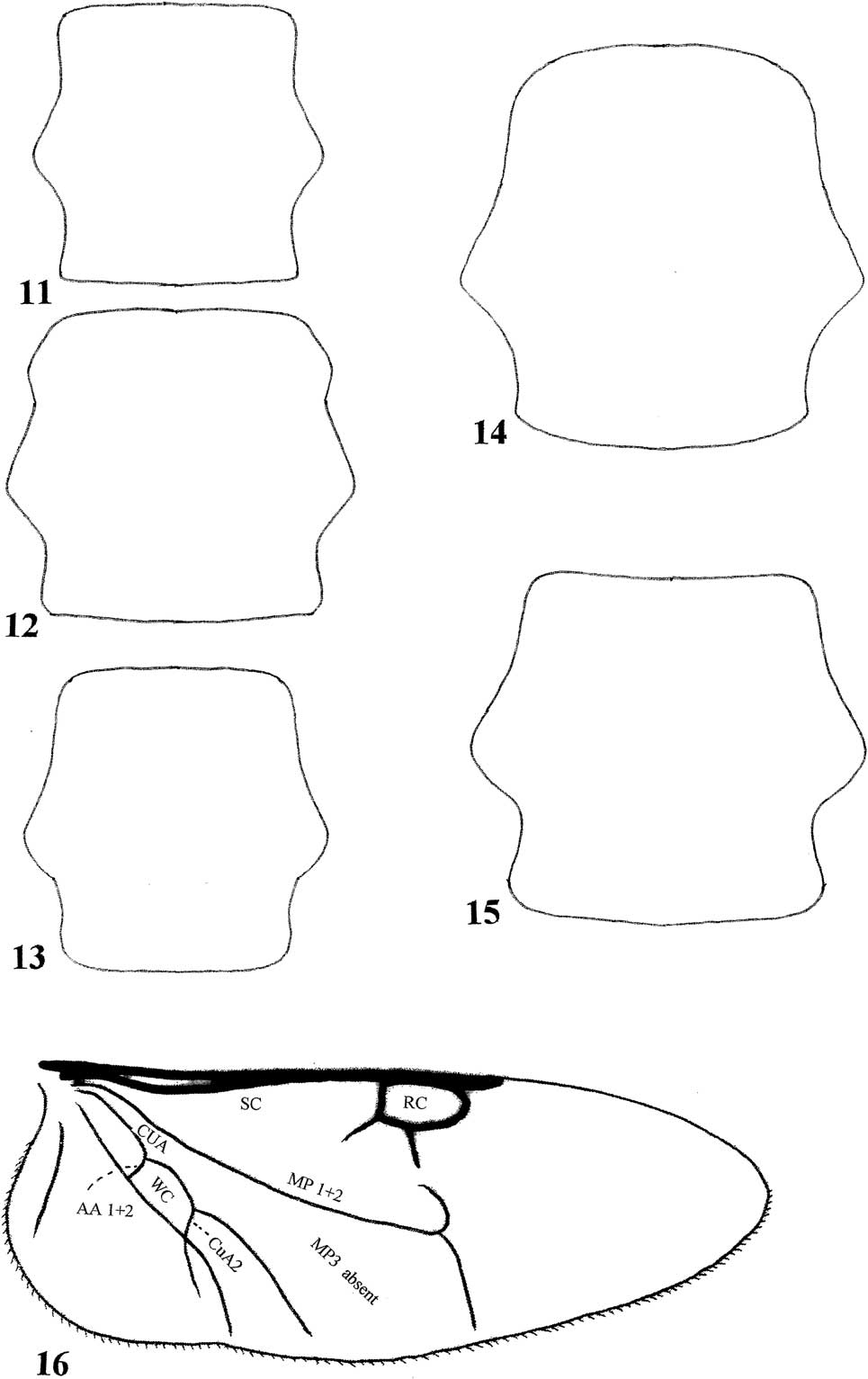
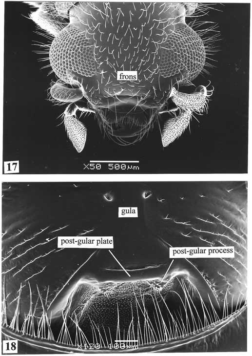
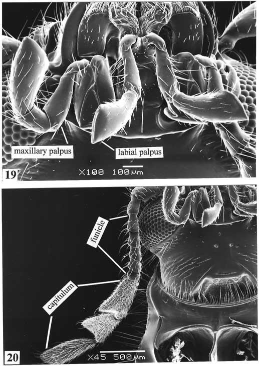
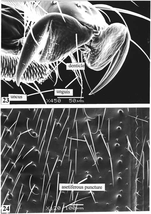
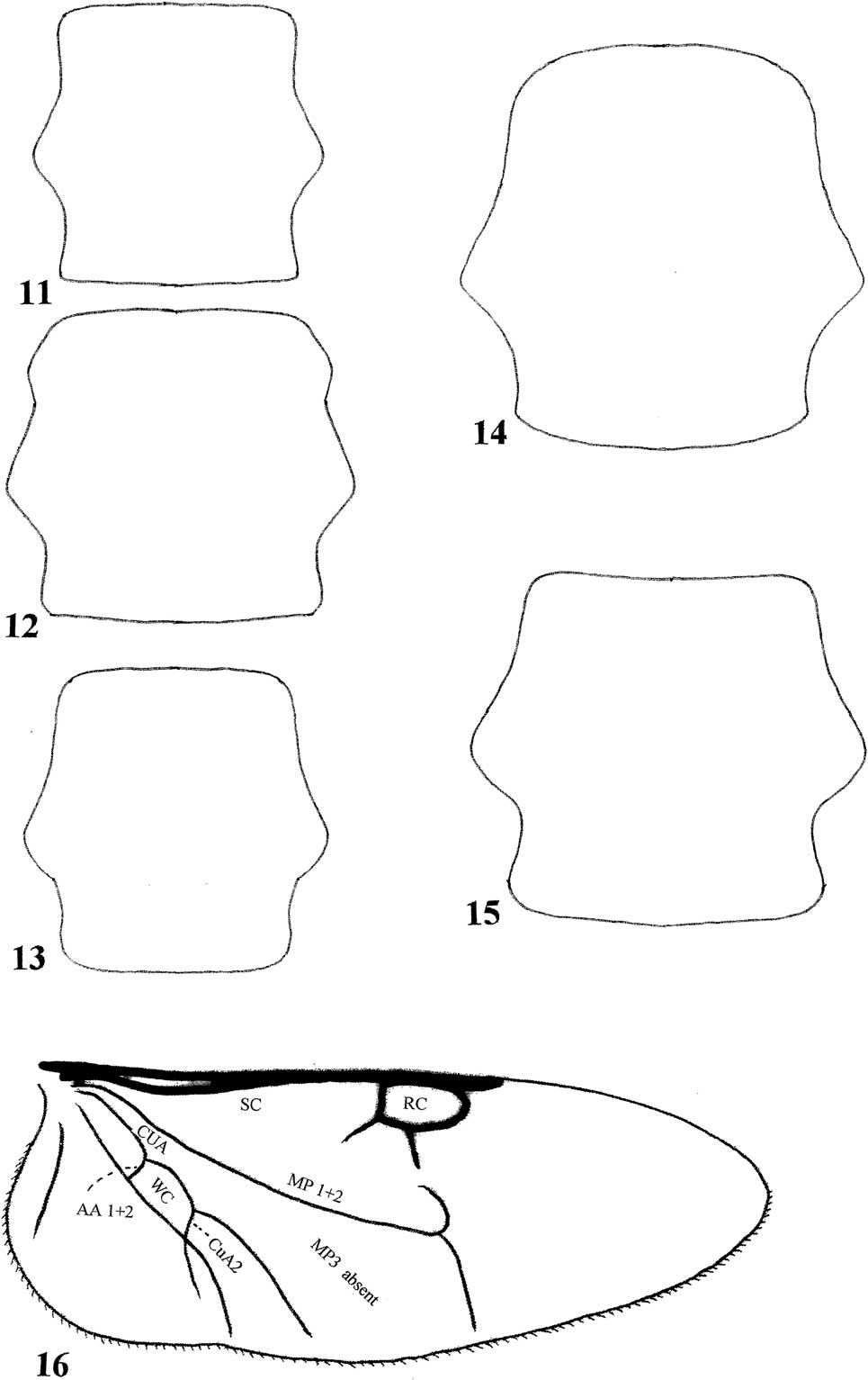
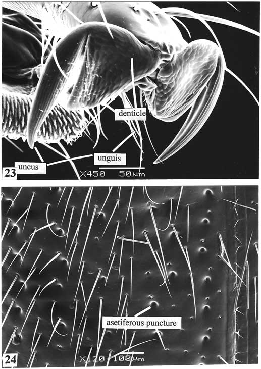
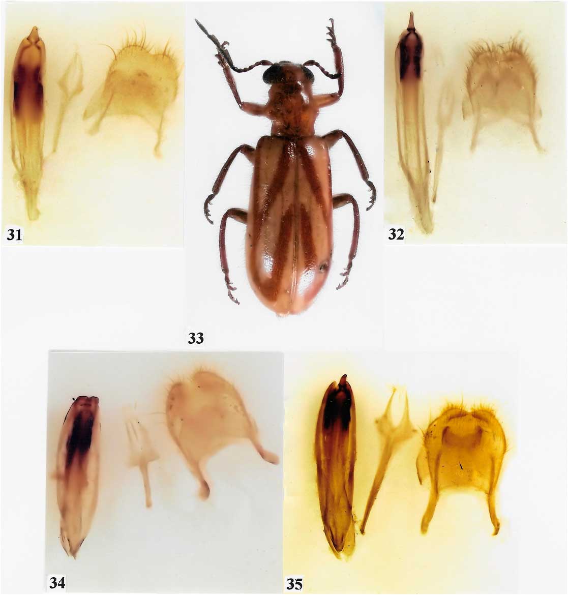
Redescription. Size : Length 8.5–11.0 mm, width 3.0–4.0 mm. Form : Oblong, robust (about 3× longer than broad), body deep. Vestiture : Dorsum profusely vested with pale pubescence, antenna moderately setose, elytra with primary and secondary setae or without primary but with secondary setae only. Head (Fig. 17): Cranium subquadrate, frons narrower or as wide as eye, profusely indented with small, setiferous punctures. Gula (Fig. 18) large, subquadrate, sutures diverging anteriorly, gula with two well-developed setose post-gular processes (Fig. 18). Labrum shallow, incised distally (Fig. 17), tormal processes (mediolateral extensions of the labral apodemes) contiguous, epipharyngeal plate small. Mandible stout, anterior dens acuminate, medial and posterior dens well developed, penicillus well developed. Maxilla (Fig. 19) with laterolacinia present as indentation, terminal palpomere securiform. Labium (Fig. 19) with ligula deeply incised, ligular lobes narrowed, terminal palpomere securiform. Antenna comprised of 10 antennomeres, capitate; capitulum long and slender (Fig. 1), rarely short (Fig. 3); funicular antennomeres subfiliform. Eyes large, coarsely faceted; ocular notch large (Fig. 17). Thorax : Pronotum (Fig. 22) slightly subquadrate, disc flat; lateral margins each with conspicuous tubercle; dorsolateral carina incomplete, anteriorly extended beyond tubercle and confluent with pronotal hem posteriorly; prointercoxal process narrow (Fig. 21), expanded distally; pronotal projections long and approximating prointercoxal process (Fig. 21). Elytron with (Fig. 24) or without asetiferous punctures; epipleural fold wide and in ventral position, tapered to elytral apex; anteriorly elytral margin not carinate. Metathoracic wing as in Fig. 16, wedge cell closed, MP3 vein absent. Metendosternite with furcal lamina, furcal anterior plate triangular. Profemora not swollen and without spines; tibial spur formula 1-2-2; tarsal pulvillar formula 3-3-3, ungues with basal denticle (Fig. 23). Abdomen : Aedeagus (Figs. 31–32, 34–35) shorter than length of abdomen, distal region of phallobase slightly lobed, lateral lobes not fimbriate, tegmen short, phallobasic struts absent, phallobasic rod absent, phallic plates wide. Spicular fork (Fig. 34) well developed, spicular apodemes fused, intraspicular plate long and linear.
Pronotum and metathoracic wing. 11. Pronotum, Akonesis pictipennis. 12. Pronotum, Akonesis privoatus. 13. Pronotum, Akonesis vittatus. 14. Pronotum, Akonesis strictus. 15. Pronotum, Akonesis ridens. 16. Metathoracic wing.
Distribution. A New World genus known from Bolivia and Brazil.

Akonesis pictipennis (Gorham, Reference Gorham1877)
Original combination. Pelonium pictipenne Gorham, Reference Gorham1877: 423.
Holotype. ♂. “Rio de Janeiro, Brazilia” (BMNH).
Catalogue. Lesne Reference Lesne1917: 149 (Galeruclerus). Corporaal Reference Corporaal1950: 283 (Cregya). Opitz Reference Opitz2014b: 415 (Akonesis).
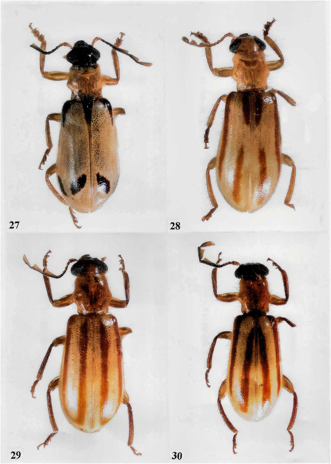
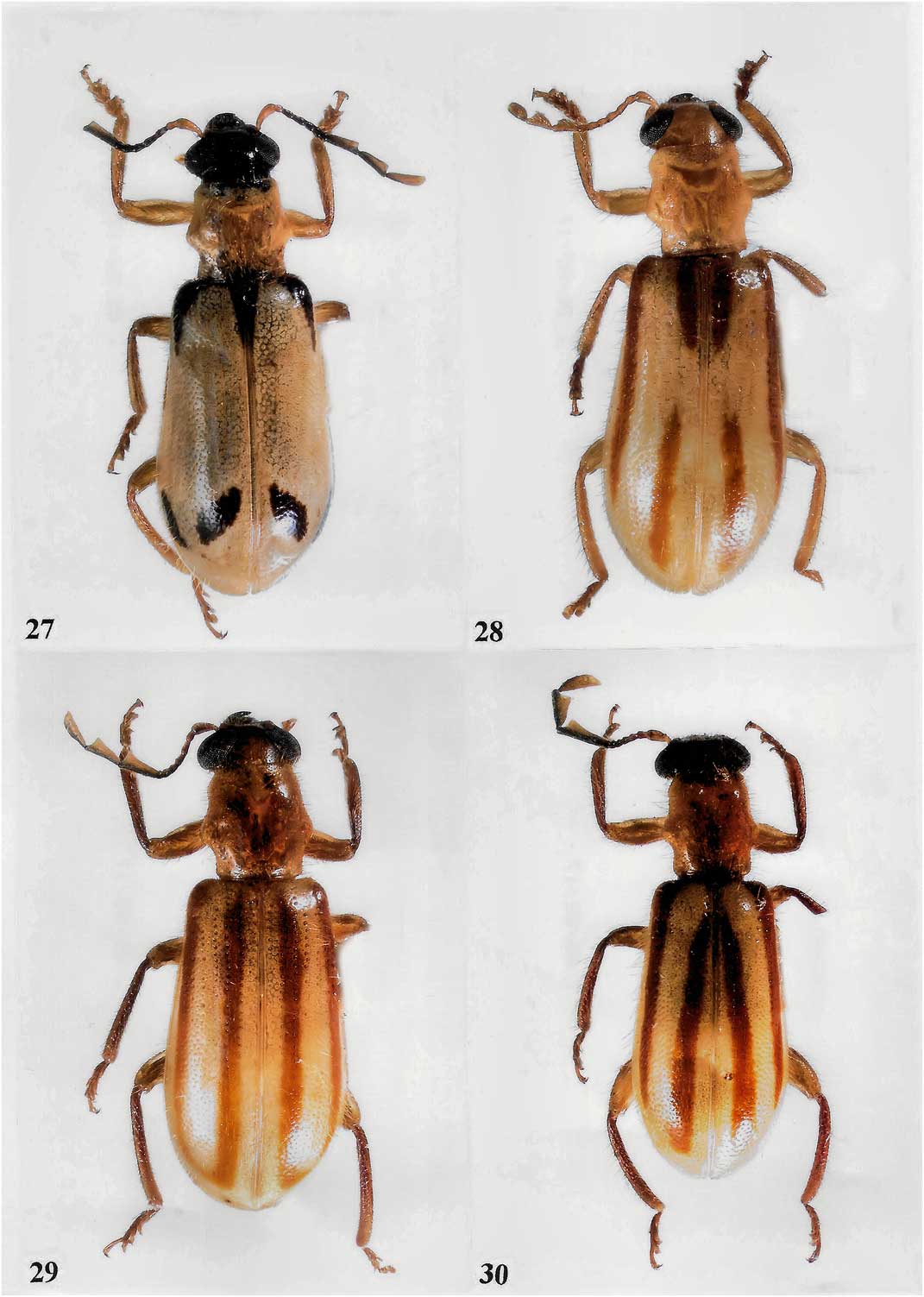
Diagnosis. The black, broad, and moderately oblique markings on the elytral disc as shown in Figures 8, 27 will distinguish the members of this species from congeners.
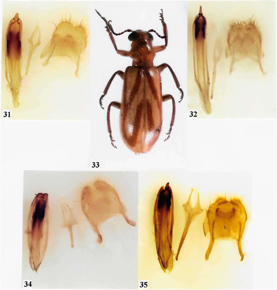
Redescription. Size : Length 10.0 mm, width 3.8 mm. Form : As in Figure 27. Colour : Mostly yellow, cranium and mouthparts light castaneous; scape and pedicel light castaneous, funicle black, posterior margin of capitular antennomeres black. Elytron mostly yellow; humerus, basal margin black with two preapical black, broad and moderately oblique maculae (Figs. 8, 27). Tibiae and tarsi brown, abdomen brown. Head : Funicular antennomeres subfiliform, capitular antennomeres expanded (Fig. 2); frons as wide as eye (eye width/frons width 26/26). Thorax : Pronotum (Fig. 11) quadrate (pronotal width/pronotal length 70/70), side margins each with well-developed tubercle, disc subglabrous finely punctate; elytra without asetiferous punctures (elytron length/elytron width 220/65). Abdomen : Pygidium scutiform (Fig. 35), posterior margin incised; 6th visible sternite broadly concave distally; aedeagus as in Figure 35.
Variation. Size : Length 9.0–11.0 mm, width 3.2–4.0 mm. Other than body size, the available beetles are quite homogeneous.
Natural history. Specimens were collected during March–May, and in December.
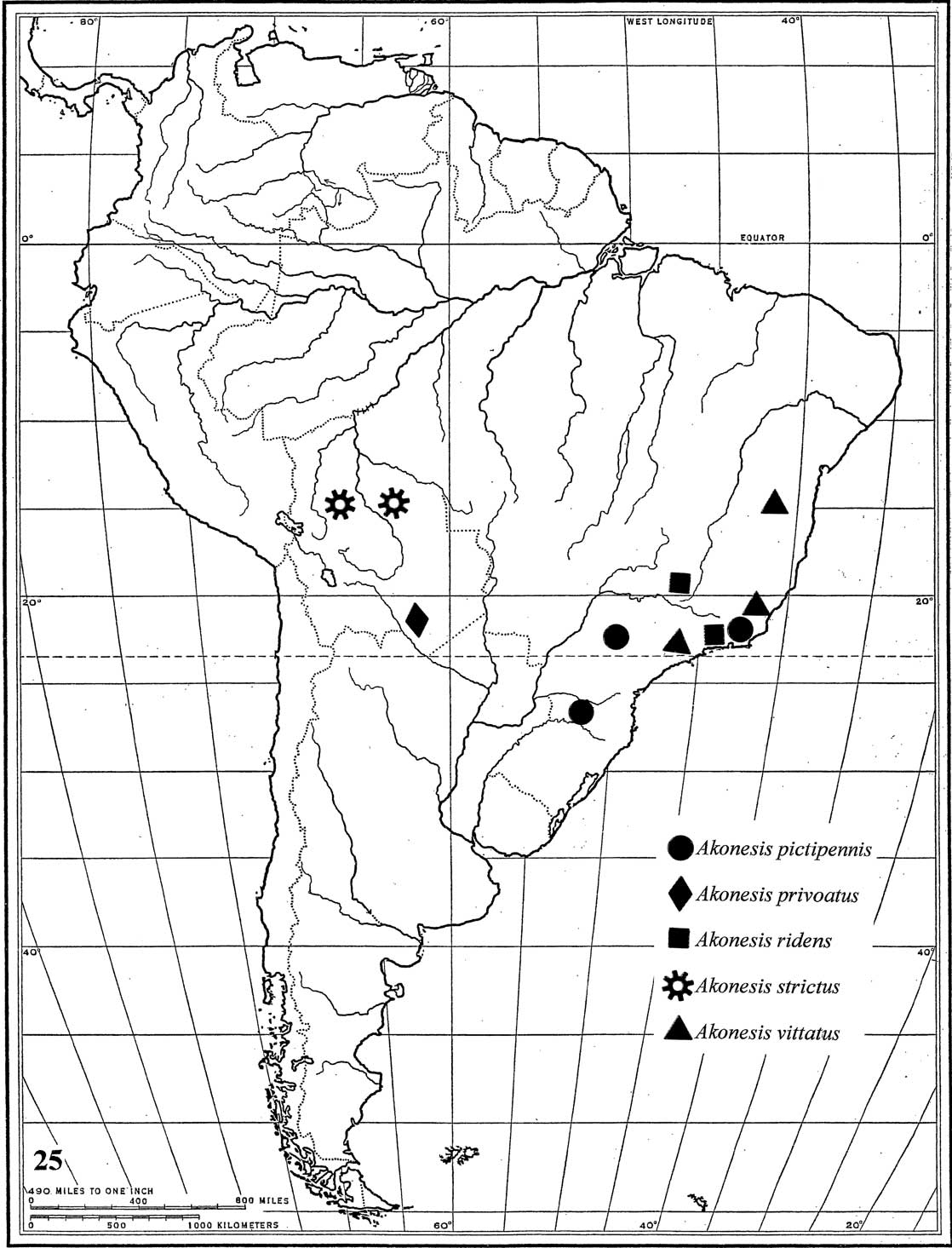
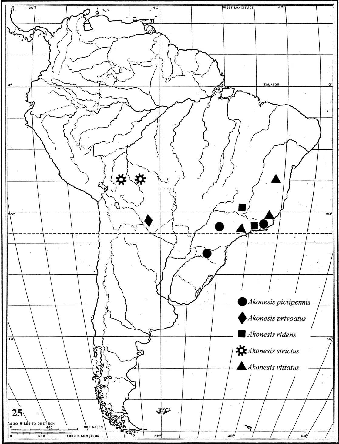
Distribution. In addition to the holotype, I examined five specimens from Brazil: Estado do Rio de Janeiro, x.1844, de Castelnau; Estado do Rio de Janeiro, Nova Freburgo, iv–v.1884, P. Germain; Estado do São Paulo, Cantereira, 1.III.1963; Estado do Santa Catarina, Nova Teutonia (Fig. 25). Specimens are deposited in BMNH, MNHN, and WOPC.
Akonesis privoatus Opitz, new species
Holotype. ♀. “Chaco (Bolivia), Garlepp” (MNHN). Paratypes. Two specimens. Bolivia, Chaco (WOPC); Chaco, Yungas, 3000 m, G. Garlepp (WOPC).
Diagnosis. The disconnection in the sutural vitta on the elytral disc (Figs. 9, 28) will distinguish members of this species from congeners.
Holotype description. Size : Length 11.0 mm, width 3.6 mm. Form : As in Figure 28. Colour : Mostly flavotestaceous; elytron with reddish brown vittae, sutural vitta comprised of two components (Fig. 9). Head : Funicular antennomeres subfiliform, capitular antennomeres not expanded (Fig. 3); frons as wide as eye (eye width/frons width 20/20). Thorax : Pronotum (Fig. 12) quadrate (pronotal width/pronotal length 65/65); side margins each with well-developed tubercle; disc subglabrous, very finely punctate. Elytra without asetiferous punctures (elytron length/elytron width 260/80). Abdomen : Pygidium scutiform (Fig. 31), posterior margin incised; 6th visible sternite incised distally; aedeagus as in Figure 31.
Variation. Size: Length 9.5–11.0 mm, width 3.3–5.0 mm. Other than body size, the available beetles are quite homogeneous.
Natural history. One specimen was collected at 3000 m.
Distribution. This species is known from Bolivia (Fig. 25).
Etymology. The specific epithet, privoatus, is a Latin adjective that stems from privo (=separate). I refer to the fractured sutural vitta on the elytral disc.
Akonesis ridens (Gorham, Reference Gorham1877)
Original designation. Pelonium ridens Gorham, Reference Gorham1877: 422.
Holotype. ♀. “Rio de Janeiro, Brazil” (BMNH).
Catalogue. Corporaal Reference Corporaal1950: 284 (Cregya).
Diagnosis. The oblique vitta on the elytral disc, which connects to the epipleural vitta (Fig. 10), will distinguish members of this species from congeners.
Description. Size : Length 9.0 mm, width 3.5 mm. Form : As in Figure 33. Colour : Mostly flavotestaceous; antennal posterior margin black; elytron with reddish-brown vittae, epipleural vitta complete, posteriorly confluent with partial sutural vitta, oblique vitta connects to middle of epipleural vitta and extends to mesoscutellum (Fig. 10). Head : Funicular antennomeres subfiliform, capitular antennomeres expanded (Fig. 1); frons slightly wider than width of eye (eye width/frons width 20/25). Thorax : Pronotum (Fig. 15) slightly transverse (pronotal width/pronotal length 70/65), side margin with well-developed tubercle, disc subglabrous, very finely punctate; elytra without asetiferous punctures (elytron length/elytron width 210/60). Abdomen : Pygidium scutiform.
Variation. Size : Length 9.0–13.0 mm, width 3.5–5.0 mm. Other than body size, the available beetles are quite homogeneous.
Distribution. In addition to the holotype, I examined three specimens from (Fig. 25): Brazil: Estado do Minas Gerais, Caraça, 1884, P. Germain. Specimens are deposited in BMNH, MNHN, and WOPC.
Akonesis strictus Opitz, new species
Holotype. ♀. “BOLIVIA: Santa Cruz, Buena Vista vic, Flora & Fauna Hotel, 17-20/X/00, R. Morris” (FSCA). Paratypes. Ten specimens. Bolivia: Departamento de Cochabamba, Cochabamba, Germain (MNHN, 1; WOPC, 1); Departamento de Cochabamba, Rio Juntas, Garlepp (MNHN, 1); Departamento de Santa Cruz, Santa Cruz: 3.7 km SSE Buena Vista, Hotel Flora & Fauna, 25–26.x.2000, 430 m, M.C. Thomas (FSCA, 1); Departamento de Santa Cruz, Santa Cruz: 3.7 km SSE Buena Vista, Hotel Flora & Fauna, 14–28.x.2000, B.K. Dozier (FSCA, 1); Departamento de Santa Cruz, Santa Cruz: 3.7 km SSE Buena Vista, Hotel Flora & Fauna, 23–25.x.2000, R. Morris (RFMC, 1); Departamento de Santa Cruz, Santa Cruz: 3.7 km SSE Buena Vista, Hotel Flora & Fauna, 10–11.xi.2003, J. Wappes, B. Dozier (ACMT, 1); Departamento de Santa Cruz, Santa Cruz: 3.7 km SSE Buena Vista, Hotel Flora & Fauna, 22–31.x.2002, J. Wappes, B. Dozier (ACMT, 1); 4 km. N Bermejo, 11–17.xii.2012, J. Wappes, Skillman (ACMT, 1); Refugio Los Volcanes, 9–12.xii.2011, 1350 m, Morris, Wappes (RFMC, 1).
Diagnosis. The epipleural and sutural vittae on the elytral disc are mostly parallel (Figs. 6, 30). This characteristic will distinguish the members of this species from congeners.
Description. Size : Length 9.0 mm, width 3.5 mm. Form : As in Figure 29. Colour : Mostly flavotestaceous; posterior margin of antennomeres infuscate; elytron with reddish-brown vittae, epipleural and sutural vittae parallel and confluent posteriorly (Fig. 6). Head : Funicular antennomeres subfiliform, capitular antennomeres expanded (Fig. 4); frons as wide as eye (eye width/frons width 23/23). Thorax : Pronotum (Fig. 14) quadrate (pronotal width/pronotal length 77/77), side margin with well-developed tubercle, disc subglabrous, very finely punctate; elytra without asetiferous punctures (elytron length/elytron width 220/60). Abdomen : Male pygidium scutiform, incised in posterior margin, 6th visible sternite incised in posterior margin; aedeagus as in Figure 32.
Variation. Size : Length 9.0–14.0 mm, width 3.5–5.0 mm. Other than body size, the available beetles are quite homogeneous.
Natural history. Specimens were collected from October through December, some at altitudes of 430 and 1350 m.
Distribution. This species is known from Bolivia (Fig. 25).
Etymology. The specific epithet, strictus, is a Latin adjective meaning straight. I refer to the linear vitta on the elytral disc.
Akonesis vittatus (Spinola, Reference Spinola1844)
Figures 5, 7, 13, 16–25, 30, 34.
Head of Akonesis vittatus. 17. Frontal view. 18. Ventral view.
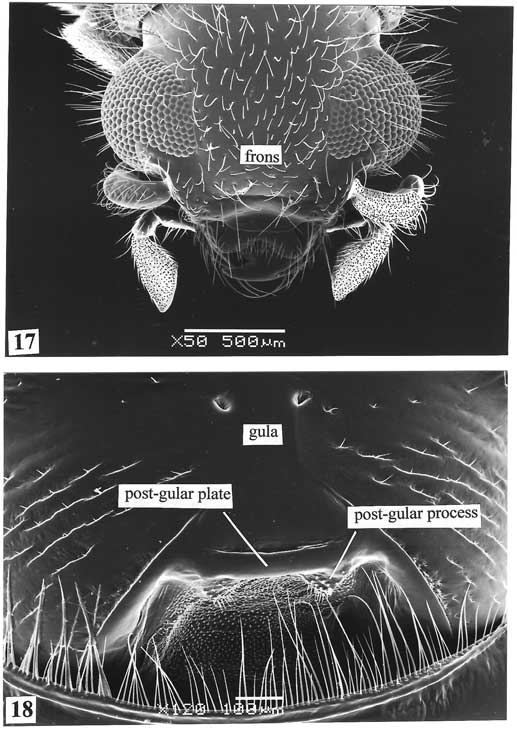
Mouthparts and antenna of Akonesis vittatus. 19. Mouthparts. 20. Antenna.
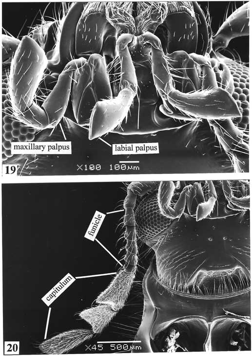
Prothorax of Akonesis vittatus. 21. Ventral view. 22. Pronotum.
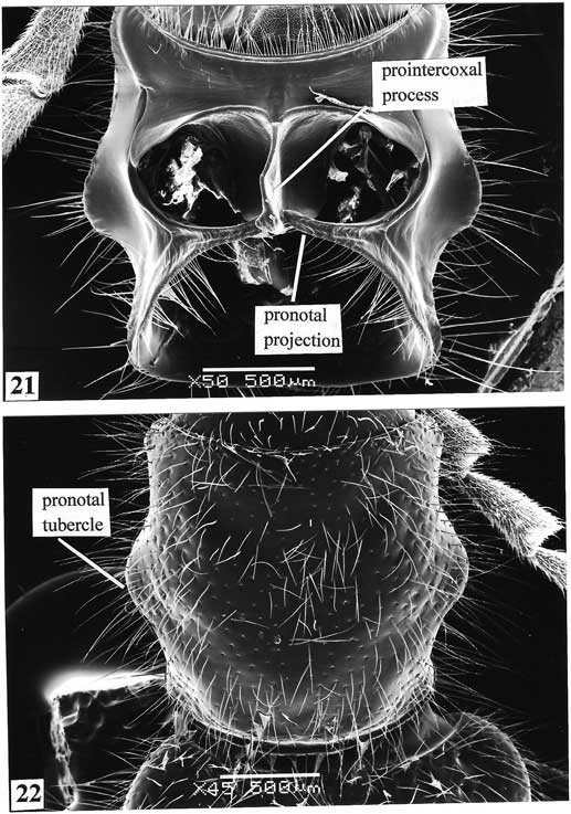
Ungues and elytral surface of Akonesis vittatus. 23. Ungues. 24. Elytral surface.
Distributions of Akonesis species as noted.
Original combination. Pelonium vittatum Spinola, Reference Spinola1844: 159.
Holotype. ♀. Brazil (MRSN). Corporaal Reference Corporaal1950: 284. (Cregya). Ekis Reference Ekis1975: 54 (Cregya). Opitz Reference Opitz2014b: 415.
Diagnosis. The sutural vitta on the elytral disc is slightly oblique and does not extend to the mesoscutellum (Figs. 7, 30). This characteristic will distinguish members of this species from congeners.
Redescription. Size : Length 10.0 mm, width 3.5 mm. Form : As in Figure 30. Colour : Mostly flavotestaceous, posterior margin of capitular antennomeres black; elytron mostly yellow, with epipleural and sutural vittae, sutural vitta is slightly oblique and does not reach mesoscutellum (Fig. 7). Head : Funicular antennomeres subfiliform, capitular antennomeres expanded (Figs. 5, 20); eye narrower than frons (eye width/frons width 20/25). Thorax : Pronotum (Fig. 13) quadrate (pronotal width/pronotal length 70/70), side margins each with well-developed tubercle, disc subglabrous, very finely punctate; elytra with very small asetiferous punctures (elytron length/elytron width 260/80). Abdomen : Male pygidium scutiform, posterior margin incised (Fig. 34); 6th visible sternite broadly concave distally; aedeagus as in Figure 34.
Natural history. Specimens were collected during August and December.
Variation. Size : Length 10.0–12.0 mm, width 3.5–4.0 mm. Other than body size, the available beetles are quite homogeneous.
Distribution. In addition to the lectotype, I examined eight specimens from (Fig. 25): Brazil : Estado do Bahia, Cachimbo, Ch Pujol, 1890; Estado do Rio de Janeiro, Guanabara, Foresta dos Macacos, 30.viii.1961, Moacir Alvarenga; Estado do Rio de Janeiro, Tijuca, E. Gounellem, xii,1884; Estado do São Paulo, Valle de Rio Pardo, E. Gounellem, xii.1898. Specimens are deposited in BMNH, FMNH, MNHN, and WOPC.
Notes. I examined the holotype during a visit to the Tassarolo Castle, in February 1974. This species description is based on a specimen compared with the holotype (Ekis Reference Ekis1975: 54).
Discussion
Morphological evidence from mouthparts, alimentary canal, and leg structure suggest that the species involved in this work are predatory. The mandibles are stoutly built, fit for grasping and crushing prey. The alimentary canal shows a well-developed proventriculus reminiscent of other predatory checkered beetles (Ekis Reference Ekis1977: 4). The leg morphology, with particular reference to the tarsal pads, is similar to that of other highly predaceous Cleridae such as members of the genus Enoclerus (Rifkind Reference Rifkind2016: 2). Also, colour patterns of the elytra and observations during field experience suggest that the members of Diutius and Akonesis are involved in a Batesian mimicry ring with Chrysomelidae (Coleoptera), and in particular with galerucine leaf beetles (Opitz Reference Opitz2014b: 412).
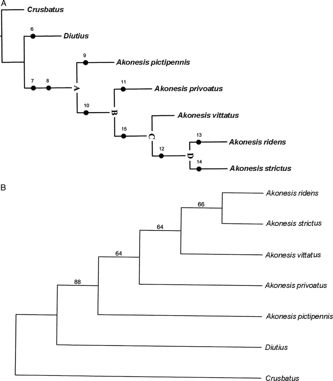
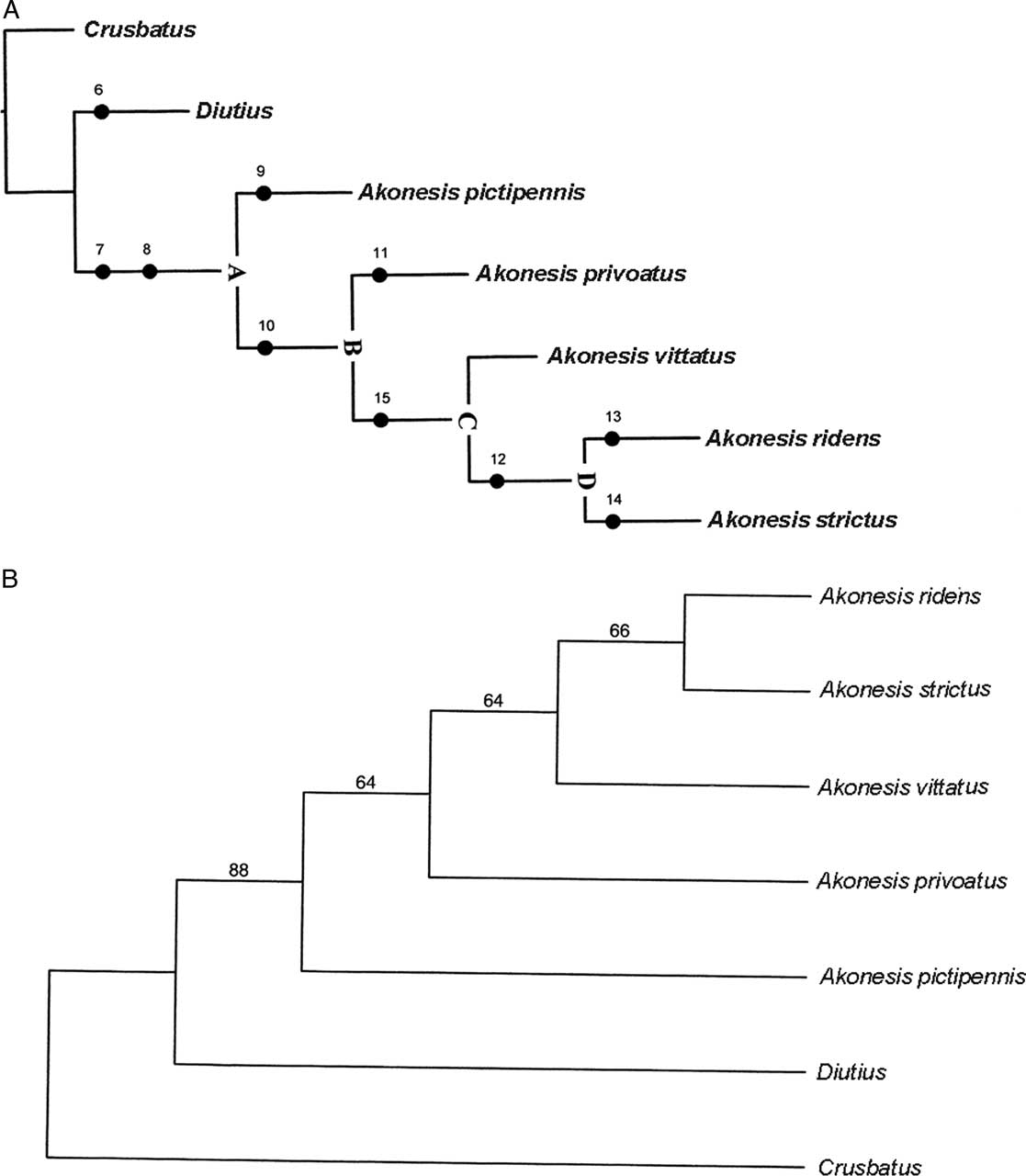
Opitz (Reference Opitz2014b: 427) presented the evolutionary relationships among the three genera treated in this paper. The additional knowledge that has been acquired about these genera supports the phylogenetic conclusions provided in that work. There is additional strong confirmatory evidence, ingrained in the phylogenetic tree (Fig. 26), to suggest that the genera Crusbatus, Diutius, and Akonesis form a monophyletic group within Peloniinae.
A, Proposed phylogeny of Crusbatus, Diutius, and Akonesis species. B, Bootstrap consensus tree of Crusbatus, Diutius, and Akonesis species.
Habitus of Akonesis species. 27. Akonesis pictipennis. 28. Akonesis privoatus. 29. Akonesis strictus. 30. Akonesis vittatus.
Male terminalia, aedeagus, and habitus of Akonesis species. 31. Terminalia and aedeagus of Akonesis privoatus. 32. Terminalia and aedeagus of Akonesis strictus. 33. Habitus of Akonesis ridens. 34. Terminalia and aedeagus of Akonesis vittatus. 35. Terminalia and aedeagus of Akonesis pictipennis.
Figures 25 presents the approximate South American geographic distributions of the Akonesis species. Species of Crusbatus and Diutius also are exclusively South American. It seems reasonable to conclude that the ancestral stock of these genera, and its eventual progression into three distinct evolutionary lines, occurred somewhere in the forested biomes of South America. For Akonesis species, this might have occurred in the “Brazilian Highlands” (sensu, Opitz Reference Opitz2005: 112), where most of the known extant species are found.
A hypothesis of phylogeny for the taxa included in this work is presented in Figures 26A–B. Based on an analysis of the plesiotypic characteristics within this lineage of genera, I postulate that the progenitor of Akonesis (Ancestor A, Fig. 26) was characterised as follows: eyes large and coarsely facetted, antennal capitulum expanded, pronotum subquadrate, pronotal tubercle well developed, legs fairly slender with tarsal pads well developed, elytral disc avittate, MP 3 vein is lost, pygidium large scutiform and incised on posterior margin, and phallobase without independent struts. Ancestor A diverged to produce Akonesis pictipennis, in which the 6th visible abdominal sternite became notably modified into a cup-like structure. This progenitor also produced ancestral species B, in which the elytral disc acquired a vittate character. Evolution proceeded from progenitor B to evolve A. privoatus, characterised by a short capitulum, which is considered herein an evolutionary reversal. Ancestor B diversified to produce progenitor C, which then led to A. vittatus and ancestral species D. In this ancestor, the epipleural and sutural vitta became confluent posteriorly, and it evolved A. ridens, characterised by showing an oblique elytral vitta, and A. strictus, in which the two elytral vittae became mostly parallel.
Acknowledgements
I am very grateful to the curators, listed in the “Repositories of specimens” section, for entrusting me with collections in their charge. A special thanks to Jaques Rifkind for his review of the manuscript. My thanks to Derek Sikes and James Hayden for making me aware of, and helping with, the bootstrap consensus programme. I also thank my wife Galena for technical assistance, Paul E. Skelley for departmental courtesies, and the Florida Department of Agriculture, for institutional affiliation and support.