A myocardial bridge is a congenital cardiac anomaly involving a band of overlying myocardial tissue that compresses a coronary artery during ventricular systole, potentially impairing coronary blood flow and precipitating sudden cardiac death.Reference Michael and Cheng-Han1 However, myocardial perfusion predominantly occurs during diastole. Myocardial bridges also appear to be quite common and are found incidentally at autopsy in patients with other medical conditions.Reference Corban, Hung and Eshtehardi2 While the exact prevalence of a myocardial bridge remains unknown in the paediatric population, its prevalence should mimic the adult population given that it is a congenital anomaly.Reference Erol3 In the paediatric population, myocardial bridges are most common in patients with hypertrophic cardiomyopathy (incidence ∼ 28%) and may be associated with adverse cardiac events.Reference Yetman, McCrindle, MacDonald, Freedom and Gow4
Recent advances in imaging have demonstrated the anatomy and dynamic behaviour of a myocardial bridge more precisely, findings that can influence management strategies. We describe a stepwise diagnostic/management approach to a previously healthy 12-year-old male who presented after resuscitated ventricular fibrillation cardiac arrest, in whom imaging demonstrated a myocardial bridge involving the mid-portion of the left anterior descending coronary artery. Ultimately, the myocardial bridge was determined to be incidental, not the cause for the patient’s cardiac arrest, and was not referred for surgical unroofing.
Case report
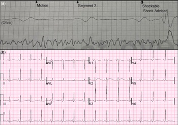
A 12-year-old previously healthy male with unremarkable personal and family medical history presented to the emergency department after being found unresponsive in his room after his parents heard a thud. Cardiopulmonary resuscitation was initiated within 2 minutes, and on arrival, emergency medical services recorded ventricular fibrillation and defibrillated his rhythm to sinus within 10 minutes of the event (Fig. 1). He was brought to the emergency department sedated and intubated, with a core temperature of 98.6 F, heart rate of 92 beats per minute, and blood pressure of 106/72 mmHg. Twelve-lead electrocardiogram demonstrated sinus tachycardia with non-specific ST-T wave abnormalities (Fig. 1), and transthoracic echocardiogram showed dyskinetic left ventricular wall motion and decreased systolic function (ejection fraction of 45%), and no evidence of hypertrophic cardiomyopathy. The coronary artery origins were not anomalous, had normal proximal courses, and were of normal calibre. Serum troponin was 0.387 ng/mL (normal < 0.03 ng/mL), normalising over the next 2 days; B-type natriuretic peptide was normal at presentation.
( a ) Automated External Defibrillator (AED) strip demonstrating ventricular fibrillation at the time of sudden cardiac arrest in a previously healthy 12-year-old male; ( b ) initial post-resuscitation 12-lead electrocardiogram demonstrating sinus tachycardia and non-specific ST-T wave abnormalities.

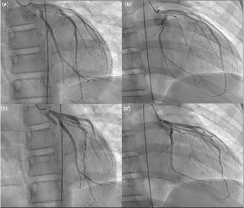
Initial cardiac catheterisation recorded normal haemodynamics, and coronary angiography showed dynamic obstruction in the mid-portion of the left anterior descending artery suggestive of a myocardial bridge (Fig. 2). Cardiac MRI showed no evidence of scar by late gadolinium enhancement. To further clarify the haemodynamic significance of the myocardial bridge, cardiac catheterisation was repeated using intravascular ultrasound and fractional flow reserve. Minimal coronary compression was noted by intravascular ultrasound at rest, with no significant enhancement during graded administration of IV dobutamine. At maximum dobutamine administration (40 mcg/kg/min), the diastolic fractional flow reserve of the left anterior descending was normal at 0.89 (values < 0.75 indicate impaired flow).Reference Pijls5 As we were not convinced this myocardial bridge would cause coronary insufficiency and thereby be responsible for this patient’s clinical presentation, surgical unroofing was not pursued and a transvenous dual-chamber implantable cardioverter-defibrillator was implanted. Post-operative course was uneventful, and he was discharged home on nadolol 40 mg twice daily. Mexiletine (150 mg three times/day) was added 2 weeks later because of symptomatic monomorphic non-sustained ventricular tachycardia at rest, suppressing future recurrences. Follow-up treadmill stress test on nadolol and mexiletine showed a blunted peak heart rate and no evidence of myocardial strain, ischaemia, or arrhythmias. A comprehensive inherited arrhythmia genetic panel ultimately reported a variant of unknown significance in MYH7 c.4283T>C (p. Leu1428Ser). The same variant was identified in the father and older brother, who have been asymptomatic and have had normal cardiac testing including imaging and electrocardiograms.
Selective left coronary artery angiogram in straight anterior posterior ( a , c ) and 30 degrees right anterior oblique ( b , d ). (a,b) A widely patent left coronary artery system with no obvious stenosis, aneurysm, or ectasia. (c,d) The same left coronary artery system in systole revealing discrete compression in the mid left anterior descending artery (*) due to the myocardial bridge.

Discussion
This report describes our experience with a previously healthy 12-year-old boy presenting after resuscitated cardiac arrest due to ventricular fibrillation, and initial evaluation found a myocardial bridge of the left anterior descending artery, raising concern for an acute coronary event. However, subsequent focused cardiac testing confirmed the bridge was unlikely to have caused coronary insufficiency and ischaemic arrhythmias, and the transient troponin elevation at presentation was attributable to systemic hypotension during the cardiac arrest.Reference Agusala, Khera, Cheeran, Mody, Reddy and Link6 An implantable cardioverter defibrillator was placed for secondary prevention, subsequent genetic testing revealed a MYH7 variant of unknown significance, and he has been clinically stable on nadolol/mexiletine for 10 months after hospital discharge.
The presentation of sudden cardiac arrest in a healthy child presents special diagnostic challenges, and management is directed at the likely underlying aetiology to prevent and/or treat possible future events. In our patient, a review of event chronology and personal and family medical history did not suggest an acquired or heritable cardiac channelopathy or cardiomyopathy. His physical exam and laboratory testing, serial 12 lead electrocardiograms, and echocardiogram also did not help to establish a diagnosis. Initial cardiac catheterisation with coronary angiography suggested a myocardial bridge involving the left anterior descending coronary artery. Genetic testing was requested, but the results were weeks away.
The finding of this bridge posed a clinical conundrum with respect to causation of this patient’s cardiac arrest. Whereas most myocardial bridges are usually considered benign, specific anatomical characteristics and the presence of hypertrophic cardiomyopathy influence the likelihood that it could cause symptoms/events.Reference Roberts, Charles and Ang7,Reference Konen, Goitein, Sternik, Eshet, Shemesh and Di Segni8 Myocardial bridges are quite prevalent, with 15–85% reported at autopsy, 0.5–16% by coronary angiography, and 3.5–38.5% by coronary cardiac tomographic angiography.Reference Michael and Cheng-Han1 A meta-analysis showed an average prevalence of 33%,Reference Roberts, Charles and Ang7 the highest prevalence reported in the autopsy series. A lower prevalence detected in the general population could reflect limitations of imaging modalities, or limited provider interest to closely look for them in asymptomatic patients.
There is limited information on myocardial bridges in children. Maeda et al. reported the largest series of children (n = 14) with structurally normal hearts with symptoms/events attributed to left anterior descending myocardial bridges.Reference Maeda, Schnittger and Murphy9 The most common symptom was chest pain, and three patients had syncope/sudden cardiac arrest. Transthoracic echocardiogram demonstrated septal dyskinesia with apical sparing and CT angiography confirmed the presence of a myocardial bridge. Intravascular ultrasound was utilised to document coronary compression at resting heart rates, and stress haemodynamic data revealed abnormal fractional flow reserve (0.59 ± 0.13). All patients had improved symptoms after surgical unroofing.Reference Maeda, Schnittger and Murphy9 In a meta-analysis of 115 studies, only 3 were paediatric, and these patients also had hypertrophic cardiomyopathy, a population where myocardial bridges appear to be common.Reference Michael and Cheng-Han1,Reference Roberts, Charles and Ang7 When a myocardial bridge is suspected to cause symptoms or events, coronary angiography with intravascular ultrasound and fractional flow reserve more precisely describes haemodynamic details, shedding light on the clinical relevance.Reference Ge, Jeremias and Rupp10 In adults, a fractional flow reserve < 0.75 during dobutamine stress is reported to represent clinically significant coronary compression due to myocardial bridges.Reference Pijls5 This approach in our patient confirmed the bridge was unlikely to cause coronary insufficiency and ischaemia, and surgical unroofing was therefore not indicated.
Having ruled out the myocardial bridge as the culprit, our patient seemed at risk for future adverse events, perhaps due to a yet elusive cardiac channelopathy/cardiomyopathy. Advances in genetic testing have improved the detection of diverse variants responsible for previously unexplained sudden cardiac arrest in patients; however, there remains no definitive diagnosis in 40–50% of cases.Reference Visser, van der Heijden, Doevendans, Loh, Wilde and Hassink11 Contemporary comprehensive genetic screening identified only a variant of unknown significance in the MYH7 gene, also found in two asymptomatic first-degree relatives. This missense variant has been observed in individual(s) with hypertrophic cardiomyopathy, is quite rare in population databases, and is expected to disrupt MYH7 protein function. Therefore, we were concerned our case may represent a so-called pre-structural phase of a cardiomyopathy, with the first manifestation being an arrhythmia.Reference Olivotto, Cecchi, Poggesi and Yacoub12 Given the likely predisposition to future events, an implantable cardioverter defibrillator was placed for secondary prevention, and future management will include continuing nadolol and mexiletine, non-invasive imaging at regular intervals to monitor for the development of cardiomyopathy, and potential adjustments to anti-arrhythmic therapy pending clinical course.
Conclusion
This case presents a case of sudden cardiac arrest in an otherwise healthy 12-year-old, in whom initial testing revealed a myocardial bridge involving the left anterior descending coronary artery. Close evaluation of the coronary anomaly confirmed this was most likely incidental, obviating referral for surgical unroofing. Genetic testing has been inconclusive to date, and so the current diagnosis is idiopathic ventricular fibrillation, likely due to either a cardiac channelopathy or a pre-structural phase of a cardiomyopathy. He has been clinically stable after treatment with nadolol/mexiletine and placement of an implantable cardioverter defibrillator.
Acknowledgements
None.
Financial support
This research received no specific grant from any funding agency, commercial or not-for-profit sectors.
Competing interests
None.
Ethical standard
The authors assert that all procedures contributing to this work comply with the ethical standards of the relevant national guidelines on human experimentation (please name) and with the Helsinki Declaration of 1975, as revised in 2008, and have been approved by the institutional committees (please name).