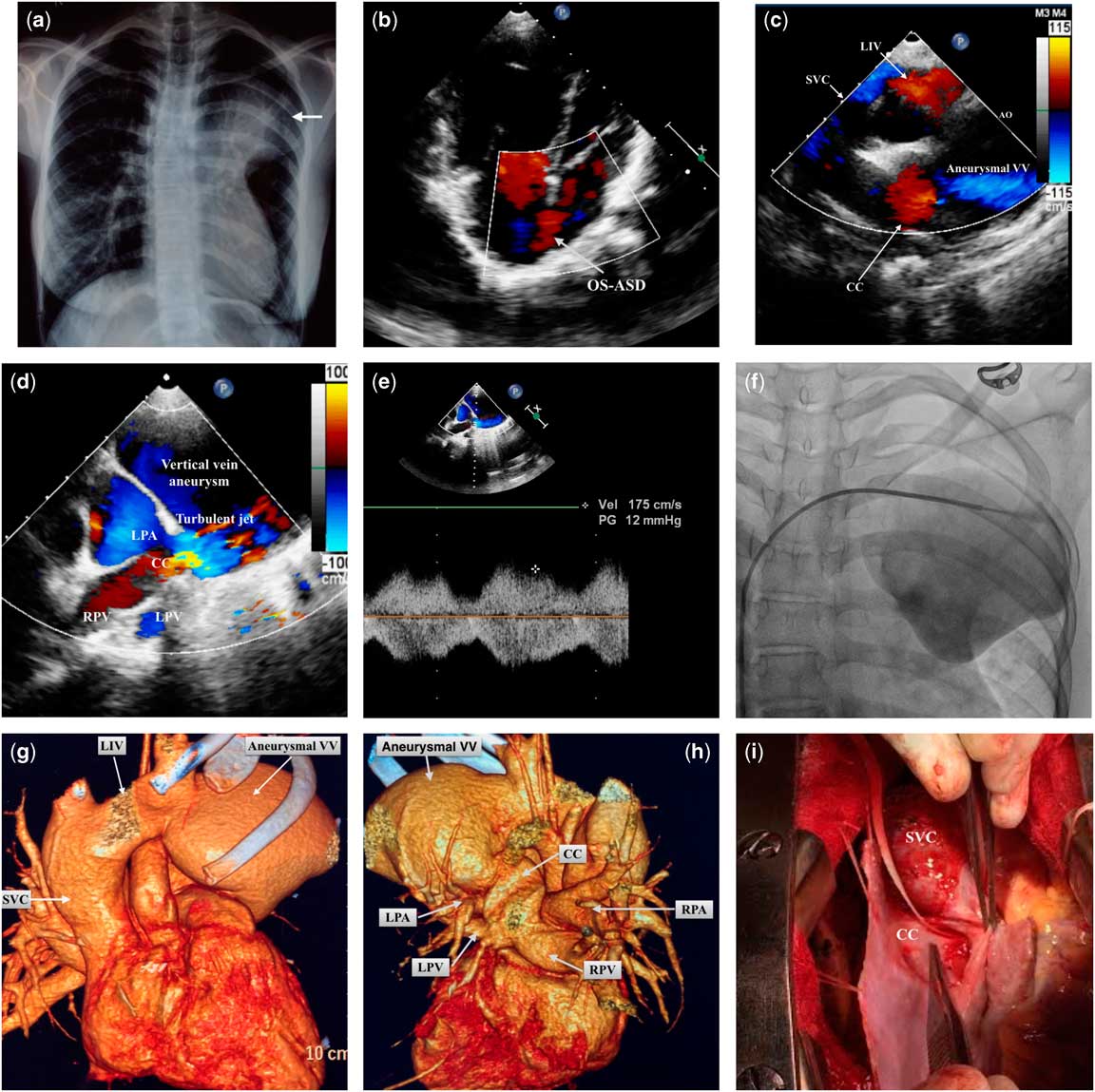
A 17-year-old female with a previously uneventful history presented with insidious onset dyspnoea on exertion. Examination revealed an oxygen saturation of 91%, with a loud pulmonary component of the second heart sound, and a Grade II/VI short ejection systolic murmur. Chest radiography (Fig 1a) showed mild cardiomegaly with a large mass in continuity with the left upper para-cardiac border. Echocardiography revealed enlarged right-sided chambers, a large ostium secundum-atrial septal defect with bidirectional shunting (Fig 1b, Supplementary video 1), moderate tricuspid regurgitation, and severe pulmonary hypertension, with pulmonary artery systolic pressure by tricuspid regurgitation jet of 60 mmHg. All four pulmonary veins were seen draining into a common chamber opening into an aneurysmally dilated left vertical vein (size 6.9×7.9 cm) (Fig 1c, Supplementary videos 2 and 3), which drained into the left innominate vein and then into the right superior caval vein. There was evidence of significant obstruction (mean gradient of 12 mmHg) of the vertical vein due to compression between the left pulmonary artery and the left bronchus (Figs 1d and e). These findings were confirmed on cardiac computed tomography (Figs 1g and h). Diagnostic cardiac catheterisation with selective angiography in the right pulmonary artery (Supplementary video 4), left pulmonary artery (Supplementary video 5), and the aneurysm (Supplementary video 6, Fig 1f) confirmed the findings. The systolic, diastolic, and mean pulmonary arterial pressures on catheterisation were 70, 34, and 46 mmHg, respectively. The mean pulmonary artery pressure was about half of the mean systemic pressure (83 mmHg). The pulmonary-to-systemic blood flow ratio was 3.7:1; pulmonary vascular resistance was 2.59 wood units. At surgery, an anastomosis was created between the common chamber and the left atrium with patch closure of the atrial septal defect (Fig 1i). The postoperative stay was uneventful and the patient was discharged on the sixth postoperative day. Postoperative echocardiogram showed a common chamber draining into the left atrium without any obstruction and reduced flow through the vertical vein. Pulmonary artery systolic pressure by tricuspid regurgitation jet was 38 mmHg.
( a ) Chest radiography in the postero-anterior view, demonstrating mild cardiomegaly with a large mass in continuity with the left upper para-cardiac border. ( b ) Two-dimensional echocardiogram, apical four-chamber view, showing a large ostium secundum-atrial septal defect (OS-ASD) with bidirectional shunting. ( c ) Two-dimensional echocardiogram, modified high parasternal short-axis view, demonstrating the pulmonary veins draining into a common chamber (CC) opening into an aneurysmally dilated left vertical vein, which drains into the left innominate vein (LIV) and then into the right superior caval vein (SVC). ( d and e ) Two-dimensional echocardiogram showing evidence of significant obstruction of the vertical vein due to compression between the left pulmonary artery (LPA) and the left bronchus. ( f ) Cineangiogram with a multipurpose catheter in the aneurysm of the vertical vein. ( g and h ) Cardiac computed tomography with three-dimensional reconstruction showing the pulmonary veins draining into a CC opening into an aneurysmally dilated left vertical vein, which drains into the LIV and then into the right superior caval vein. ( i ) Intra-operative image showing the common chamber behind the left atrium. LPV=left pulmonary vein; RPA=right pulmonary artery; RPV=right pulmonary vein; VV=vertical vein.

Discussion
Survival until the second decade is unusual in patients with unrepaired total anomalous pulmonary venous connection. We speculate that the major factors that contributed to the long-term survival in this patient could have been the supracardiac type, a large inter-atrial connection, and maintenance of pulmonary vascular resistance. The present case is remarkable for the presence of a large vertical vein aneurysm. The possible mechanism for formation of the aneurysm could be an extreme form of post-stenotic dilatation after severe obstruction to the vertical vein when it passes between the left pulmonary artery and the left bronchus – the so-called vascular vice.Reference Seo, Lee, Choi and Lee 1
Acknowledgement
None.
Financial Support
This research received no specific grant from any funding agency, commercial, or not-for-profit sectors.
Conflicts of Interest
None.
Supplementary Material
To view supplementary material for this article, please visit http://dx.doi.org/10.1017/S1047951116000111