Introduction
Cystic echinococcosis (CE), caused by infection with larval Echinococcus granulosus, has public health importance not only in areas of endemicity but also in countries or regions without endemicity, due to the migration of infected people (Schantz et al., Reference Schantz, Chai, Craig, Eckert, Jenkins, MacPherson, Thakur, Thompson and Lymbery1995; Craig et al., Reference Craig, Rogan and Allan1996) and livestock exchanges. The parasite is recognized as one of the world's relevant zoonoses, affecting both humans and their domestic animals (McManus & Smyth, Reference McManus and Smyth1986). Movement of infected livestock also raises the potential for transmission, creating new areas of endemicity. Immunodiagnosis of CE is commonly performed as a primary screening test with crude antigens and a confirmatory test with specific antigens (Lightowlers & Gottstein, Reference Lightowlers, Gottstein, Thompson and Lymbery1995). For preparation of such crude or purified antigens, most researchers collect hydatid cyst fluid (HCF) from naturally infected intermediate hosts, such as sheep, cattle, horses, camels, pigs or moose, or from CE patients (Ito et al., Reference Ito, Ma, Schantz, Gottstein, Liu, Chai, Abdelhafez, Altintas, Joshi, Lightowlers and Pawloski1999; Margutti et al., Reference Margutti, Ortona, Vaccari, Barca, Rigano, Teggi, Muhschlegel, Frosch and Siracusano1999; Irabuena et al., Reference Irabuena, Nieto, Ferreira, Battistoni and Ferragut2000). The nature and quality of the antigens are variable among the host species. This may be one of the reasons why different laboratories obtain different results for the detection of anti-CE antibodies with antigens prepared from different host species (Poretti et al., Reference Poretti, Felleisen, Grimm, Pfister, Teuscher, Zuercher, Reichen and Gottstein1999).
The outcome of infection in livestock is hydatid cyst development in the lung, liver or other organs (Jenkins et al., Reference Jenkins, Romig and Thompson2005). The disease is characterized by steadily growing, fluid-filled, unilocular cysts, surrounded by a two-layered hydatid cyst wall, in the host internal organs, mostly liver and lungs. Large hydatid cysts in the liver and lungs of sheep and cattle can result in significant economic loss to the meat industry through condemnation of the infected organs (Lightowlers et al., Reference Lightowlers, Rickard, Honey, Obendorf and Mitchell1984). The infection data of hydatidosis in livestock are most commonly collected at slaughterhouses. If those animals destined for slaughter did not necessarily represent the population, the reliability of such data would be hampered. Abattoirs which are strictly regulated can often provide acceptable prevalence data. However, in many areas where the disease is endemic, home slaughter is practised and few abattoirs have adequate veterinary supervision. In addition, post-mortem diagnosis of the disease is of little use in areas of low prevalence, where segregation of infected animals is essential for disease control (Moro et al., Reference Moro, Verastagui, Gilman, Falcon, Bernal, Gavidia, Gonzalez, Malqui, Moro and Dueger1997).
In Ethiopia, the status of hydatidosis in animals is not well documented and explored. According to Jobre et al. (Reference Jobre, Labag, Tirone, Abebe and Dorchies1996) the prevalence rates of hydatidosis were 46.5%, 25.7% and 24.3% in cattle in abattoir-based surveys conducted in Debre Zeit, South Omo and Gondar, respectively. The total annual economic losses from organ condemnation and carcass weight loss due to ruminant hydatidosis at Adama, Hawassa municipal abattoir and Tigray were estimated at 52,828 Ethiopian Birr (US$4085.7) (US$1 = 12.93 Ethiopian Birr (ETB)), 1,791,625.89 ETB and 25,608 ETB, respectively. In another abattoir-based study, the prevalence of hydatidosis in cattle was 52.69%, 16.0%, 32.1% and 34.05% in Hawassa, Wolaita Sodo, Mekelle and Bahir Dar, respectively (Minas et al., Reference Minas, Biluts, Bekele and Alemie2007; Berhe, Reference Berhe2008; Kebede et al., Reference Kebede, Mekonnen, Wossene and Tilahun2008, Reference Kebede, Hagos, Girma and Lobago2009; Getaw et al., Reference Getaw, Beyene, Ayana, Megersa and Abunna2010; Ragassa et al., 2010).
Echinococcus granulosus is considered the major cause of human hydatid cysts, especially in rural communities of Ethiopia. Significant cases of hydatidosis in humans have also been identified in research conducted using serological survey and hospital-based scrutiny (Ali et al., Reference Ali, Biluts and Gulilat2005). In a study conducted using clinical examinations, a filter-paper indirect haemagglutination test (IHA) and a hydatid skin test, a hyperendemic focus of hydatid disease (E. granulosus) was found in the Dassanetch and Nyangatom tribes of southwestern Ethiopia (Fuller & Fuller, Reference Fuller and Fuller1981). Moreover, patients with hydatid cyst of the ovary have been reported from Ethiopia (Gaym et al., Reference Gaym, Abebe and Degefe2002).
A variety of serological tests has been carried out to diagnose the disease in domestic animals (Blundell-Hasell, Reference Blundell-Hasell1969). However, few serological tests have been applied in practical diagnosis, because most of the tests gave rise to false-negative and false-positive reactions. These false reactions are thought to result from the poor productivity of anti-E. granulosus antibody in naturally infected animals (Hamel & Ris, Reference Hamel and Ris1982). Serological tests using crude antigens for diagnosis of E. granulosus are sensitive; however, their specificity is not satisfactory (Sadjjadi et al., Reference Sadjjadi, Abidi, Sarkari, Izadpanah and Kazemian1986). Currently, enzyme-linked immunosorbent assays (ELISA), IHA, indirect immunoelectrophoresis (IEP), counter immunoelectrophoresis (CIEP) and double diffusion (DD) are used in the serodiagnosis of hydatidosis, but they have some disadvantages, such as cross-reactions with some Taeniid cestodes (Sbihi et al., Reference Sbihi, Rmigui, Maria, Orduna, Rodrigues-Torres and Osuna2001). Each, to varying degrees, has been shown to give false results, with considerable variation between laboratories (Sbihi et al., Reference Sbihi, Rmigui, Maria, Orduna, Rodrigues-Torres and Osuna2001; Nasrieh & Abdel-Hafez, Reference Nasrieh and Abdel-Hafez2004). There are several conflicting reports on the suitability of ELISA for the diagnosis of hydatidosis (Farag et al., Reference Farag, Bout and Capron1975; Iacona et al., Reference Iacona, Pini and Vicari1980), some researchers showed that it may be adapted successfully for the serological diagnosis of hydatidosis. The sensitivity and accuracy of ELISA depend on the composition, concentration and stability of the antigen used.
Early diagnosis of human hydatid disease by detecting the specific antibodies in patients' sera is considered an important step in the treatment of infection. But the diagnostic efficiencies of assays depend greatly on the characteristics of the antigen and the various conditions used. The development of a sensitive, specific and reproducible serological assay for livestock would provide useful epidemiological data for the antemortem study and control of hydatid disease (Moro et al., Reference Moro, Verastagui, Gilman, Falcon, Bernal, Gavidia, Gonzalez, Malqui, Moro and Dueger1997). As HCF contains many antigenic components, it seems to be a superior source of diagnostic antigen. This study was designed to evaluate the diagnostic ability of ELISA and IHA using hydatid cyst fluid antigens.
Materials and methods
Study site and animals
The study was conducted at the Addis Ababa municipality abattoir from March to June 2007. About 800 cattle, 300 sheep and 85 goats are slaughtered per day. Even though it was difficult to trace back precisely the geographical origins of all animals slaughtered and relate the findings to a particular locality, sources are usually claimed to be adjacent regions (Oromia, Amhara, and South Nations and Nationalities).
Post-mortem examination
A total of 502 cattle were examined for hydatid cyst infection. Thorough meat inspection was carried out on the different organs of each of the slaughtered animals, particularly lung, liver, spleen, kidney, heart and the muscles. Each organ was examined visually and manually (palpation and incision) and, where necessary, one or more incisions were made in order to detect small hydatid cysts. The infected organs from each positive animal were collected and recorded for hydatid cyst characterization. The total numbers of hydatid cysts were counted and recorded per infected organ. Of the collected hydatid cysts, each individual cyst was carefully incised and examined for protoscoleces, which look like white dots on the germinal epithelium; such cysts were characterized as fertile cysts. Fertile cysts were subjected to a viability test. A drop of the sediment containing the protoscoleces was placed on a microscope glass slide, covered with a cover slip and observed with a × 40 objective lens for amoeboid-like peristaltic movements. Furthermore, infertile cysts were further classified as sterile or calcified. Sterile hydatid cysts were characterized by their smooth inner lining, usually with slightly turbid fluid content. Typical calcified cysts produce a gritty sound/feeling on incision (Soulsby, Reference Soulsby1982).
Collection of serum samples
Blood samples (10 ml) were obtained from the jugular vein at the time of slaughter and collected in disposable plastic tubes which were marked with the animal's identification number. Serum was obtained by centrifugation, refrigerated at 4°C and stored in 1 ml aliquots at 20°C until subsequent use. Each serum sample was numbered and labelled as positive or negative for CE according to the post-mortem examination result. Sera from CE-infected cattle (173) were carefully registered as hydatidosis positive, while those from non-CE-infected cattle (329) were identified as hydatidosis negative.
Preparation of hydatid cyst fluid antigens (HCFAgs)
Hydatid cyst antigens were prepared from hydatid cyst fluid collected from E. granulosus cysts in the livers and lungs of sheep slaughtered at the Addis Ababa abattoir by the method described by Moose & Abdel-Hafez (Reference Moose and Abdel-Hafez1994). Briefly, hydatid-infected livers and lungs of naturally infected sheep were collected in a plastic bag and transported to the Microbiology, Immunology and Parasitology Department of the Medical Faculty, Addis Ababa University. After disinfecting the cyst surfaces, hydatid cyst fluid was drawn aseptically from the cysts and centrifuged at 1000 g for 30 min at 4°C to sediment protoscoleces (PSCs), brood capsules and membranes. Only fertile hydatid cysts were selected for the study. The fluid from several fertile cysts was pooled. The supernatant was collected as crude HCF antigen and stored at − 70°C in 1 ml aliquots after determination of protein content by Lowry's method (Lowry et al., Reference Lowry, Rosebrough, Farr and Randall1951) using bovine serum albumin (BSA) as a reference standard.
Enzyme linked immunosorbent assay
Conditions for ELISA were optimized by performing checkerboard titrations. ELISA standardization included determination of the optimal working dilution (and concentration) for the antigen, sera and anti-species immunoglobulins or conjugates using checkerboard titration from serial dilutions with known positive and negative serum samples (Voller et al., Reference Voller, Bidwell and Bartlett1976). Dilution of the conjugate and antigen concentration at which positive and negative controls yielded the maximum optical density (OD) value were chosen for the subsequent testing of the samples. The reference serum samples used in this standardization were from animals confirmed to have heavy infection with CE as assessed at post-mortem inspection and identified to have high IHA titre. In addition, negative control serum samples, essentially parasite free, were purchased from a supplier (New Zealand, lot numbers 5936716D, 3095883D). An ELISA cut-off value for the antigen was based on the mean OD values of all serum samples from the negative control plus 2 standard deviations above the mean. The specificity and sensitivity of ELISA were calculated using this positive–negative cut-off value. Data above this threshold were considered positive and those below as negative. The optimized ELISA procedures were as follows.
Test procedures
ELISA was performed essentially as described by Hira et al. (Reference Hira, Bahr, Shweiki and Behbehani1990) with slight modification. Briefly, high-binding polystyrene microtitration plates were coated by adding 100 μl/well of crude hydatid antigens prepared in carbonate–bicarbonate buffer, pH 9.6 at a concentration of 5 μg/ml and kept overnight at 4°C. After thorough washing (phosphate-buffered saline (PBS) containing 0.05% Tween-20), non-specific binding sites were blocked by addition of 100 μl of 1% BSA to each well. After 2 h incubation at 37°C the plates were washed four times to remove the blocking solutions. After addition of 100 μl of the tested sera diluted at 1:200 in PBS, the plates were incubated for 2 h at room temperature (RT). The serum samples were removed and plates were washed as described above at least four times and 100 μl of conjugate – peroxidase-labelled antibodies against bovine IgG (anti-bovine IgG peroxidase conjugate, Sigma Chemicals, St. Louis, Missouri, USA) – were dispensed to each well. The optimum dilution of the conjugate was found to be 1:3000, as determined by checkerboard titration. After 2 h incubation at RT and further washing to remove unbound conjugate, the amount of enzyme bound was assayed by adding 100 μl/well of o-phenylene diamine (OPD) (Sigma) containing hydrogen peroxide (20% H2O2) in citrate/phosphate buffer as substrate. Conveniently, results were obtained after 30 min incubation at RT. ODs were recorded using an automatic ELISA reader.
From the absorbance values of the 15 serum samples from a non-infected newborn calf, means and standard deviation (SD) were calculated. OD cut-off was calculated by addition of 2SD to mean of OD values of the 15 negative sera. Values above this cut-off were recorded as positive and below and/or equal as negative. Absorbance values were read at 450 nm using an ELISA reader. Positive and negative serum controls were included in each plate. Samples were tested in duplicate and the experiments were repeated twice.
Indirect haemagglutination test
Formalinization
Fifty millilitres of fresh sheep blood were collected aseptically by bleeding into a flask containing equal volume of Alsever's solution, prepared by modification of the method described by Bakantz et al. (Reference Bakantz, Rein and Rent1946). The fresh red cells in Alsever's solution were washed five times with PBS (pH 7.2) and packed. Twenty-five millilitres of the packed cells were suspended with 200 ml PBS and placed in a 500 ml conical flask. About 50 ml of 40% commercial formalin was introduced into a length of dialysis tubing. The tube was tied off so that it became air-free and only two-thirds full. This tube was then submerged into the red cell suspension in the flask. The flask was then placed into a shaker and gently agitated at 1000 rpm at room temperature. After 3 h of shaking, the swollen dialysis sac was punctured and removed so as to allow the formalin to escape out of the tube. Gentle shaking was then continued overnight. The dark-brown suspension produced was filtered through gauze and washed five times (PBS, pH 7.2) to remove the formalin and kept at 4°C until use.
Sensitization and coating
One hundred microlitres of 25% glutaraldehyde were mixed with 0.2 ml of 50% packed sheep red blood cells in 5 ml PBS (pH 7.2) and incubated at 37°C in a water bath for 20 min. After centrifugation at 1800 rpm for 5 min at 4°C to remove the supernatant, the sheep red blood cells (SRBCs) were re-suspended in 5 ml PBS (pH 7.2). Coating involved gentle mixing of 250 μl HCF antigen at a concentration of 0.1 mg/ml to 0.2 ml 50% sensitized SRBCs in 4 ml PBS (pH 6.4) and incubation at 37°C in a water bath for 30 min after shaking twice. The mixture was centrifuged and washed three times with PBS (pH 7.2) containing 1% fetal calf serum (FCS) and eventually suspended in 4 ml FCS. This gave a 2.5% suspension ready for use.
Test procedures
Fifty microlitres PBS (pH 7.2) were distributed in a v-shaped microplate, apart from the second column. Tested sera, including positive and negative controls, were diluted in a twofold dilution series in PBS (1:8, 1:16, 1:32 … 1:2048). Of each diluted serum sample 50 μl were added to microplate wells. To the second column, 100 μl of test sera diluted at 1:8 in PBS (pH 7.2) were added. Serial twofold dilution was made by transferring 50 μl serum to the next wells.
Following these steps, 50 μl of 0.2% sensitized and coated SRBCs were added to the respective wells and kept on the bench undisturbed. After incubation from 2 h to overnight at room temperature, the total antibody titre in the test serum was recorded as one dilution before that which yielded a clear, sharp, dark spot similar to those in the negative control wells. An IHA titre less than or equal to 32 was considered negative as determined by several pooled negative serum samples. Moreover, the IHA results were also evaluated with different sets of cut-off titres (1:16, 1:32, 1:64).
Data analysis
The data were analysed using SPSS version 16 (SPSS Inc., Chicago. Illinois, USA) and a significance value determined with 95% confidence interval. The sensitivity, specificity, diagnostic efficacy and predictive value of ELISA and IHA were computed to the result of the presumed ‘gold standard’, post-mortem findings. Agreements of serological tests and post-mortem examination were determined.
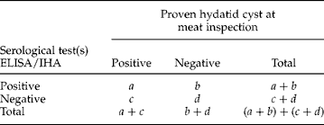
The statistical method used for assessment of the validity of serological test(s) for the diagnosis of hydatidosis is as follows.
Diagnostic sensitivity:

Diagnostic specificity:
Positive predictive value:
Negative predictive value:
Diagnostic efficacy:




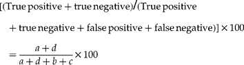
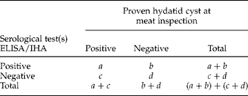
Assuming a = true positive; b = false positive; c = false negative; and d = true negative:
Results
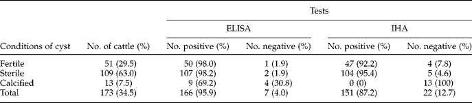
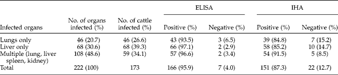
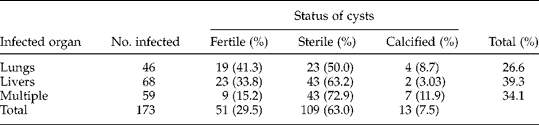
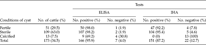
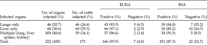
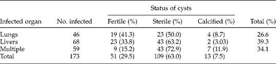
Of 502 cattle examined, hydatid cysts were seen in 34.5% (95% confidence interval (CI) 30.0–38.8). All cattle examined were more than 4 years of age (data not shown). Among 222 cysts counted, 63.0% were infertile and conditions of cysts are depicted in table 1. There was a significant association between cyst fertility and positivity rates of ELISA (χ2 = 25.9; P < 0.001) and IHA (χ2 = 96.8; P < 0.001). Single infection to multiple organ involvement was identified. The distributions of hydatid cysts with respect to organs are described in table 2. Though statistically not significant, there was a slight difference in cyst location and positivity rates seen in ELISA (χ2 = 1.005; P = 0.605) and IHA (χ2 = 1.5; P = 0.482). Of 222 hydatid cysts counted, 114 (51.4%) were found in a single organ and the remaining 108 (48.6%) had multiple organ involvement (table 2). Most fertile cysts were found in the liver and rates of cyst fertility with respect to organ sites are shown in table 3.
Conditions of cysts and their reactivity towards ELISA and IHA in cattle slaughtered in the Addis Ababa abattoir.
Distribution of hydatid cysts by organs in cattle slaughtered in the Addis Ababa abattoir.
Fertility rates of the hydatid cysts with respect to organ sites.
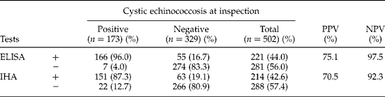
In general, 4.0% (7/173) of hydatidosis cases diagnosed in the slaughterhouse were considered non-hydatid by ELISA. On the other hand, 16.7% (55/309) of those diagnosed as healthy were found to be infected with hydatidosis. In IHA, 12.7% (22/173) of hydatidosis cases diagnosed in the slaughterhouse were considered non-hydatid and 19.1% (63/309) of those diagnosed as healthy were found to be infected with hydatidosis. Positive and negative predictive values of ELISA were 75.1 and 97.5%, respectively. ELISA had better diagnostic performance than IHA and the sensitivity, specificity, positive and negative predictive values of both tests are shown in table 4.
Diagnostic performances of ELISA and IHA in comparison with meat inspection findings.
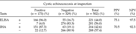
NPV, negative predictive value; PPV, positive predictive value.
The diagnostic efficacy of ELISA was 87.6% and its specificity was 83.3% (CI 78.8–87.2). ELISA false negative and positive values were 4.0 and 16.7%, respectively (table 4). ELISA had a sensitivity of 96.0% (CI 91.8–98.4). The sensitivity and specificity of ELISA and IHA were statistically significant (χ2 = 504.1; P < 0.001). Sensitivity of IHA was 87.3% (CI 81.4–91.9) at cut-off point ≥ 1:64 as determined by pooled negative sera. Positive and negative predictive values for IHA were 70.5 and 92.3%, respectively. IHA false negative and positive values were 12.7 and 19.1%, respectively. Specificity of IHA was 80.9% (CI 76.2–85.0) (table 4).
Antibody titre of sera from 173 CE-infected cattle ranged from as low as ≥ 1:16 (4.6%) to as high as ≤ 1:512 (3.5%). The antibody titre of sera from 329 non-CE-infected cattle is also displayed in table 4. Diagnostic efficacy of IHA was 83.6%. The spectra of antibody titre of CE-infected and non-infected cattle are shown in table 5. All the negative control sera (15) from a newborn calf, otherwise proven echinococcosis free, tested negative (100%) (data not shown). Cattle with fertile cysts were more reactive to ELISA and IHA than those with infertile and calcified cysts, and performance of both tests in relation to conditions of the cyst, is displayed in table 1.
Spectrum of antibody titres of sera from cattle with cystic echinococcosis (CE) and non-CE in an indirect hemagglutination test (IHA).

Discussion
In Ethiopia, hydatidosis in livestock is endemic, with the magnitude of records ranging from low, medium to high rates of occurrence. The distribution of hydatid disease in animals and humans is affected by a variety of factors. The highest incidence rates in human are often seen in areas where there is a close association between humans and domestic livestock, often using dogs as working dogs and feeding them on viscera of home-butchered livestock (Moro et al., Reference Moro, Verastagui, Gilman, Falcon, Bernal, Gavidia, Gonzalez, Malqui, Moro and Dueger1997). In Ethiopia, backyard slaughtering of ruminants is not very uncommon and, especially during holidays, most cattle and almost all small ruminants are slaughtered in backyards and roadsides. Thus, an artificial system (deliberate feeding of infected livestock or wildlife offal to dogs) is more efficient than the natural system in perpetuating the life cycle of hydatidosis in Ethiopia. Similar experiences seen in most parts of the country are considered to encourage the maintenance and further propagation of hydatid disease.
Hydatidosis in humans is also common in Ethiopia, as can be revealed in certain hospital-based surveys. In a study conducted from 1995 to 2005, 234 patients were admitted to Tikur Anbessa hospital for surgical management (Minas et al., Reference Minas, Biluts, Bekele and Alemie2007). In another study done from 1993 to 2003 in the same hospital, 72 patients were diagnosed to have thoracic hydatid disease and treated surgically (Ali et al., Reference Ali, Biluts and Gulilat2005). Based on clinical examinations, filter-paper IHA test and hydatid skin test, a hyperendemic focus of hydatid disease was found in southwestern Ethiopia. Out of 1342 individuals from the Dassanetch and Nyangatom tribes in the lower Omo River Valley, 4.8% had palpable abdominal cysts and 15% had hepatomegaly (Fuller & Fuller, Reference Fuller and Fuller1981).
Prevalence data on hydatidosis in livestock is usually collected passively during meat inspection in abattoir and/or slaughterhouse. Abattoir-based studies usually suffer from imperfect diagnostic features. As a result, it has proved difficult to establish accurate prevalence data of hydatidosis in Ethiopia. In areas far from town centres, where there are no butcheries or slaughter slabs, and where home slaughter is usually carried out, information on the status of the disease could not be obtained (Baldock et al., Reference Baldock, Arthur and Lawrence1985; Njoroge et al., Reference Njoroge, Mbithi, Gathumaa, Wachira, Gathura, Magamboc and Zeyhle2002). Thus, an alternative approach to studying hydatidosis prevalence in livestock is needed, given the high public health significance and economic consequences of hydatidosis, both in animals and humans in Ethiopia. In order to evaluate the specificity and sensitivity of the immunological diagnosis in both naturally and experimentally infected animals, panels of serum samples have been classified as negative or positive according to macroscopic analysis at necropsy (Kittelberger et al., Reference Kittelberger, Reichel, Jenner, Heath, Lightowlers, Moro, Ibrahem, Craig and O'Keefe2002). As little research has been done, either at national or international level, evaluation of serological test applicability and subsequent comparison of results in cattle remain imperfect. In this study, therefore, the applicability of serological tests in cattle was appraised.
In this study the prevalence of hydatidosis in cattle was 34.5%. This is lower than the prevalence values obtained from Hawassa (52.69%) and higher than values from Wolaita Sodo (16.0%), Mekelle (32.1%) and Bahir Dar (35.05%) (Minas et al., Reference Minas, Biluts, Bekele and Alemie2007; Berhe, Reference Berhe2008; Kebede et al., Reference Kebede, Mekonnen, Wossene and Tilahun2008, Reference Kebede, Hagos, Girma and Lobago2009; Getaw et al., Reference Getaw, Beyene, Ayana, Megersa and Abunna2010; Ragassa et al., 2010). Comparison of serological test results is hardly possible in animals due to the fact that no such studies have been done before in Ethiopia. In this study the sensitivity, specificity, positive and negative predictive values of ELISA were 96.0, 83.3, 75.1 and 97.5%, respectively. This is in agreement with Larrieu et al. (Reference Larrieu, Costa, Cantoni, Alvarez, Cavagion, Labanchi, Bigatti, Raya, Herrero, Mancini and Cabrera2001) where ELISA sensitivity, specificity, positive and negative predictive values using cyst fluid antigens were 89.2, 89.5, 67.9 and 97.7%, respectively, in ovine echinococcosis. Ibrahem et al. (Reference Ibrahem, Craig and Mcvie1996) attained sensitivity and specificity of 36 and 93%, respectively, using antigen from sheep hydatid liquid and 71 and 96% using antigens from camel hydatid liquid in ELISA. These are lower sensitivity and specificity values than the present findings. According to Khan et al. (Reference Khan, Afzal and Ali1990) sensitivity, specificity and efficiency of IHA and ELISA were low and hydatidosis was seen in 38.90% of cattle, 33.06% of buffaloes and 58.9% of camels slaughtered at a local abattoir. The diagnostic sensitivity and specificity of serological tests may also be related to technical factors, such as the source and quality of cyst fluid antigen purification, and laboratory techniques being used. Those animals which were negative at necropsy but showed serology results above the cut-off level (possibly false positive) need to be tested further using a more sensitive and specific assay so as reach a certain conclusion regarding their infection status.
In this study, IHA predictive values of positive and negative were 70.5 and 92.3%, respectively. The sensitivity and specificity of IHA were 87.3 and 80.9%, respectively. This shows a better diagnostic performance than similar tests performed elsewhere using the same antigen source but on different species of animals. An IHA using crude hydatid cyst fluid antigen was reported to have 72% sensitivity for camel CE in Nigeria (Dada et al., Reference Dada, Adegboys and Mohammed1981), and Ibrahem et al. (Reference Ibrahem, Rafiei, Dar, Azwai, Carter and Craig2002) obtained 43% sensitivity and 69% specificity with IHA for CE in Sudanese camels. A much lower sensitivity (27%) with IHA for sera from camel CE was reported from Egypt (Ibrahem et al., Reference Ibrahem, Craig and Mcvie1996). Serological tests for ovine CE, for instance, showed a range of specificities and sensitivities (40–90%) using sera from sheep experimentally or naturally infected with CE (Craig, Reference Craig, Andersen, Ouhelli and Kachani1997). Most cattle with calcified hydatid cysts were identified as being less reactive to both ELISA and IHA than cattle with fertile cysts.
The likely explanations for those animals negative at necropsy but positive at serology could be multiple. A positive antibody response was seen in three sheep infected experimentally with a low infestation (ten eggs) of E. granulosus but hydatid cysts could not be found at necropsy. Serological conversion 10–14 days after inoculation confirms that animals with recent infections would be classified as positive in serological tests and as negative at necropsy (Yong et al., Reference Yong, Health and Knapen1984).
In a study conducted in the Province of Rio Negro, 37.2% of false-positive diagnoses identified at necropsy were caused by unspecific granulomas, pseudo-tuberculosis, emphysema and fatty degeneration, while 1.1% of false-negative diagnoses were due to small intra-parenchymal cysts (Larrieu et al., Reference Larrieu, Costa, Cantoni, Alvarez, Cavagion, Labanchi, Bigatti, Raya, Herrero, Mancini and Cabrera2001). This information coincides with research carried out in Uruguay (Cabrera et al., Reference Cabrera, Haran, Benavidez, Valledor, Perera, Lloyd, Gemmell, Baraibar, Morana, Maissonave and Carballo1996), in which 26.1% of the sheep that were classified as positive at necropsy were not identified as having hydatidosis at histology, but caseous lymphadenitis, larval stage of Taenia hydatigena, white spot or abscesses were found. Small intra-parenchymal cysts, especially in the lungs, can be unnoticeable at sanitary inspection. As a result, these cases are falsely classified as negative at necropsy.
In this study ELISA showed 4.0% ‘false-negative’ and 16.7% ‘false-positive’ values, while the corresponding rates in IHA were 12.7 and 19.1%, respectively. With respect to the cross-reaction, we assumed that the so-called negative sera used had contained antibodies to hydatid cyst and/or to antigenically cross-reacting parasites such as Taenia spp. and Fasciola, since we carried out only macroscopic examination in detection of infection with hydatid cysts. Those animals may also have been at early stages of infection with these parasites so that necropsy failed to diagnose the infection.
The presence of other parasites was not systematically registered and computed for this purpose, and this could be considered as the limitation of this study. Moreover, Gatti et al. (Reference Gatti, Rosa Alvarez, Araya, Mancini, Herrero, Santillan and Larrieu2007) identified intra-parenchymal oncospheres through histology, and cysts of recent development which were considered negative at macroscopic inspection (false-negative at necropsy). This confirms the limitations of macroscopic diagnosis at necropsy and the validity of the histological test as a reference test for the diagnosis of cystic echinococcosis. In this study, the false-positive results may also have arisen from similar reasons of necropsy shortcomings mentioned above, even though the status of cattle identified as false positive and negative were not systematically registered for analysis.
Echinococcosis is rife in rural communities of Ethiopia especially in cattle/sheep-raising regions. The economic repercussions seen in the meat industry as a result of this cestode parasite are significant for poor countries like Ethiopia. As a result, the determination of hydatidosis prevalence in livestock and initiation of control measures are needed. This demands the development and application of serological tests under field conditions which would, in turn, pave the way for subsequent instigation of a control strategy to reduce the economic and public health consequences of hydatidosis. Information concerning the serological diagnosis of hydatid disease in animals is either scanty or non-existent. Locally prepared crude hydatid cyst fluid antigens have shown a promising result in detecting anti-echinococcus antibodies in cattle. ELISA and IHA are proved adequate for the detection of the presence of infected cattle on a flock basis, for surveillance and evaluating control programmes.
Acknowledgements
The authors would like to thank Addis Ababa University School of Graduate Studies and Microbiology, Immunology and Parasitology Department of the Medical Faculty for funding this project.