Introduction
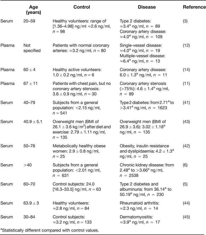
Angiopoietin-like 2 (angptl2) belongs to the angiopoietin-like family, a family of eight (angptl1-8) members of glycoproteins. Their structure is similar to that of angiopoietins, they possess a typical N-terminal helical coiled-coil domain, a short linker peptide and a C-terminal globular fibrinogen-like domain (Ref. Reference Kim1), except for angptl8, which does not have a fibrinogen-like domain (Ref. Reference Quagliarini2). Angptl2 was cloned, expressed and characterised for the first time in 1999 (Ref. Reference Kim1); it is a glycosylated protein of 493 amino acids, of 57 kDa (64 kDa with the glycosylations) (Ref. Reference Kim1) and a secreted circulating protein (Refs Reference Kim1, Reference Tabata3, Reference Kadomatsu, Tabata and Oike4). Circulating levels of angptl2 in human healthy volunteers range from ≈1 to 3 ng/ml (with the exception of (Ref. Reference Li5)) (Table 1), with similar levels between males and females (Refs Reference Tabata3, Reference Usui6). Overall, the quantitative differences in circulating levels of angptl2 in human appear to be rather small (Table 1). Very recently, it has been reported that angptl2 could be cleaved into domain fragments by the tolloid-like 1 protease and be inactivated, at least in vitro (Ref. Reference Odagiri7). Very few studies describe the specific roles of the N-terminal coiled-coil domain and the C-terminal fibrinogen-like domain of angptl2. While the helical domain is supposed to form dimeric or trimeric coiled-coils and enhance survival and replating capacity of hematopoietic cells (Ref. Reference Broxmeyer8), the fibrinogen-like domain is thought to be a putative receptor binding site (Ref. Reference Kubota9).
Circulating levels of angptl2 (ng/ml) in humans

aStatistically different compared with control values.
Unlike angiopoietins, angptl proteins (including angptl2) do not bind Tie 1 and Tie 2 receptors (Refs Reference Kim1, Reference Kubota9, Reference Zheng10). Angptl proteins were in fact considered until recently as orphan ligands. Some candidates, however, have been proposed: the team of Dr Oike, leader in the field of angptl2, proposed that integrins α5β1 could be considered as receptors for angptl2 in adipocytes and endothelial cells, as effects of recombinant angptl2 were blocked by integrins α5β1 neutralising antibodies (Refs Reference Tabata3, Reference Horio11). In addition, they hypothesised that Toll-like receptor 4 (TLR4) could be another potential receptor for angplt2 in endothelial cells and monocytes, since angplt2 possesses a fibrinogen-like domain and that fibrinogen acts as an intrinsic TLR4 ligand (Ref. Reference Oike and Tabata12). On the other hand, the immune-inhibitory receptor human leucocyte immunoglobulin-like receptor B2 (LILRB2) and its mouse orthologue paired immunoglobulin-like receptor (PIRB) have been reported to bind several angptl proteins (Reference Kim1, Reference Quagliarini2, 5 and Reference Odagiri7), especially angptl2 and angptl5, in hematopoietic cells (Ref. Reference Zheng10). To date, this is the only, but may be not unique, identified angptl2 receptor. It might be restricted to hematopoietic cells and platelets (Ref. Reference Fan13), since minimal LILRB2 mRNA expression was observed in human aortic endothelial cells, in contrast to robust integrins α5β1expression (Ref. Reference Horio11). On the other hand, we observed minimal expression of integrins α5β1 in murine freshly isolated endothelial cells (Ref. Reference Farhat14). With this limited knowledge, no specific antagonist or inhibitor of angptl2 receptor has been designed, and only antisense (Ref. Reference Kubota9), small or micro interference RNA (Refs Reference Odagiri7, Reference Toyono15, Reference Richardson16) or genetically modified angptl2 knockout mice (Refs Reference Tabata3, Reference Toyono15, Reference Aoi17, Reference Aoi18, Reference Mederle19, Reference Tazume20, Reference Tian21, Reference Yu22, Reference Yu23) approaches are used in preclinical studies.
Angptl2 is a multifaceted protein, displaying both physiological and pathological functions. Its physiological functions are not well understood, as recently reviewed (Ref. Reference Kadomatsu24), but angptl2 was first acknowledged for its pro-angiogenic (Refs Reference Kim1, Reference Oike, Yasunaga and Suda25, Reference Hato, Tabata and Oike26) and antiapoptotic capacities (Ref. Reference Kubota9). In addition, angptl2 can be considered a growth factor since it increases survival and expansion of hematopoietic stem and progenitor cells (Refs Reference Broxmeyer8, Reference Zheng10, Reference Oike, Yasunaga and Suda25, Reference Akhter27, Reference Zhang28, Reference Deng29, Reference Kubota30) and may contribute to vasculogenesis (Ref. Reference Richardson16). Angptl2 has an important, but largely unrecognised, physiological role: in the cytosol, angptl2 binds to type 1A angiotensin II (AT1) receptors and induces AT1A receptors recycling, with recovery of the receptor signal functions (Refs Reference Guo31, Reference Guo32, Reference Guo33). Finally, it has recently been reported that angptl2 displays antithrombotic properties (Ref. Reference Fan13). Despite these important physiological properties, angptl2 is better acknowledged for its deleterious pro-inflammatory properties (for review (Ref. Reference Kadomatsu24) and its contribution in multiple chronic diseases such as cancer (Refs Reference Odagiri7, Reference Zheng10, Reference Aoi17, Reference Aoi18, Reference Endo34, Reference Sasaki35, Reference Szarc Vel Szic36, Reference Teicher37, Reference Endo38, Reference Gao39, Reference Zhou40), diabetes (Refs Reference Tabata3, Reference Li5, Reference Doi41), atherosclerosis (Refs Reference Tabata3, Reference Horio11, Reference Oike and Tabata12, Reference Farhat14), metabolic disorders (Refs Reference Tabata3, Reference Oike and Tabata12, Reference Meng42, Reference Muramoto43) and other chronic inflammatory diseases such as rheumatoid arthritis (Ref. Reference Okada44), dermatomyositis (Ref. Reference Ogata45), corneal inflammation (Ref. Reference Toyono15), chronic kidney disease (Ref. Reference Usui6), aortic aneurysms (Ref. Reference Tazume20), neointimal hyperplasia (Ref. Reference Tian21) and fibrosis and hypertrophy of the ligamentum flavum in lumbar spinal canal (Ref. Reference Nakamura46). Of note, the potent pro-inflammatory property of angptl2 was implicated in all these diseases. In this review, we aim at presenting a most recent update of both the beneficial and deleterious biological properties of angptl2, in addition to its molecular signalling pathways and its transcriptional regulation. Other available reviews focused on the main pathological features of each (Ref. Reference Santulli47) or some (Refs Reference Kadomatsu, Tabata and Oike4, Reference Oike and Tabata12, Reference Oike, Yasunaga and Suda25, Reference Hato, Tabata and Oike26, Reference Morisada48) members of the angptls family.
Physiological roles of angptl2
Angiogenesis
Because angptl2 belongs to the angiopoietin family, it is not surprising that its first identified property concerned angiogenesis. Angptl2 was reported to induce endothelial cells sprouting, demonstrating for the first time its pro-angiogenic properties (Ref. Reference Kim1). These in vitro studies were later confirmed in vivo: in transgenic mice overexpressing angptl2 under the control of the keratinocyte-specific promoter K14 in the epidermis of skin tissues (K14- angptl2 transgenic mice), an increase in the number of blood vessels was observed (Ref. Reference Hato, Tabata and Oike26), suggesting that angptl2 promotes angiogenesis in vivo. The same team, however, reported that in K14-angptl2 transgenic mice, this angiogenic effect was less important than the pro-inflammatory effect of angptl2 (Refs Reference Tabata3, Reference Oike and Tabata12). In zebrafish, knockdown of both angptl1 and angptl2 with antisense produced severe vascular defects due to increased apoptosis of endothelial cells at the sprouting stage, and the antiapoptotic capacities of angptl2 were further confirmed in vitro (Ref. Reference Kubota9). In addition, it has been suggested that angptl2 could interfere with the apoptotic pathway via the prolyl-4-hydrolase domain 3 (PDH3), a pro-apoptotic protein that decreases angptl2 expression in macrophages (Ref. Reference Swain49). Very recently, angptl2 (through its coiled-coil domain) was shown to positively regulate vasculogenesis by promoting cell migration in isolated human cord blood derived endothelial colony forming cells, with no effect on either cell proliferation or apoptosis (Ref. Reference Richardson16). Angptl2 is acknowledged as an angiogenesis-related gene in various contexts such as pregnancy (Ref. Reference Mitko50), parturition (Ref. Reference Haddad51) and organogenesis (Ref. Reference Niki, Katsu and Yokouchi52). Recently, angptl2 (among other genes) was found to be up-regulated in the angiogenic process occurring after stroke in young rats (Ref. Reference Buga53). Angptl2 is also required for hematopoiesis: it has been questioned whether angptl2 could maintain the self-renewal of hematopoietic stem cells (Ref. Reference Oike, Yasunaga and Suda25) and demonstrated that angptl2 increased the survival and the expansion of bone marrow-derived hematopoietic stem cells (Refs Reference Zheng10, Reference Akhter27, Reference Zhang28, Reference Deng29, Reference Kubota30) and cord blood-derived hematopoietic stem cells (Ref. Reference Broxmeyer8). Accordingly, low angptl2 expression due to hypermethylation of angptl2 gene (among many other genes) was associated with a decrease in hematopoietic stem cell number and development of refractory anemia with excess blasts in myelodysplasic syndrome (Ref. Reference Lee54). The relationship between the angiogenic activity of angptl2 and its implication in the development of chronic inflammatory diseases is still not clear, with the exception of cancer (Refs Reference Odagiri7, Reference Aoi17, Reference Endo34, Reference Endo38). Altogether, these data suggest that angptl2 is a significant growth and survival factor.
Type 1A angiotensin II receptor recycling
Angptl2 has an important role in AT1A receptors recycling, but it is largely unrecognised because the team of Guo et al. (Refs Reference Guo31, Reference Guo32, Reference Guo33) that identified it did not use the name angptl2 but ARAP1, for angiotensin II receptor associated protein 1. More confusing is the fact that ARAP1 is also the abbreviated name for Centaurin-δ2 (Arf-GAP, ρ-GAP, ankyrin repeat and pleckstrin homology domain-containing proteins 1), which is entirely different from ARAP1 identified by Guo et al. (Ref. Reference Guo31). ARAP1 is therefore another name for angptl2, but it is only used in the field of angiotensin II receptors. Of note, ARAP1 is only one of several other AT1 receptor-associated proteins (Refs Reference Castrop55, Reference Higuchi56); for example, the protein ATRAP also modulates AT1 receptor surface expression, but it decreases the sensitivity of the target cell for angiotensin II, in contrast to ARAP1, which increases the sensitivity to angiotensin II (Refs Reference Castrop55, Reference Higuchi56). Two years after first identification of angptl2, employing yeast two-hybrid screening of a mouse embryo cDNA library with the carboxyl-terminal cytoplasmic domain of the murine AT1A receptor as bait, Guo et al. isolated a novel protein that they called ARAP1, that contained a coiled-coil domain and a fibrinogen-like domain, with 493 amino acid residues and a calculated molecular mass of 57 kDa (Refs Reference Guo32, Reference Guo33), and apparently identical to angptl2 (Ref. Reference Guo31). However, although angptl2 and ARAP1 are encoded by the same gene, this does not necessarily mean that the corresponding proteins are truly identical. Indeed, splicing of the gene and/or post-translational modifications may lead to different proteins. Nevertheless, Guo et al. demonstrated that ARAP1/angptl2 binds to the intracellular C-terminal domain of AT1A receptors in HEK-293 cells and that it specifically promotes AT1A recycling to the plasma membrane, with concomitant recovery of AT1A receptor signal functions (Refs Reference Guo32, Reference Guo33). Interestingly, renal proximal tubule specific overexpression of ARAP1 in mice leads to an increase in blood pressure, which is salt sensitive and can be reversed by perindopril (angiotensin converting enzyme inhibitor) or losartan (AT1 receptor antagonist), suggesting that renal ARAP1/angptl2 plays a role in the regulation of blood pressure via the renin-angiotensin system (Ref. Reference Guo31). Recently, ARAP1/angptl2 expression in the kidney was shown to be suppressed by angiotensin II, indicating that the renin-angiotensin system controls renal ARAP1/angptl2 expression (Ref. Reference Doblinger57). The down-regulation of renal ARAP1/angptl2 by angiotensin II, however, might be specific to the kidney, since its expression in the heart and aorta was not affected by angiotensin II (Ref. Reference Doblinger57). The implication of ARAP1/angptl2 in AT1A receptor recycling was also illustrated by the demonstration that ARAP1/angptl2 is down-regulated during sepsis, causing reduced renal sensitivity to angiotensin II and thus contributing to hypotension (Ref. Reference Mederle19). Because there is no clear consensus that ARAP1 is angptl2, more investigation is necessary to confirm the potential role of angptl2 in the regulation of blood pressure. Interestingly, while overexpression of ARAP1/angptl2 in renal proximal tubule leads to hypertension (Ref. Reference Guo31), angptl2−/− mice are not hypotensive, likely due to a compensatory higher plasma renin concentration that could maintain a normal mean arterial blood pressure (Ref. Reference Mederle19). Hence, when searching in the literature databases, there are two separate worlds, that of ARAP1 (i.e. angptl2 in AT1A receptor function) and that of angptl2 (i.e. the role of angptl2 in general, except in the renin angiotensin system). Only one study combines the two worlds, although without specifying it, using ARAP1−/− mice (Ref. Reference Mederle19), which are the angptl2−/− mice developed by Dr Oike's group (Ref. Reference Kadomatsu24).
Circadian angptl2 expression
Interestingly, angptl2 expression shows pulsatility and a circadian rhythmicity (Ref. Reference Kitazawa58), and is strictly controlled and regulated by a complex molecular clock (CLOCK/BMAL1) (Ref. Reference Kadomatsu59). This periodicity has been proposed to be related to the physiological functions of angptl2, while a disruption in this rhythmicity may underlie chronic inflammation and diseases associated with high and sustained expression of angptl2 (Ref. Reference Kadomatsu59). Further studies are necessary to understand the exact impact of this circadian expression in the physiological functions of angptl2.
Angptl2 is antithrombotic
Very recently, it has been reported that angptl2 is highly expressed in human platelets and that angptl2 binding is partly dependent on LILRB2 (Ref. Reference Fan13), further confirming that LILRB2 is a receptor for angptl2. In addition, the authors demonstrated that recombinant purified angptl2 inhibited the aggregation of human platelets stimulated with various agonists, revealing for the first time that angptl2 possesses antithrombotic functions (Ref. Reference Fan13).
Pathological roles of angptl2
Cancer
Numerous studies reported a deleterious role for angptl2 in various types of cancer. Nevertheless, the first study published concerning the role of angptl2 in cancer suggested that angptl2 was a putative tumour suppressor (Ref. Reference Kikuchi60). Indeed, in some ovarian cancer cell lines and in primary ovarian cancer tissues, hypermethylation of angptl2 promoter was observed, leading to a loss-of-function that could contribute to abnormal cell growth and carcinogenesis (Ref. Reference Kikuchi60). In contrast, all the other studies proposed that angptl2 promotes or is associated with carcinogenesis and metastasis (Refs Reference Odagiri7, Reference Zheng10, Reference Aoi17, Reference Aoi18, Reference Endo34, Reference Sasaki35, Reference Szarc Vel Szic36, Reference Teicher37, Reference Endo38, Reference Gao39, Reference Kenney61, Reference Stockwin62). The first experimental evidence demonstrating that angptl2 contributes to both carcinogenesis and metastasis was provided by the team of Dr Oike in Japan, using a mouse model of chemically induced skin cancer in both K14-angptl2 transgenic mice and angptl2-knockout mice (Ref. Reference Aoi17). While overexpression of angptl2 per se is not sufficient to induce carcinogenesis, chemical treatment accelerated the formation and the number of skin papillomas in K14-angptl2 transgenic mice compared to wild-type mice (Ref. Reference Aoi17). Accordingly, papilloma formation was slowed and their number reduced in chemically treated angptl2−/− mice (Ref. Reference Aoi17). In addition, the authors demonstrated that angptl2-associated chronic inflammation in skin cancer cells increased metastasis to lymph nodes and distant secondary organs, by promoting epithelial-to-mesenchymal transition and tumour angiogenesis and lymphoangiogenesis (Ref. Reference Aoi17). The critical role of angptl2 in driving metastasis was confirmed in human lung (Refs Reference Endo34, Reference Sasaki35), breast (Ref. Reference Endo38) and liver (Ref. Reference Gao39) cancer and in osteosarcoma cell lines (Ref. Reference Odagiri7). It has been proposed that angptl2 increased tumour cell metastasis through integrins α5β1, p38 mitogen activated protein kinase (MAPK) and matrix metalloproteinase (MMP)-9 activation (Ref. Reference Odagiri7). In addition, it was shown that in osteosarcoma cells, the expression of tolloid-like 1 protease, that could cleave angptl2 into inactive domain fragments, was weak, maintaining high levels of pro-inflammatory and metastatic angptl2 (Ref. Reference Odagiri7). The same team recently published that in their experimental model of skin cancer, angptl2 induced inflammation and oxidative stress, creating a microenvironment that favours methylation of gene coding for DNA repair enzymes (such as Msh2), thus lowering their expression and leading to DNA mutations and cancer (Ref. Reference Aoi18). Similarly, in human sun-exposed squamous carcinoma skin tissues, DNA repair enzyme Msh2 levels were inversely correlated with angptl2 expression (Ref. Reference Aoi18). In human with lung cancer, the tumour-derived angptl2 levels correlated with poor disease-free survival (Ref. Reference Endo34) and angptl2 mRNA expression correlated with metastasis (Ref. Reference Sasaki35). Similarly, demethylation of angptl2 gene promoter, leading to an increase in angptl2 expression, was observed in osteosarcoma cells (Ref. Reference Odagiri7). In sarcoma, up-regulated genes related to angiogenesis included angptl2 (Refs Reference Teicher37, Reference Kenney61, Reference Stockwin62). It has been suggested that the detection of higher angptl2 levels in blood samples may be useful for cancer patients (Ref. Reference Sasaki35), in particular in metastatic breast cancer patients (Ref. Reference Endo38), and serum angptl2 levels seem to have strong potential to predict diagnosis, recurrence and poor prognosis in human gastric and colorectal cancer (unpublished data from the team of Dr Kusunoki M., abstracts #009 and #417 from the 2014 Gastrointestinal Cancer Symposium). Finally, the binding of angptl2 to its LILRB2 receptor promotes leukaemia development, likely through inhibiting differentiation of acute myeloid leukaemia cells (Ref. Reference Zheng10). Paradoxically, angptl2 binding to LILRB2 also supports hematopoietic stem cells repopulation, maintaining a balance in normal stemness (Ref. Reference Zheng10). Altogether these data demonstrate the critical role for angptl2 in induction of cancer and metastasis, making angptl2 one of the multiple pro-inflammatory cancer hallmarks (Refs Reference Szarc Vel Szic36, Reference Endo38) and a therapeutic target.
Diabetes, obesity and obesity-related insulin resistance
In 2009, the team of Dr Oike published an outstanding paper demonstrating for the first time that angptl2 is a key adipocyte-derived inflammatory mediator, linking obesity to insulin resistance (Ref. Reference Tabata3). They proposed that in human, visceral fat might be one of the major sources of circulating angptl2. In human, circulating angptl2 levels were elevated in overweight or obese subjects (Refs Reference Tabata3, Reference Meng42, Reference Muramoto43) (Table 1), and correlated with inflammation (Ref. Reference Tabata3) and with the levels of insulin resistance (Refs Reference Tabata3, Reference Meng42). In addition, circulating levels of angptl2 decreased after a weight reduction in overweight men (Ref. Reference Muramoto43) or after 3 months antidiabetic treatment with pioglitazone in obese type 2 diabetic patients (Ref. Reference Tabata3). In mice, exposure to a high-fat diet increased the levels of circulating angptl2 (Ref. Reference Tabata3), indicating that angptl2 reflects adiposity (Ref. Reference Muramoto43). Adipose tissue secretes various pro-inflammatory mediators implicated in insulin resistance, and adipocyte-derived angptl2 seems to play a role in the pathogenesis of diabetes. In angptl2−/− mice fed with a high-fat diet, insulin resistance and glucose tolerance were improved, although not normalised to levels seen in wild-type mice fed a normal chow diet; in contrast, in mice overexpressing angptl2 in adipose tissue under the control of aP2, an adipose tissue-specific promoter (aP2-angptl2 transgenic mice), inflammation and systemic insulin resistance were strong (Ref. Reference Tabata3). In addition, in liver and skeletal muscle cells from angptl2−/− mice, insulin-signalling pathway was exacerbated, while in cells from aP2-angptl2 transgenic mice, this pathway was significantly diminished (Ref. Reference Tabata3). Angptl2 secreted by macrophages, endothelial cells and adipocytes was shown to induce local inflammation, to increase the vascular permeability and infiltration of macrophages (Refs Reference Tabata3, Reference Horio11). The data of Tabata et al. were the first to demonstrate the critical role of angptl2 in adipose tissue inflammation and systemic insulin resistance in mice (Ref. Reference Tabata3). The authors concluded that angptl2 probably influenced systemic insulin sensitivity by exacerbating adipose tissue inflammation (Ref. Reference Tabata3). This thesis of low-grade chronic inflammation is still accepted today (Ref. Reference Tateya, Kim and Tamori63). In contradiction with this conclusion, Kitazawa et al. reported that in diabetic mice treated with recombinant angptl2, insulin sensitivity, glucose tolerance and diabetes were improved, suggesting that angptl2 might be an anti-diabetic factor (Ref. Reference Kitazawa58). The discrepancies between the findings could reside in the distinct disease state, that is developing diabetes in the study of Tabata et al. and established diabetes in the study of Kitazawa et al.; angptl2 could play different roles at different stages of the pathogenesis of diabetes (Ref. Reference Kitazawa58). However, in a large study including 2164 Japanese volunteers without diabetes followed up for 7 years and an average of five times a year (the Hisayama Study), it was reported that the risk of developing diabetes was higher in the subjects in the highest serum angptl2 quartile (Table 1), demonstrating that serum angptl2 level is an independent risk factor for the development of type 2 diabetes (Ref. Reference Doi41). Accordingly, as circulating levels of angptl2 in diabetic patients correlated with albumin/creatinine ratio (Table 1), it has been proposed that levels of angptl2 could identify patients with diabetes and nephropathy at a very early point in its course (Ref. Reference Li5). This concords with the first observation of increased angptl2 levels in microvascular lesions of diabetic glomerulopathy (Ref. Reference Sun64). Altogether, these data suggest that pro-inflammatory angptl2 reflects adiposity and enhances insulin resistance, predicting the development of diabetes. The suppression of angptl2 has thus been proposed to be a strategy against obesity and obesity-related insulin resistance (Refs Reference Kadomatsu, Tabata and Oike4, Reference Oike and Tabata12).
Atherosclerosis and cardiovascular diseases
It has been said that angptl2 has the unique capacity of inducing an inflammatory response in blood vessels (Ref. Reference Kadomatsu, Tabata and Oike4), and endothelial dysfunction associated with inflammation is considered an early feature of atherosclerosis. We previously reported a significant 4-fold increase in angptl2 mRNA expression in endothelial cells isolated from arteries of active smokers with severe coronary artery disease (CAD) compared with non-smokers (Ref. Reference Farhat65). In addition, circulating levels of angptl2 were higher in Japanese (Refs Reference Tabata3, Reference Horio11) and Caucasian (Ref. Reference Farhat14) patients with CAD than in healthy subjects (Table 1), and angplt2 levels correlated with the severity of the disease (Ref. Reference Oike and Tabata12) (Table 1). Accordingly, it has been reported that plasma angptl2 levels were correlated with coronary artery stenosis in patients with CAD (Refs Reference Horio11, Reference Horio66) (Table 1) and that circulating levels of angptl2 increased with Framingham risk scores, suggesting that angptl2 concentrations could predict the development of cardiovascular disease (Ref. Reference Horio67). Very recently, the same team reported that in older subjects without cardiovascular disease history, circulating levels of angptl2 positively correlated with vascular inflammation status and with carotid intima-medial thickness and the presence of plaque, suggesting that angptl2 could predispose to future cardiovascular events (Ref. Reference Horio11). However, while it is established that angptl2 induces vascular inflammation, including in endothelial cells (Refs Reference Tabata3, Reference Horio11, Reference Farhat14), its contribution to the development of chronic endothelial/vascular inflammation leading to atherosclerosis is less understood. Only very recently, Horio et al. demonstrated that endothelial cell-derived angptl2 accelerated vascular inflammation by activating integrins α5β1/nuclear factor kappa B (NFκB) signalling and increasing macrophages infiltration, leading to endothelial dysfunction, atherogenesis and plaque instability (Ref. Reference Horio11). In their seminal study, Tabata et al. clearly demonstrated that angptl2 promoted an inflammatory response in cultured endothelial cells via the integrins α5β1/Rac1/NFκB pathway, stimulated monocytes adhesion and both endothelial cells and monocytes migration (Ref. Reference Tabata3). We also recently reported that recombinant angptl2 induced a pro-inflammatory response in freshly isolated endothelial cells and triggered leucocyte adhesion onto the native endothelium of pre-atherosclerotic severely dyslipidaemic mice (LDLr−/−; hApoB100+/+ mice), but not of healthy mice (Ref. Reference Farhat14). We also demonstrated that endogenous angptl2 was abundant in the blood and in the atherosclerotic lesion that spontaneously developed with age in these mice and that chronic administration of recombinant angptl2 to LDLr−/−; hApoB100+/+ mice strongly induced the expression of endothelial pro-inflammatory cytokines and adhesion molecules, and accelerated the formation of atherosclerotic plaques (Ref. Reference Farhat14). In addition, we reported that recombinant angptl2 reduced acetylcholine-mediated dilation of isolated mouse arteries; accordingly, in angptl2 knockdown mice, endothelial function was greater than in wild-type mice (Ref. Reference Yu23). Furthermore, after a 3-month high-fat diet, NO production was reduced in arteries isolated from high-fat diet-fed wild-type mice, whereas NO release was preserved in angptl2 knockdown mice (Ref. Reference Yu23). This suggests that angptl2 regulates endothelial function. Interestingly, although angptl2 is not acknowledged to have an impact on lipid metabolism, unlike angptl3 and angptl4 (Refs Reference Hato, Tabata and Oike26, Reference Santulli47, Reference Miida and Hirayama68), we observed that chronic administration of recombinant angptl2 to LDLr−/−; hApoB100+/+ mice also significantly increased total cholesterol and LDL-cholesterol levels, an effect that could synergistically contribute to the accelerated atherogenesis (Ref. Reference Farhat14). Accordingly, we observed that angptl2-knockdown mice were protected against high-fat diet-induced hypercholesterolemia and endothelial dysfunction (Refs Reference Yu22, Reference Yu23). On the other hand, in angptl2−/− mice fed a high-fat diet compared with wild-type mice under the same diet, the lipid profile was not different (Refs Reference Tabata3, Reference Horio11) and the endothelial dysfunction associated with high expression of angptl2 in endothelial cells (Tie2-angptl2 transgenic mice) was independent from dyslipidaemia (Ref. Reference Horio11). Altogether, our data (Refs Reference Farhat14, Reference Yu22, Reference Yu23) and other reports (Refs Reference Tabata3, Reference Horio11) strongly suggest that angptl2 causally contributes to the pathogenesis of atherosclerosis through induction of vascular inflammation and endothelial dysfunction.
In addition to its implication in atherosclerosis, angptl2 has been reported to be involved in the progression of abdominal aortic aneurysm (Ref. Reference Tazume20) and in neointimal hyperplasia after endovascular injury (Refs Reference Tian21, Reference Shimizu and Walsh69). Angptl2-derived from either macrophages (Ref. Reference Tazume20) or from perivascular adipose tissues (Ref. Reference Tian21) induced vascular chronic inflammation by activating an NFκB-dependent cascade, leading to pathological vascular remodelling. Thus, angptl2 could be a new target in preventing and treating cardiovascular diseases (Refs Reference Horio11, Reference Oike and Tabata12, Reference Tazume20, Reference Tian21) and/or be a biomarker of endothelial dysfunction (Ref. Reference Santulli47), an early feature of atherosclerogenesis.
Kidney disease
Using the same large cohort to demonstrate that angptl2 was an independent risk factor for diabetes (Ref. Reference Doi41), it has been reported that elevated circulating angptl2 levels were also associated with chronic kidney disease in a general Japanese population (Ref. Reference Usui6) (Table 1). In the latter study, serum angptl2 were mainly related to albuminuria (Ref. Reference Usui6), which concords with a Chinese study where high levels of angptl2 were associated with parallel rise in albuminuria in patients with diabetic nephropathy (Ref. Reference Li5) (Table 1). Accordingly, angptl2 was increased in microvascular lesions of diabetic glomerulopathy (Refs Reference Sun64, Reference Dei Cas and Gnudi70). In mice, overexpression of angptl2 in renal proximal tubules led to hypertension and kidney hypertrophy (Ref. Reference Guo31). Angptl2 may itself contribute to kidney disease via local inflammation and local endothelial dysfunction (Ref. Reference Usui6).
Chronic inflammation in the eyes
Although there is numerous evidence showing that angptl2 mediates chronic inflammation, the first evidence reporting an acute pro-inflammatory effect of angptl2 was demonstrated in the eye (Ref. Reference Kanda71). The authors showed that angptl2 was ubiquitously expressed in the retina and increased by endotoxin treatment, leading to endotoxin-induced uveitis, associated with leucocyte adhesion to the retina vessels and leucocyte infiltration in the vitreous; all these inflammatory manifestations, which were mediated by the integrins α5β1/NFκB pathway, were significantly reduced in angptl2−/− mice (Ref. Reference Kanda71). In addition, in a mouse model of suture-induced inflammatory corneal neovascularisation, up-regulation of angptl2 was shown to potently induce haemangiogenesis and lymphangiogenesis (Ref. Reference Toyono15). This in vivo corneal inflammation and angiogenesis were increased in K14-angptl2 transgenic mice and reduced in angptl2−/− mice or after angptl2–siRNA subconjunctival treatment (Ref. Reference Toyono15). Unpublished data from Dr Oike's team show that the vitreous concentration of angptl2 is increased in patients with diabetic retinopathy and that angptl2 causes chronic inflammation in retinal endothelial cells (Ito Y. et al, abstract from the 2013 European Association for Vision and Eye Research Conference). All these data led to the conclusion that anti-angiogenic therapy targeting angptl2 might be useful for treating corneal transplant rejection, macular degeneration and proliferative diabetic retinopathy (Ref. Reference Ambati72).
Other chronic inflammatory diseases
In addition to its contribution in cancer, diabetes, atherosclerosis, kidney disease and corneal/retinal inflammation, angptl2 has been reported to be a key player in the chronic inflammation observed in rheumatoid arthritis (Ref. Reference Okada44), dermatomyositis (Ref. Reference Ogata45) and in the hypertrophy of the ligamentum flavum in patients with lumbar spinal canal stenosis (Ref. Reference Nakamura46). Angptl2 concentration in synovial fluid, but not in the serum (Table 1), was significantly higher in patients with rheumatoid arthritis compared with osteoarthritis (Ref. Reference Okada44). However, angptl2 circulating levels correlated with rheumatoid arthritis disease activity score, and angptl2 increased the chemotactic activity of rheumatoid arthritis synovial fluid-derived monocytes/macrophages and endothelial cells, promoting synovial inflammation (Ref. Reference Okada44). Similarly, an increase in angptl2 circulating levels was reported in patients with dermatomyositis (Table 1) and K14-angptl2 transgenic mice exhibited dermatomyositis-like manifestations and chronic inflammation in the keratinocytes through the NFκB cascade (Ref. Reference Ogata45). Finally, angptl2 may accelerate tissue fibrosis through activation of the transforming growth factor β (TGFβ)/Smad signalling cascade, leading to remodelling of elastin and collagen fibres and contributing to ligamentum flavum hypertrophy and degeneration (Ref. Reference Nakamura46). This was demonstrated using ligamentum flavum tissue, and isolated fibroblasts from patients with lumbar spinal canal stenosis exposed to mechanical stress and/or recombinant angptl2 (Ref. Reference Nakamura46). In these cells, mechanical stress-induced angptl2 expression and angptl2-siRNA prevented TGFβ expression (Ref. Reference Nakamura46). Because mechanical stress contributes to various diseases including heart diseases, and because angptl2 is largely expressed in cardiac tissues, the authors asked the pertinent question whether mechanical stress-induced angptl2 contributes to the progression of heart diseases (Ref. Reference Nakamura46), an unresolved issue.
Molecular pathways of angptl2 signalling
In cancer cells, angptl2 promotes epithelial to mesenchymal transition, a critical step for tumour invasive properties and metastasis, via the activation of the TGFβ–Smad pathway (Ref. Reference Aoi17). Indeed, in chemically induced cancer cells from K14-angptl2 transgenic mice, an increase in TGFβ1, TGFβ2, TGFβR1 and TGFβR2 mRNA was observed and associated with the phosphorylation of Smad, a TGFβ effector (Ref. Reference Aoi17). Similarly, angptl2 was shown to contribute to the acceleration of tissue fibrosis in ligamentum flavum through the activation of the TGFβ–Smad pathway (Ref. Reference Nakamura46). In the latter study, the following cascade has been proposed: mechanical stress activates mechano-sensitive ion channels, leading to increased intracellular calcium concentration and activation of calcineurin phosphatase, stimulating the dephosphorylation and nuclear translocation of the transcription factor nuclear factor of activated T cells (NFATc) and in turn, angptl2 expression in ligamentum flavum fibroblasts (Ref. Reference Nakamura46). Then, and by an unknown pathway, angptl2 activates the TGFβ–Smad cascade, an effect prevented by both angptl2-siRNA and an inhibitor of calcineurin (Ref. Reference Nakamura46).
In endothelial cells, angptl2 could exert anti-apototic effects via activation of the phosphatidyl inositol-3 kinase (PI3K)/Akt pathway: stimulation of HUVEC with angptl2 led to phosphorylation of extracellular signal-regulated kinase 1 and 2 (ERK1/2) and Akt, with no effect on p38 MAPK and cJun N-terminal kinase (JNK) (Ref. Reference Kubota9). In contrast, angptl2 enhanced the invasion of osteosarcoma cells by promoting p38 MAPK activity, with no effect on ERK1/2 or JNK (Ref. Reference Odagiri7). In isolated endothelial colony forming cells, angptl2 silencing (siRNA) decreased cell migration and JNK phosphorylation, but not ERK1/2, reducing in vitro vasculogenesis (Ref. Reference Richardson16). In adipocytes, angptl2 increased insulin sensitivity by stimulating insulin-dependent phosphorylation of Akt and its substrates (Ref. Reference Kitazawa58). Treatment of adipocytes with angptl2 siRNA inhibited insulin-induced phosphorylation of Akt and its substrates including forkhead box protein O1 (FoxO1) and AS160 (Ref. Reference Kitazawa58). Accordingly, treatment with angptl2 increased insulin-induced phosphorylation of Akt, FoxO1, AS160 and increased glucose uptake (Ref. Reference Kitazawa58). The authors further demonstrated that angptl2 regulated Akt activity by reducing the expression of the specific Akt inhibitory protein, tribbles homologue 3 (Trib3) (Ref. Reference Kitazawa58).
Angptl2 induces endothelial cells adhesion and migration through activation of Rac1, a small Rho-GTPase known to play a pivotal role in cell migration, via an integrin α5β1-dependent pathway: angptl2 binds to integrins α5β1, activates NFκB translocation to the nucleus by increasing inhibitor of kappa B (IκB) degradation (Ref. Reference Tabata3), resulting in NFκB-dependent inflammatory gene expression (such as tumour necrosis factor α (TNFα), interleukin (IL)-6, IL1β) in endothelial cells and chronic vascular inflammation (Refs Reference Tabata3, Reference Horio11). Similar signalling pathway is involved in chronic inflammation in keratinocytes (Ref. Reference Ogata45), adipocytes (Ref. Reference Tabata3), macrophages (Ref. Reference Tazume20), cells from the sinovial fluid (Ref. Reference Okada44), and corneal (Ref. Reference Toyono15) and cancer (Ref. Reference Odagiri7) cells. The integrin α5β1/NFκB-dependent inflammatory pathway seems therefore to be ubiquitously distributed. In endothelial cells, in addition to inducing formation of inflammatory cytokines, angptl2 also promotes, through NFκB, the synthesis of adhesion molecules such as Endothelial- and Platelet-selectin, intercellular adhesion molecule 1 (ICAM-1) and vascular cellular adhesion molecule 1 (VCAM-1) (Refs Reference Tabata3, Reference Horio11, Reference Farhat14), contributing to the adhesion of monocytes on the endothelium and to the process of atherogenesis (Refs Reference Horio11, Reference Farhat14). Furthermore, endothelial nitric oxide synthase (eNOS) inactivation by angptl2 has been demonstrated to occur in endothelial cells, contributing to endothelial dysfunction, the hallmark of atherosclerosis (Ref. Reference Horio11).
Angptl2 promotes not only inflammation but also oxidative stress, effects that likely synergise to increase the susceptibility for carcinogenesis (Ref. Reference Aoi18). Angptl2 derived from inflammatory cells such as activated macrophages and neutrophils, and angptl2-induced activation of NFκB-dependent pathway could be sources of reactive oxygen species (ROS) leading to oxidative stress (Ref. Reference Aoi18). Furthermore, ROS activated the phosphorylation of the transcription factors cJun and activated transcription factor 2 (ATF2), thereby increasing the expression of their target genes, including angptl2 itself (Refs Reference Aoi17, Reference Aoi18, Reference Endo34). Thus, angptl2 promotes a vicious circle of inflammation and oxidative stress, where angptl2 induces ROS that in turn promotes angptl2 expression. This creates a deleterious microenvironment, favouring carcinogenesis (Refs Reference Aoi17, Reference Aoi18). Whether angptl2-induced oxidative stress also contributes to cardiovascular diseases and atherogenesis remains to be demonstrated. It could be speculated that angptl2-induced oxidative stress is at least partially responsible for the eNOS inactivation observed in endothelial cells (Ref. Reference Horio11); we indeed observed that endothelial dysfunction induced by recombinant angptl2 could be reversed by an antioxidant (Ref. Reference Yu23). Furthermore, we reported that the expression of angptl2 in human endothelial cells from CAD patients was reduced by a treatment with the antioxydant N-acetyl-cystein (Ref. Reference Farhat14) and that angptl2 expression was increased in a pro-oxidative environment such as smoking (Ref. Reference Farhat65), suggesting that angptl2 expression in endothelial cells is sensitive to oxidative stress.
Angptl2 also contributes to vascular remodelling by enhancing vascular inflammation and extracellular matrix degradation, through activation of MMPs, particularly MMP-9, in infiltrating macrophages (Ref. Reference Tazume20). The combination of the pro-inflammatory effects of angptl2 and the activation of MMP-9 contributes to the development of abdominal aortic aneurysms (Ref. Reference Tazume20). Interestingly, MMP-9 expression is regulated by NFκB, which is activated by angptl2 (Ref. Reference Tabata3). MMP dysregulation-induced tissue remodelling is involved in various diseases such as cancer, as observed recently in osteosarcoma cells (Ref. Reference Odagiri7). In these cancer cells, angptl2 activated MMP-9 under the control of p38 MAPK (but not NFκB) through integrins α5β1 (and not LILRB2) (Ref. Reference Odagiri7).
In summary, angptl2 activates molecular pathways leading to inflammation, oxidative stress and tissue remodelling (summarised in Fig. 1), where the integrins α5β1/NFκB-dependent pathway seems to play a key role in a multitude of cell types and pathologies.
Molecular pathways induced by angptl2. eNOS, endothelial nitric oxide synthase; E-selectin, endothelial selectin; ICAM1, intercellular adhesion molecule 1; IL, interleukin; LILRB2, leucocyte immunoglobulin-like receptor B2; MMP, matrix metalloproteinase; NFκB, nuclear factor kappa B; p38 MAPK, p38 mitogen-activated protein kinase; PI3 K, phosphatidyl inositol-3 kinase; ROS, reactive oxygen species; TGFβ, transforming growth factor β; TNFα, tumour necrosis α; VCAM1, vascular cellular adhesion molecule 1.
Transcriptional regulation of angptl2
Recently, some of the mechanisms underlying the transcriptional regulation of angptl2 have been elucidated (summarised in Fig. 2). On angptl2 promoter, potential binding sites for ATF/CREB (cAMP-response element-binding protein) and for NFATc were identified in cancer cell lines (Ref. Reference Endo34), suggesting that angptl2 is a target gene of these transcription factors in cancer cells. The role of NFATc in inducing angptl2 expression is also reported in ligamentum flavum fibroblasts (Ref. Reference Nakamura46) and endothelial cells (Ref. Reference Horio11). In addition, putative binding sites for NFκB were identified in the angptl2 promoter in cancer cells (Ref. Reference Endo34), suggesting a vicious circle between angptl2 and NFκB: the transcription factor promotes angptl2 expression and angptl2 activates NFκB pathway, inducing pro-inflammatory cytokines production, such as TNFα (Ref. Reference Tabata3). In adipocytes, TNFα per se up-regulates angptl2 mRNA expression via increased transcriptional activity of FoxO1 (Ref. Reference Zheng73). Binding sites for insulin-responsive elements, where FoxO1 binds, were indeed identified on angptl2 gene (Ref. Reference Zheng73). Of note, in cultured mesenglial cells, the combination of TNFα, INFγ and ILβ-1 reduced angptl2 expression (Ref. Reference Mederle19), contrasting with the stimulating effects observed in adipocytes (Ref. Reference Zheng73). In macrophages and differentiated adipocytes, angptl2 expression was induced by TGFβ through a Smad3-dependent mechanism: TGFβ activated Smad3 that binds to the Smad-binding element region located on the angptl2 promoter (Ref. Reference Lee74). This represents another vicious circle where TGFβ–Smad activates angptl2 expression while angptl2 also activates the TGFβ–Smad pathway (Refs Reference Aoi17, Reference Nakamura46). Mel-18, a mammalian polycomb gene, also regulates the expression of angptl2 in endothelial cells, among other angiogenic genes (Ref. Reference Jung75). Finally, clock genes such as CLOCK/BMAL1 control the circadian expression of angptl2, through E box-binding sites on the angptl2 promoter (Ref. Reference Kadomatsu59).
Conditions and drugs increasing or inhibiting angptl2 expression. ATF2, activated transcription factor 2; DMBA, 7, 12-dimethylbenzanthracene; IBMX, isobutylmethylxanthine; FOXO1, forkhead box protein O1; LPS, lipopolysaccharide; NFATc, nuclear factor of activated T cells; NFκB, nuclear factor kappa B; p38 MAPK, p38 mitogen-activated protein kinase; PI3 K, phosphatidyl inositol-3 kinase; PMA, phorbol 12-myristate 13-acetate; TGFβ, transforming growth factor β; TNFα, tumour necrosis α; IL1β, interleukin-1 beta; INFγ, interferon gamma.
In cancer cells, it has also been reported that epigenetic DNA methylation could regulate the transcription of angptl2: in ovarian cancer cell lines and in primary ovarian cancer tissues, hypermethylation of angptl2 promoter was observed, leading to a loss-of-function that could contribute to carcinogenesis (Ref. Reference Kikuchi60). In contrast, demethylation of angptl2 gene promoter, leading to an increase in angptl2 expression, was observed in osteosarcoma cells (Ref. Reference Odagiri7). The impact of the epigenetic regulation of angptl2 expression in cancer is therefore unclear and deserves further investigation.
Different chemicals were found to induce angptl2 expression, such as palmitate and thapsigargin to promote endoplasmic reticulum stress in adipocytes (Ref. Reference Tabata3), phorbol 12-myristate 13-acetate (PMA) combined with 7, 12-dimethylbenzanthracene (DMBA) to promote cancer in skin epidermal cells (Ref. Reference Aoi17), lipopolysaccharide (LPS) to promote uveitis in retinal cells (Ref. Reference Kanda71), and dexamethasone combined with isobutylmethylxanthine and insulin in pre-adipocytes to induce their differentiation (Ref. Reference Kitazawa58). Of note, in cultured glomerular mesanglial cells, PMA (Ref. Reference Doblinger57) or LPS (Ref. Reference Mederle19) reduced angptl2 expression. In general, stresses such as endoplasmic reticulum stress (Refs Reference Tabata3, Reference Endo34), chronic hypoxia (Refs Reference Tabata3, Reference Endo34, Reference Ogata45), oxidative stress (Ref. Reference Aoi17), high-fat diet (Ref. Reference Tabata3), smoking (Ref. Reference Farhat65), mechanical stress (Ref. Reference Nakamura46) and UV light (Refs Reference Aoi17, Reference Aoi18, Reference Ogata45) were found to induce angptl2 expression in various cell types. However, one study reported that in brown fat, high-fat diet and cold negatively affected angptl2 expression (Ref. Reference Fu76).
With the exception of PMA, angiotensin II, LPS and the combination of TNFα, INFγ and ILβ-1, that reduced angptl2 expression specifically in renal cells (Refs Reference Mederle19, Reference Doblinger57), very few studies reported negative translational regulation of angptl2. Recently, however, it was demonstrated that in isolated adipocytes, epinephrine through β-adrenergic receptors reduced angptl2 expression (Ref. Reference Meng42). In addition, the latter study reported that in metabolically healthy but obese women, high-serum angptl2 levels were negatively correlated with epinephrine levels (Ref. Reference Meng42). Since physical exercise decreased angptl2 levels in overweight men (Ref. Reference Muramoto43), and since exercise is associated with an activation of the sympathetic system, it is possible that the beneficial effect of exercise on angptl2 levels is mediated by epinephrine. This hypothesis remains to be tested.
Research in progress and outstanding research questions
The most important unanswered question in the field of angptl2 research concerns the identity of angptl2 receptor. Since neither integrins α5β1, TLR4 nor LILRB2 have been clearly identified as angptl2 receptors, to date, no antagonist or inhibitor for angptl2 has been developed. Are there only one or multiple receptors for angptl2? Is angptl2 receptor cell-type specific? Considering the implication of angptl2 in the development and progression of diseases such as cancer, diabetes and atherosclerosis, the question concerning the nature of angptl2 receptor must be addressed. The clinical implication of an angptl2 receptor antagonist is major. Lifestyle intervention combining 3 months of physical exercise and diet counselling was the only successful clinical approach to lower angptl2 circulating levels (Ref. Reference Muramoto43). However, the overweight subjects in this study were healthy, and overweight patients with diabetes, hypertension, metabolic disorders or dyslipidaemia were excluded. It would be of great interest to test whether lowering circulating angptl2 levels would be beneficial in patients with cardiovascular risk factors.
Another unanswered question is the exact role of angptl2 on lipid metabolism. There is no evidence that angptl2 is directly linked to lipid metabolism, although visceral fat might be the primary source of angptl2 in the circulation in humans (Ref. Reference Oike and Tabata12). Angptl2 does not inhibit lipoprotein lipase (LPL), unlike angptl3 and angptl4 (Refs Reference Miida and Hirayama68, Reference Li77, Reference Mattijssen and Kersten78). In addition, no sequence variants in angptl2 gene have been associated with alteration in plasma lipoprotein levels (Ref. Reference Mattijssen and Kersten78), in contrast to angptl3 and angptl4. However, indirect effects of angptl2 on lipid metabolism cannot be excluded: administration of recombinant angptl2 to diabetic mice significantly reduced serum triglycerides and free fatty acids (Ref. Reference Kitazawa58) while recombinant angptl2 enhanced cholesterol levels in pre-atherosclerotic mice (Ref. Reference Farhat14). In addition, since angptls are highly homologous to each other, they could function, either cooperatively like angpl2 and angptl4 in rheumatoid arthritis (Ref. Reference Okada44), or competitively through a common receptor (Ref. Reference Zheng10). It has been proposed that angptls undergo oligomerisation and that they are subjected to proteolytic cleavage to generate N- and C-terminal fragments with specific properties (Refs Reference Odagiri7, Reference Broxmeyer8, Reference Mattijssen and Kersten78). Angptl2 could therefore form heterodimers with other angptls and/or be cleaved, and thus contribute in lipid metabolism. The answers to these questions are unresolved.
Finally, angptl2 could have a role inside the cells by regulating AT1A receptor recycling (Refs Reference Mederle19, Reference Guo31, Reference Guo32, Reference Guo33, Reference Doblinger57). The physiopathological consequences of this intracellular function of angptl2 remain to be addressed, but by increasing AT1A receptor recycling, angptl2 likely contributes to sensitising the cells to angiotensin II, known to be pro-inflammatory and pro-oxidative (Ref. Reference Savoia79). Therefore, the contribution of angptl2 in various chronic inflammatory diseases could be related, at least in part, to sensitisation of the target cells to angiotensin II.
In conclusion, angptl2 expression is controlled by different transcription factors, by pro-inflammatory pathways and by various stresses (Fig. 2). Up-regulation of angptl2 expression then induces pro-inflammatory cytokine production, inflammatory molecular pathways and oxidative stress, leading to chronic inflammatory diseases (Fig. 1). High levels of angptl2, in tissues and blood, are therefore associated with aging (Refs Reference Horio11, Reference Farhat14), hypercholesterolemia (Refs Reference Farhat14, Reference Tian21), obesity (Refs Reference Tabata3, Reference Muramoto43), diabetes (Refs Reference Tabata3, Reference Doi41) and cancer (Refs Reference Aoi17, Reference Aoi18, Reference Endo34, Reference Endo38), making angptl2 a relevant clinical biomarker and a new therapeutic target for these medical conditions in high therapeutic needs.
Financial support
This work is supported by the Canadian Institutes for Health Research (MOP14496 to E.T.) and by the Montreal Heart Institute Foundation for research.
Conflict of interest
None.