Introduction
Carcinoma of the uterine cervix is the most common site for intracavitary treatment. For locally advanced cervical cancer, a combination of intracavitary brachytherapy and external beam radiotherapy (EBRT) can be successfully employed. The morbidity of the treatment is acceptable, being serious in 2–6% of patients in most published series related to bladder and rectum.Reference Atahan, Yildiz and Ozyar 1 , Reference Okkan, Atkovar and Sahinler 2 The morbidity of the treatment is closely related to total radiation dose to rectum and bladder. There is extensive experience in conventional 2D planning and dose calculations by plain orthogonal radiographs.Reference Weeks and Montana 3 However, despite this experience and use as a reference for dose recommendations for intracavitary applications, it is obvious that plain radiographs cannot precisely identify anatomical structures and cannot show any image distortion. computed tomography (CT)-based 3D brachytherapy gives us the opportunity to see exact dose distributions.
Recently many studies have suggested the use of a 3D planning system for intracavitary management of cervical cancer.Reference Cengiz, Gurdalli and Selek 4 – Reference Cengiz, Selek and Genc 6 These support the accuracy of dose distribution both in tumour and surrounding critical structures in 3D plans, and the uncertainty of dose estimation in conventional intracavitary plans.Reference Pelloski, Palmer and Chronowski 7 Recently, many centres have used CT-based 3D plans and both The Groupe Européen de Curiethérapie-The European Society for Radiotherapy and Oncology (GEC-ESTRO) and American Brachytherapy Society (ABS) published recommendations.Reference Nag, Gardenes and Chang 8 , Reference Pötter, Haie-Meder and Limbergen 9
To our knowledge there is no evidence-based consensus on patient position during delivery of intracavitary brachytherapy. Patients feel more comfortable if the legs are flexed, as in the frog leg position. Steenbakkers et al.Reference Steenbakkers, Duppen and Betgen 10 studied the optimal leg position for external beam radiation of prostate. They reported that prostate and rectum move backward due to knee support. As rectum moves further backwards than prostate, knee support leads to dose reduction in rectum. We hypothesised that organ or applicator movement might be an issue for leg position changes during brachytherapy. We therefore aimed to determine the effect of leg position on critical structures and target during intracavitary high-dose rate (HDR) brachytherapy.
Patients and Methods
In total, 11 patients with stage II-B cervical cancer who had been referred to the Radiation Oncology Department at Hacettepe University (Ankara, Turkey) for radical radiotherapy were enrolled in this dosimetric comparison study. All patients were assigned to concomitant chemotherapy and EBRT followed by an intracavitary brachytherapy boost.
All patients were prescribed an external beam dose of 50·4 Gy in 28 fractions using the 4-field box technique along with 40 mg/m2 cisplatin weekly, and after the delivery of the first 45 Gy of the total dose, a mid-line block was introduced and the remaining 5·4 Gy were delivered using A–P opposing fields.
Brachytherapy was completed in four fractions prescribed to point A (four times 7 Gy/fraction/week), initiated at week 3 of the external beam treatment and performed on Fridays. HDR brachytherapy was delivered by Ir-192 via microSelectron HDR remote afterloading system (Nucletron B.V., Veenendaal, The Netherlands). EBRT and cisplatin were not administered on intracavitary application days. All patients received diazepam and morphine sulphate injection for sedation and analgesia before implantation. Folley catheter was used for bladder catheterisation and all patients were implanted with CT/magnetic resonance imaging compatible applicators (Nucletron, Veenendaal, The Netherlands) according to the guidelines of ABS.Reference Nag, Erickson and Thomadsen 11 This applicator system was composed of tandem and ovoids with dummy sources. Radio opaque gauze was used for packing in order to keep the walls of bladder and rectum away from the applicator.
Patients were transferred to a CT Unit (Somatom® Emotion® Duo; Siemens AG, Munich, Germany). Two sets of CT scans with 5 mm slices were taken. One set was taken while the patient’s legs were straight and the second set was taken subsequently with legs flexed by knee support. A radiation oncologist determined the clinical target volume as the whole cervix containing the gross tumor volume (GTV) and an additional 1 cm safety margin superiorly and inferiorly towards the vagina and the uterus. Dose prescription was done to point A.
Other structures within the pelvis including uterus, vagina, urethra, rectum, bladder, sigmoid colon and small intestines were outlined on CT images.Reference Chao, Cengiz and Herzog 12 Rectum, bladder, sigmoid colon and small intestines were contoured with their contents. The Plato brachytherapy planning system V14·1 (Nucletron) was used to generate a 3D treatment plan with the participation of a medical physicist. During delivery of the HDR brachytherapy, rectum and bladder doses were measured in both leg positions during treatment by the Scanditronix in vivo dosimetry system (IBA Dosimetry Inc., Schwarzenbruck, Germany).
The doses to and the volumes of target and critical organs were compared via the Wilcoxon signed-rank test by using Statistical Package for the Social Sciences (SPSS) 11·5 statistical software (SPSS Inc., Chicago, IL, USA). The median, maximum doses and dose–volume histograms of all structures were calculated. The maximum dose is described as the 1 cc volume receiving the maximum recorded dose. We also calculated maximum dose to a 2 cc volume, which has previously been reported by the GEC-ESTRO group to correlate with late side effects.Reference Pötter, Haie-Meder and Limbergen 9 We also monitored the dose to a 5 cc volume, as this has previously been reported as the minimum volume required for fistula formation.Reference Schoeppel, LaVigne and Martel 13 Comparison of empty and full bladder was done by Wilcoxon signed-rank test for all doses and volumes.
Results
Leg position did not make any significant change in dose distribution on the target. However, target coverage of the 3D plans showed inferior coverage of the prescription dose in both leg positions. In total, 95% isodose covered only 57·1% of the target in flexed leg and 52·5% in straight leg positions. The maximum (Dmax) and minimum (Dmin) doses within the target were also similar across the groups.
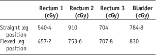
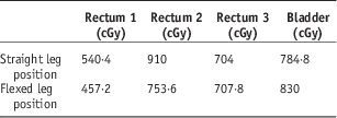
Critical organ maximum doses to volumes of 1, 2 and 5 cc doses, and in vivo dosimetric measurements, are summarised in Tables 1 and 2, respectively. No significant difference in the maximum doses on sigmoid colon, rectum or bladder due to leg position was observed either on 3D planning or for in vivo dose measurements.
Median maximum doses specified on 1, 2, 5 cc volumes of critical structures

Median in vivo measured doses of rectum and bladder
Dose–volume measurements also did not show any significant change for target, bladder and sigmoid colon doses (Table 3). However 25 and 50% isodoses that cover percentage of rectum volume showed that higher volumes received significantly higher doses in the flexed knee position. In the straight leg position, smaller volumes of rectum were covered by 25% isodose (29·6 versus 39·6%, p=0·04), and 50% isodose (9·2 versus 12%, p=0·05). V75 and V100 also appeared to be less in the straight leg position, but the difference was not significant.
Volumes (cc) of critical structures covered by reference isodoses

*p<0·05.
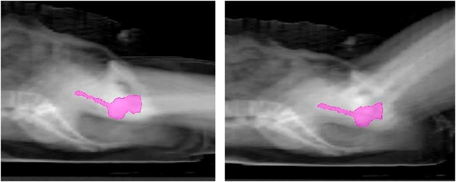
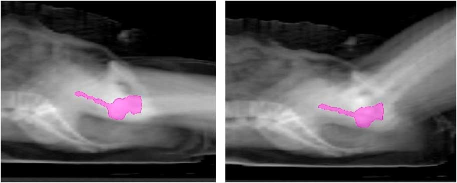
We checked applicator position to detect applicator movement, and we detected in all patients posterior rolling of the applicators with leg flexion (Figure 1). The tip of the brachytherapy applicator moved closer to sacrum (median 8·4 mm, range 4·3–14 mm). We hypothesise that pelvic muscle relaxation displaces the applicators posteriorly closer to rectum and that this may yield a small but important rise in rectum doses, although Dmax did not change.
Lateral reconstructed images of applicator in straight and flexed leg positions.
Discussion
To our knowledge, this is the first study to investigate the importance of leg position during HDR brachytherapy. In relation to leg position, we found significant changes in radiation dose to rectum but not to other critical structures or the target.
Thomadsen et al.Reference Thomadsen, Shahabi and Stitt 14 and Pham et al.Reference Pham, Chen and Rouby 15 previously noticed applicator movement and resulting dose changes during HDR brachytherapy. These authors recommended immobilisation of the patient together with applicator fixation in order to avoid dose uncertainty at point A and critical structures. These two studies are based on conventional brachytherapy planning, and leg position was not investigated. Our study is a 3D CT-based dose calculation on both target and critical structures. Although the clinical importance might be small, our data showed a significant increase in rectum dose with leg flexion on 3D plans. This increase may be due to the posterior rolling of applicators towards the rectum, which we observed during leg flexion.
The significance of anterior rectal wall dose for late rectal toxicity in HDR brachytherapy has been previously shown.Reference Cheng, Peng and Chen 16 The ABS also recommends keeping the cumulative rectum reference dose to <75 GyReference Nag, Erickson and Thomadsen 11 to prevent rectal toxicity. A recent Cochrane review of cervical brachytherapy showed 1·8% bladder and 3·1% rectosigmoid Grade 3 or 4 toxicities.Reference Wang, Liu and Yank 17 Manoeuvres that keep rectum dose low, such as straight leg position as investigated in the present study, are therefore important.
Our in vivo measurements did not show any significant change in rectum dose, although 3D dosimetric calculations showed a difference. This is likely due to the limitations of in vivo dosimetry. Usla et al.Reference Usla, Wabersieb, Potter and Georg 18 previously showed the considerable difference between calculated dose and in vivo measurements. They reported a mean 11% difference for rectum and a 4% difference for bladder. Dose is underestimated by a mean 29% for rectum and 58% for bladder the International Commission on Radiation Units and Measurements (ICRU) references.
Conclusion
The evidence from this and previous studies shows that it is possible to manipulate the dose distributions of critical structures by certain manoeuvres without changes in dose to the target.Reference Cengiz, Gurdalli and Selek 4 This could be performed during brachytherapy for selected patients. However, we recommend extended leg position for at least HDR brachytherapy because of lower rectal dose during a short treatment course.
Acknowledgement
This research was supported by Hacettepe University, Scientific Research Projects Coordination Unit.