INTRODUCTION
The coronate jellyfish (class Scyphozoa, order Coronatae) are composed of five families, three of which, the Atollidae, Paraphyllinidae and Periphyllidae, are considered to exclusively comprise deep sea species (Arai, Reference Arai1997; Jarms et al., Reference Jarms, Tiemann and Båmstedt2002). Of the two other families, the Linuchidae are found in shallow water only, while the Nausithoidae have jellyfish in both the deep sea and shallow waters, including caves (see Jarms et al., Reference Jarms, Båmstedt, Tiemann, Martinussen and Fosså1999; Morandini & Silveira, Reference Morandini and da Silveira2001).
Of the deep sea jellyfish, Periphylla periphylla (Péron & Lesueur, 1810) and Atolla spp. are the most well-known. Periphylla periphylla is widely distributed at mesopelagic depths in several oceans from the Arctic to Antarctic (e.g. Mauchline & Harvey, Reference Mauchline and Harvey1983; Larson et al., Reference Larson, Mills and Harbison1991; Mianzan & Cornelius, Reference Mianzan, Cornelius and Boltovskoy1999; Donnelly et al., Reference Donnelly, Sutton and Torres2006; Osborn et al., Reference Osborn, Silver, Castro, Bros and Chavez2007; Gershwin & Zeidler, Reference Gershwin and Zeidler2008; Youngbluth et al., Reference Youngbluth, Sørnes, Hosia and Stemmann2008). Abundance estimates from net tows and ROVs are in the region of 1 ind 1000 m−3 to 2 ind 100 m−3 (Pagès et al., Reference Pagès, White and Rodhouse1996; Dalpadado et al., Reference Dalpadado, Ellersten, Melle and Skjoldal1998; Youngbluth et al., Reference Youngbluth, Sørnes, Hosia and Stemmann2008). Permanent and highly abundant populations up to 2–3 orders of magnitude greater than open ocean environments have also been observed in several Norwegian fjords, and research into these populations has greatly increased our understanding of the biology and ecology of this species (e.g. Youngbluth & Båmstedt, Reference Youngbluth and Båmstedt2001; Riemann et al., Reference Riemann, Titelman and Båmstedt2006; Kaartvedt et al., Reference Kaartvedt, Klevjer, Torgersen, Sørnes and Rostad2007; Soetje et al., Reference Soetje, Tiemann and Båmstedt2007; Sørnes et al., Reference Sørnes, Hosio, Båmstedt and Aksnes2008). Like P. periphylla the genus Atolla is widely distributed in many of the world's oceans at mesopelagic and bathypelagic depths (e.g. Larson et al., Reference Larson, Mills and Harbison1991; Lindsay et al., Reference Lindsay, Furushima, Miyake, Kitamura and Hunt2004; Raskoff et al., Reference Raskoff, Purcell and Hopcroft2005; Donnelly et al., Reference Donnelly, Sutton and Torres2006; Osborn et al., Reference Osborn, Silver, Castro, Bros and Chavez2007; Youngbluth et al., Reference Youngbluth, Sørnes, Hosia and Stemmann2008). However, compared with Periphylla much less is known about its biology and ecology beyond the early descriptions of Russell (Reference Russell1959, Reference Russell1970).
Life cycles and reproductive biology in the Scyphozoa have been studied extensively in coastal species, primarily the semaeostomes (e.g. Aurelia sp., Cyanea sp., Chrysaora sp. and Pelagia sp.) and rhizostomes (e.g. Cassiopea sp., and Rhizostoma sp.) (Eckelbarger & Larson, Reference Eckelbarger and Larson1988; Rottini-Sandrini & Avian, Reference Rottini-Sandrini and Avian1991; Arai, Reference Arai1997; Pitt & Kingsford, Reference Pitt and Kingsford2000; Lucas, Reference Lucas2001). In the coronates, sexual reproduction has been described for the coastal species Nausithoe aurea (Morandini & Silveira, Reference Morandini and da Silveira2001), Nausithoe atlantica and Linuche unguiculata (Eckelbarger & Larson, Reference Eckelbarger and Larson1992). However, in deep sea jellyfish (including coronates) reproduction is poorly described, primarily due to low sampling frequency typical of deep-sea research and the relatively low abundances encountered at mesopelagic and bathypelagic depths. Nevertheless, we know that adaptations to life in the deep-sea include holopelagic life histories that may involve reduced and/or parasitic polyps or direct development with a complete absence of the benthic polyp (Russell, Reference Russell1953; Kramp, Reference Kramp1961; Osborn, Reference Osborn2000; Lucas & Reed, Reference Lucas and Reed2009). Recently the life cycle and development of P. periphylla has been described by Jarms et al. (Reference Jarms, Båmstedt, Tiemann, Martinussen and Fosså1999, Reference Jarms, Tiemann and Båmstedt2002). The species has a holopelagic life cycle with direct development from a large yolky egg into a medusa without either planula larva or ephyra stages, and based on preserved and fresh material, 14 developmental stages from egg to adult medusa have been identified (Jarms et al., Reference Jarms, Tiemann and Båmstedt2002).
The two studies by Jarms et al. (Reference Jarms, Båmstedt, Tiemann, Martinussen and Fosså1999, Reference Jarms, Tiemann and Båmstedt2002) have greatly advanced our understanding of the life history of deep-sea coronate medusae. However, ultrastructural descriptions of gonads and gametogenesis in coronates, and indeed other jellyfish, are extremely sparse. Apart from older texts (Haeckel, Reference Haeckel1882; Claus, Reference Claus1883; Maas, Reference Maas1897; Vanhöffen, Reference Vanhöffen1902; Widersten, Reference Widersten1965; Russell, Reference Russell1970) descriptions of ovarian morphology and oogenesis in scyphozoans are limited to those of Eckelbarger & Larson (Reference Eckelbarger and Larson1988, Reference Eckelbarger and Larson1992), Eckelbarger (Reference Eckelbarger, Wilson, Stricker and Shinn1994), Lesh-Laurie & Suchy (Reference Lesh-Laurie, Suchy, Harrison and Westfall1991) and Morandini & Silveira (Reference Morandini and da Silveira2001). Apart from older texts (e.g. Haeckel, Reference Haeckel1882; Claus, Reference Claus1883; Maas, Reference Maas1897; Vanhöffen, Reference Vanhöffen1902), published accounts of gonad structure and gametogenesis in the coronates are limited to the shallow water species L. unguiculata (Eckelbarger & Larson, Reference Eckelbarger and Larson1992) and N. aurea (Morandini & Silveira, Reference Morandini and da Silveira2001). This paper describes the structure of gonads and gametogenesis in the deep water coronates Periphylla periphylla and Atolla wyvillei (Haeckel, 1880) collected from mesopelagic depths in the Gulf of Mexico and Cape Hatteras region of the western North Atlantic.
MATERIALS AND METHODS
Study areas and sample collection
Specimens were identified according to the descriptions of Russell (Reference Russell1959, Reference Russell1970). Atolla wyvillei and Periphylla periphylla medusae were collected from Cape Hatteras (33°10′N to 35°14′N, and 74°52′W to 75°47′W) in early–mid-September 1994, and from the eastern Gulf of Mexico (running along the 1000 m depth contour between 26°24′N 84°51′W and 27°04N 85°09′W) in mid-September 1995.
At both Cape Hatteras and the Gulf of Mexico, medusae were collected using the ‘detritus sampler’ on the ‘Johnson-Sea-Link’ submersible. At Cape Hatteras, 3 specimens of P. periphylla were collected from between 400 and 686 m depth and 5 specimens of A. wyvillei were collected from between 820 and 909 m depth. Water temperature at these depths ranged between 5.5 and 6.2°C. In the Gulf of Mexico, a total of 18 P. periphylla and 7 A. wyvillei medusae were collected from depths between 638 m and 831 m. The temperature recorded at these depths ranged between 5.45 and 6.8°C. Additional specimens of P. periphylla (N = 26) and A. wyvillei (N = 7) were collected using a Tucker Trawl on the Gulf of Mexico cruise. Trawling took place primarily at night time, at depths ranging between 130 and 850 m.
Within 2 hours of collection, the bell diameters (BD, mm) of all medusae were measured using a ruler and their sex and state of maturity noted where possible (i.e. immature with no gametes visible, male and female). Undamaged individuals were then either frozen at −20°C for biochemical analysis (see Lucas, Reference Lucas2009), or preserved in 2% gluteraldehyde for histological analysis.
Histology and image analysis
Whole male and female gonads were dehydrated in graded alcohol, cleared in Histo-Clear®, embedded into 20 × 20 mm paraffin wax blocks and serial sectioned at 7 µm. Tissue sections were mounted on slides and stained with Pasini's triple stain. Image analyses of gonads containing gametes were captured using SigmaScan Pro 4 software, with feret (average) diameter used to measure the size of developing and mature oocytes.
RESULTS
Atolla wyvillei Haeckel, 1880
GENERAL MORPHOLOGY OF GONADS
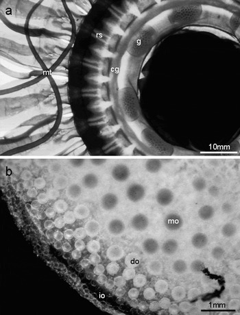
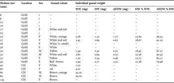
Observations on the general morphology of Atolla wyvillei gonads were based on 14 specimens from the Gulf of Mexico, 8–40 mm bell diameter (BD), and 5 specimens from Cape Hatteras, 40–145 mm BD, summarized in Table 1. Gonads were visible in specimens >17–22 mm BD, appearing as 8 oval or bean-shaped structures on the subumbrella wall of the gastrovascular sinus proximate to the coronal groove (Plate 1a). As the gonads matured, each one thickened along its periphery and in males the outer edge of the gonad was folded over. Together, mature male gonads formed an almost continuous ring around the stomach. Gonad colour varied from white-cream to tan, red or dark brown, and they appeared to be very organic-rich, as demonstrated by the very high percentage ash-free dry weights ranging from 67.6–96.5% of dry weight (Table 1). Gametes were clearly visible within the gonads (Plate 1b) even in medusae as small as 17 mm BD.
(a) Subumbrella view of 40 mm bell diameter (BD) female Atolla wyvillei from Cape Hatteras showing the position of the gonads (g) containing oocytes, coronal groove (cg), radial septum (rs) and marginal tentacle (mt); (b) gonad of female Atolla wyvillei (40 mm BD) containing oocytes in all stages of development from the immature oocytes (io) located at the gonad periphery to developing oocytes (do) and mature oocytes (mo) both unattached within the central mesoglea.

Observations on the morphology of gonads of Atolla wyvillei medusae collected from the Gulf of Mexico (GoM) and Cape Hatteras (CH) regions. WW, wet weight; DW, dry weight; AFDW, ash free dry weight; F, female; M, male; i, immature—gonads or their contents not visible.

SPERMATOGENESIS
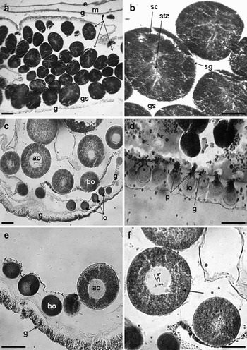
Histology was carried out on 1 or 2 gonads from each of 4 males of Atolla wyvillei. Spermatogenesis occurred within follicles arising from the gastrodermis. Many sperm follicles were present, evenly distributed throughout each gonad (Plate 2a). Each follicle was ovoid, but with some variability in shape and size; in a single male 80 mm BD where 27 follicles were measured, average (±SD) follicle size was 366 (±83.9) µm × 254 (±41.9) µm. Mature follicles, containing spermatogonia, spermatocytes, spermatids and spermatozoans (Plate 2b) were observed in individuals measuring 80 mm in Cape Hatteras and 30 mm in the Gulf of Mexico.
(a) Cross-section through a male gonad of a 40 mm bell diameter (BD) Atolla wyvillei from Cape Hatteras showing the evenly distributed sperm follicles (f). Also shown are the exumbrella surface (e), mesoglea (m), gastrodermis (g) and genital sinus of the gastrovascular cavity (gs); (b) close-up of sperm follicles from the same individual showing the spermatogonia (sg) located on the margin of the follicle. Spermatocytes (sc) mature into spermatids then spermatozoons (stz) as they move down the follicle. They are released into the genital sinus (sg); (c) cross-section through the female gonad of a 40 mm BD A. wyvillei from the Gulf of Mexico. Two areas of gastrodermis (g) are seen—thicker external and thinner internal. Small immature oocytes (io) are found at the periphery of the ovary, developing into densely-staining basophilic oocytes then lighter-staining acidophilic (mature) oocytes with a large nucleus (see Morandini & Silveira, Reference Morandini and da Silveira2001); (d) oocytes (io) arise from the gastrodermis, initially attached by a peduncle (p) (A. wyvillei 145 mm BD from Cape Hatteras); (e) basophilic (bo) and acidophilic oocytes (ao) continue their development unattached in a 40 mm BD A. wyvillei from the Gulf of Mexico; (f) mature oocytes can be identified by their large nucleus (n) and granular cytoplasm (arrow) (A. wyvillei 40 mm BD Gulf of Mexico). All scales = 100 µm.

OOGENESIS
Oocytes in different stages of development were observed in all the female medusae mature enough to have gonads. As described by Russell (Reference Russell1970) the smallest oocytes were visible around the periphery of the gonads, with the largest oocytes located in the central mesoglea region (Plates 1b & 2c). Although there was no significant relation between state of maturity and medusa size, it was noted that the largest specimen from Cape Hatteras (BD = 145 mm) had the largest gonads, measuring 11 × 7 mm, and that these contained a greater number of oocytes. In each gonad, the oocytes developed from the gastrodermis. These small pre-vitellogenic oocytes, 20–40 µm, appeared to be held to the cell wall by a peduncle (Vanhöffen, Reference Vanhöffen1902) (Plate 2d) before detaching and migrating into the mesoglea as unattached early–mid and late vitellogenic oocytes without nutritive trophocytes (Plate 2e). As the oocytes developed and grew to ~70 µm and beyond, they appeared to gain increasing quantities of organic-rich yolk material and a large nucleus was present (Plate 2f).
OOCYTE SIZE-FREQUENCY DISTRIBUTIONS
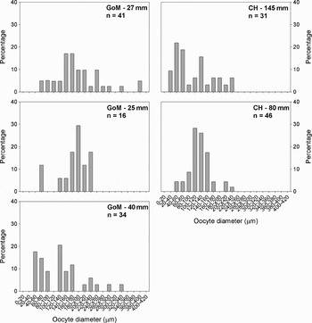
The size–frequency distribution of oocytes was measured in 5 individuals of Atolla wyvillei; 3 from the Gulf of Mexico 25–40 mm BD, and 2 from Cape Hatteras, 80 and 145 mm BD (Figure 1). All of the medusae contained both developing and mature oocytes reflected in the wide range of oocyte diameters. In the medusae from the Gulf of Mexico the smallest oocytes measured between 43 and 71 µm, increasing to maximum diameters of between 331 and 543 µm. In the larger Cape Hatteras medusae, oocyte sizes ranged from minima of 24–53 µm to maxima of 226–263 µm.
Percentage size–frequency distributions of Atolla wyvillei oocytes from the Gulf of Mexico (GoM) and Cape Hatteras (CH). Medusa size given in mm, n is number of oocytes measured.

Periphylla periphylla Péron & Lesueur, 1810
GENERAL STRUCTURE OF GONADS
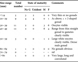
The observations on general morphology of Periphylla periphylla gonads summarized in Table 2 were based on 44 specimens 15–80 mm bell diameter (BD) collected from the Gulf of Mexico. Gonads were visible in specimens >17 mm BD, although not all larger individuals had obvious gonads (see Table 2). In total, 13 medusae were identified as females (38.8 ± 10.0 mm BD), 11 as male (46.4±12.6 mm BD), with the remaining 20 unknown (29.11±12.9 mm BD). Of these 20 individuals, 11 medusae did not have any gonads, while in the remainder the sex could not be determined because the gonads were too thin.
The number of observations on the state of maturity of gonads in 10 mm size-groups of Periphylla periphylla collected from the Gulf of Mexico. No G, no gonads; unident, gonads present but sex not identifiable; M, male; F, female; nd, no data.

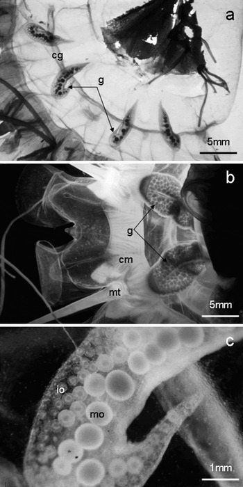
The eight gonads were located on the gastrovascular cavity attached to the subumbrella wall of the medusa. They initially appeared as thin straight structures in small medusae, 17–22 mm BD, becoming J-shaped in 24–30 mm BD medusae (Plate 3a). The distal end of the gonad was longer in medusae >35 mm BD, finally resulting in a convoluted (particularly males) U-shaped gonad with the inner margins of the adradial and interradial arms almost contiguous (Plate 3b). As with Atolla wyvillei, gametes were clearly visible in the gonads in medusae as small as 22 mm BD (Plate 3c). Gonad colour varied between cream, tan, orange and deep red, generally with darker colours in larger medusae.
(a) Exumbrella view of a 30 mm bell diameter (BD) female Periphylla periphylla from the Gulf of Mexico showing the J-shaped gonads (g) lying under the coronal groove (cg); (b) a 35 mm BD male Periphylla periphylla from Cape Hatteras with densely-packed gonads (g). Also shown are the coronal muscle (cm) and a marginal tentacle (mt); (c) close-up of a female gonad from a 30 mm BD medusa highlighting the gradient from immature oocytes (io) at the periphery of the gonad to mature gonads (mo) towards the inner margins.

SPERMATOGENESIS
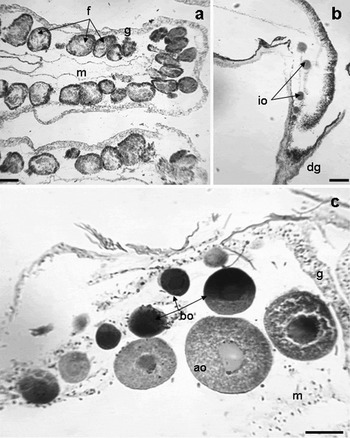
Histology was carried out on 1–2 gonads from each of 4 males of Periphylla periphylla, 33–55 mm BD. The male gonads consisted of follicles containing spermatogonia, spermatocytes, spermatids and spermatozoans, consistent with other descriptions of coronate scyphomedusae (Russell, Reference Russell1970). The sperm follicles were positioned in long convoluted rows normally one follicle thick (Plate 4a), with the follicle contents at similar stages of development. The smallest male with mature gonads had a bell diameter of 33 mm from Cape Hatteras and 40 mm from the Gulf of Mexico.
(a) Cross-section through the male gonad of a 55 mm bell diameter (BD) Periphylla periphylla from the Gulf of Mexico showing the long convoluted rows of sperm follicles (f) inside the gastrodermis (g). Also shown is the mesoglea (m); (b) immature oocytes (io) at the distal end of the gonad (dg) of a 53 mm BD P. periphylla from the Gulf of Mexico; (c) a clear gradient of increasing oocyte size and maturity in a 30 mm BD P. periphylla from the Gulf of Mexico, from basophilic oocytes (bo) nearer the gastrodermis (g) to mature acidophilic oocytes (ao) within the mesoglea (m) of the gonad. All scales = 100 µm.

OOGENESIS
Histology was carried out on 1–2 gonads from each of 9 females, 30–80 mm BD. Oocytes in different stages of development (i.e. pre-vitellogenic to late-vitellogenic) were observed in each female medusa examined; even the gonads of a 30 mm BD medusa contained oocytes ranging between 49 and 288 µm diameter. The oocytes initially appeared near the distal end of the gonad (Plate 4b). A gradient of increasing oocyte size was apparent within each gonad with the largest oocytes located in the inner region of the gonad (Plate 4c). Mature, late-vitellogenic oocytes could be recognized from dense immature oocytes by their light granular acidophilic (eosin red staining) cytoplasm.
OOCYTE SIZE-–FREQUENCY DISTRIBUTIONS
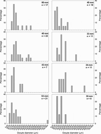
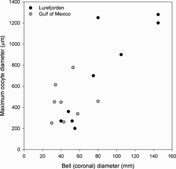
Oocyte diameters were measured in 9 individuals, 30–80 mm BD, collected from the Gulf of Mexico. As previously mentioned, 25% of medusae collected did not appear to have gonads, and in those that did have the larger J-shaped or U-shaped gonads, oocyte size–frequency distributions were variable (Figure 2), possibly biased by the small medusa and gonad sizes, and very low numbers of oocytes (N = 7 to 22) present in each gonad. For example, minimum oocyte sizes varied from between 44 and 168 µm in 30 mm and 58 mm BD medusae respectively, with no apparent trend with medusa size. The largest oocytes measured ranged between 252 µm in a 30 mm medusa and 777 µm in a 53 mm medusae (mean±SD, 431.8±164.9 µm, N = 9).
Percentage size–frequency distributions of Periphylla periphylla oocytes from the Gulf of Mexico. Medusa size given in mm, n is number of oocytes measured.

DISCUSSION
The findings presented in this paper contribute to our understanding of gonad morphology and gametogenesis in Atolla wyvillei beyond the early texts (Haeckel, Reference Haeckel1882; Claus, Reference Claus1883; Maas, Reference Maas1897; Vanhöffen, Reference Vanhöffen1902; Russell, Reference Russell1959, Reference Russell1970), and to our increased understanding of the life history and breeding of Periphylla periphylla (Mauchline & Harvey, Reference Mauchline and Harvey1983; Jarms et al., Reference Jarms, Båmstedt, Tiemann, Martinussen and Fosså1999, Reference Jarms, Tiemann and Båmstedt2002). The relatively low sample numbers, particularly for Cape Hatteras, reflect the sampling methodology (i.e. individually collected intact specimens using a submersible) as well as the relative contribution of the study species to overall jellyfish diversity at the two sites. Cape Hatteras had a high diversity of mesopelagic jellyfish comprising coronate scyphomedusae (A. wyvillei and P. periphylla), the ulmarid scyphomedusa Poralia rufescens, narcomedusae and trachymedusae (Aeginura grimaldii, Colobonema sp., Cunina peregrina, Eumedusa sp. and Halitrephes sp.), as well as a variety of mainly unidentified ctenophores. Atolla wyvillei and P. periphylla accounted for less than a quarter of all gelatinous zooplankton collected. In contrast, submersible and trawl samples from the Gulf of Mexico were dominated by P. periphylla (40% of catch), with siphonophores (Apolemia sp. and Halistemma sp.), ctenophores (Llyria sp. and Beroe sp.) and A. wyvillei also abundant (21%, 15% and 8% of catch respectively).
The bell diameters of P. periphylla sampled (16–80 mm from the Gulf of Mexico, 33–60 mm from Cape Hatteras) fall within the range of those reported in other ecosystems, e.g. the Rockall Trough (Mauchline & Harvey, Reference Mauchline and Harvey1983) and Australia (Gershwin & Zeidler, Reference Gershwin and Zeidler2008), although the biggest jellyfish sampled in this study are much smaller than those in the Lurefjord, Norway (145–170 mm) (Youngbluth & Båmstedt, Reference Youngbluth and Båmstedt2001; Jarms et al., Reference Jarms, Tiemann and Båmstedt2002), Japan (150 mm) and the Antarctic (~230 mm) (Lindsay, personal communication). Indeed, Larson (Reference Larson and Kornicker1986) has reported that P. periphylla medusae were up to 350 mm in the Southern Ocean. Regarding A. wyvillei Russell (Reference Russell1959) reported that Atolla spp. from the Bay of Biscay reached a size of 130 mm, although it was suggested that two species were present: smaller A. parva and larger A. wyvillei. Similarly, in the Rockall Trough, north-eastern Atlantic, Mauchline & Harvey (Reference Mauchline and Harvey1983) reported that A. wyvillei ranged from 9.3–79.7 mm, A. vanhoeffeni was 4.8–31.6 mm and A. parva was 5.0–46.3 mm. The largest specimens captured in this study were from Cape Hatteras (40–145 mm), whereas those from the Gulf of Mexico were between 8 and 40 mm BD. It is unclear whether variation in sizes represents sampling bias, geographical differences in food availability or breeding period (see Mauchline & Harvey, Reference Mauchline and Harvey1983). Similarly, although only one species of Periphylla is reported in the literature at present, Jarms et al. (Reference Jarms, Tiemann and Båmstedt2002) have commented that several species may exist in different regions, which may have different morphologies, sizes and developmental characteristics, as has been discovered with the common shallow water scyphozoan Aurelia (e.g. Dawson & Jacobs, Reference Dawson and Jacobs2001).
The wide range of medusa sizes and stages of maturity observed for both species in this study have been documented elsewhere. Several studies have published size–frequency distributions and stages of maturity in both Periphylla and Atolla species that indicate ongoing breeding throughout the year (Russell, Reference Russell1959; Mauchline & Harvey, Reference Mauchline and Harvey1983; Jarms et al., Reference Jarms, Tiemann and Båmstedt2002), possibly with a primary breeding period in the summer months in more northerly latitudes in open waters (Russell, Reference Russell1959; Mauchline & Harvey, Reference Mauchline and Harvey1983). Although Jarms et al. (Reference Jarms, Tiemann and Båmstedt2002) state that ‘mature medusae with ripe gonads are present (in Lurefjord) throughout the year, and a distinct spawning period does not exist’ the strong dominance of individuals <10 mm in coronal diameter in October seem to indicate that periods of relatively high recruitment do occur in the summer (see Jarms et al., Reference Jarms, Båmstedt, Tiemann, Martinussen and Fosså1999, p. 61), which has also been found in enclosed and tropical shallow water jellyfish populations (Lucas, Reference Lucas1996; Martin, Reference Martin1999). Although sample numbers are rather low, female:male sex-ratios were 3.5 (N = 14) and 1.5 (N = 5) for A. wyvillei in the Gulf of Mexico and Cape Hatteras respectively, and 1.4 (N = 24) for P. periphylla in the Gulf of Mexico.
In both A. wyvillei and P. periphylla from the Gulf of Mexico gonads were distinguishable in medusae >17 mm. In their description of development stages of P. periphylla from the Lurefjord Jarms et al. (Reference Jarms, Tiemann and Båmstedt2002) noted that gonads were first evident in ‘Stage 14B’ medusae (coronal diameter, CD >25 mm; 37.0 ± 6.8 mm), with sexual dimorphism of gonads in ‘Stage 14C’ medusae (CD >40 mm; 57.3±17.8 mm). Mature specimens, i.e. those containing eggs >1 mm, were >80 mm CD. In the Gulf of Mexico sexual dimorphism of gonads was evident at 22 mm. Using Jarms et al. (Reference Jarms, Tiemann and Båmstedt2002) criteria, maturity was not attained as oocytes did not reach 1 mm in size; indeed the maximum was 777 µm in a 53 mm medusa. However, based on histological examination we believe that the larger specimens in this study were mature, and this will be discussed further later. Russell (Reference Russell1959) identified 3 stages of maturity in Atolla spp.: I—gonads absent or just appearing, sex not distinguished; II—sexes determined but gonads not mature; and III—mature males and females, i.e. gonads full. In the Bay of Biscay mature (stage III) specimens of A. wyvillei were between 40 and 130 mm in size, while mature A. parva were 10–30 mm in size (Russell, Reference Russell1959), with similar sizes reported by Mauchline & Harvey (Reference Mauchline and Harvey1983) in the more northerly Rockall Trough.
The general structure of the gonads was very similar to previous descriptions of these and other coronate scyphomedusae (e.g. Russell, Reference Russell1970; Morandini & Silveira, Reference Morandini and da Silveira2001). In A. wyvillei the gonad walls were many cells deep in places. In one individual the thickened gonad walls appeared to contain bunches of possible organic-rich granular material with small passages visible through them—similar to that reported by Morandini & Silveira (Reference Morandini and da Silveira2001) for Nausithoe aurea.
The majority of descriptions of gametogenesis in shallow and deep water medusae have focused on oogenesis (e.g. Eckelbarger & Larson, Reference Eckelbarger and Larson1988, Reference Eckelbarger and Larson1992), with very few modern descriptions of male gonads (but see Kikinger, Reference Kikinger1992; Pitt & Kingsford, Reference Pitt and Kingsford2000; Morandini & Silveira, Reference Morandini and da Silveira2001). Nevertheless, the general structure of the testis of both study species resembled the descriptions of Widersten (Reference Widersten1965), Campbell (Reference Campbell, Giese and Pearse1974) and Morandini & Silveira (Reference Morandini and da Silveira2001). In coronates sperm follicles arise from invagination of the gastrodermis, and it is within these follicles that spermatogenesis occurs. In A. wyvillei, numerous ovoid sperm follicles were evenly distributed throughout the gonad (see Plate 2a), whereas in P. periphylla the gonads contained convoluted ribbons one follicle thick (see Plate 4a). According to Campbell (Reference Campbell, Giese and Pearse1974) release of spermatozoa occurs through the rupture of the testis wall.
In both A. wyvillei and P. periphylla oocytes in different stages of development were observed in all medusae possessing gonads mature enough for sexes to be distinguishable. In agreement with earlier descriptions of coronates (e.g. Russell, Reference Russell1970) the gonads had very distinctive and characteristic distribution of oocytes, with small oocytes at the periphery and large oocytes in the centre. Unlike rhizostome (Pitt & Kingsford, Reference Pitt and Kingsford2000) and semaeostome (Eckelbarger & Larson, Reference Eckelbarger and Larson1988) scyphozoans, early-mid vitellogenic oocytes break free from the gastrodermis, from where they arise, and continue their development freely in the mesoglea without the presence of nutritive trophocytes. In accordance with the conclusions of numerous authors (e.g. Russell, Reference Russell1959; Mauchine & Harvey, 1983; Larson, Reference Larson and Kornicker1986; Jarms et al., Reference Jarms, Båmstedt, Tiemann, Martinussen and Fosså1999, Reference Jarms, Tiemann and Båmstedt2002), the presence of oocytes in various stages of development in a wide variety of medusa sizes coupled with the apparent lack of spent gonads implies continuous and steady egg production over several years. There are no obvious relationships between oocyte size and medusa development.
Berrill (Reference Berrill1949) was the first to suggest that large eggs were an indication of direct development, and indeed Jarms et al. (Reference Jarms, Båmstedt, Tiemann, Martinussen and Fosså1999) described the holopelagic life cycle of P. periphylla, from large yolky egg to young medusa, without a polyp, planula larvae or ephyra. The eggs of P. periphylla are among the largest in the Cnidaria, being up to 10 times bigger than those for other scyphomedusae (see Jarms et al., Reference Jarms, Båmstedt, Tiemann, Martinussen and Fosså1999). The organic-rich eggs produced by P. periphylla from the Gulf of Mexico were also large (maximum 777 µm) compared with shallow water scyphozoans, but did not attain the very large sizes, up to 1.28 mm, found in P. periphylla in the Lurefjord (Jarms et al., Reference Jarms, Tiemann and Båmstedt2002) (Figure 3). Jarms et al. (Reference Jarms, Tiemann and Båmstedt2002) suggested that oocytes were not mature until they were >1 mm diameter, although they provided no histological or quantitative criterion of how they defined oocyte maturity. Nevertheless, based on the large nucleus and light, granular acidophilic staining (see Morandini & Silveira, Reference Morandini and da Silveira2001) we believe that some of the oocytes were mature. Atolla wyvillei oocytes were measured in specimens from both the Gulf of Mexico and Cape Hatteras, with maximum diameters of mature eggs ranging between 331–543 µm and 226–263 µm respectively. Given the very small sample sizes it is unclear whether the larger mature oocytes found in the smaller Cape Hatteras medusae reflects a genuine inverse relationship between maximum oocyte size and size/age of medusa, a site-specific relationship influenced by productivity of the two regions, or even molecular differences in medusae from the two regions. These ambiguities may be clarified if and when comparisons with other populations of Periphylla and Atolla occur in a global setting, but until more detailed ecological and molecular analyses are carried out on specimens, this area will remain poorly understood.
Relationship between maximum oocyte diameter (μm) and medusa size (mm) in Periphylla periphylla (bell diameter for the Gulf of Mexico, coronal diameter for the Lurefjorden). Lurefjorden data taken from Jarms et al. (Reference Jarms, Tiemann and Båmstedt2002).

ACKNOWLEDGEMENTS
Specimens were collected during the following cruises: Cape Hatteras, September 1994: NOAA/NURP UNCW9406 awarded to Dr Tom Bailey, NOAA/NURC UNCW9410 and NSF OCE9313872 awarded to Dr Tammy Frank and Dr Edith Widder; Gulf of Mexico, September 1995: NSF OCE 9313872 awarded to Dr Tammy Frank and Dr Edith Widder. C.H.L. would like to thank the above PIs for their help and support during her Harbor Branch Oceanographic Institute Fellowship, and the crew of the RV ‘Edwin Link’ and ‘Johnson-Sea-Link II’ submersible for their assistance during field operations. Finally, the authors would like to thank the anonymous referees for their invaluable comments.