The developmental origins of disease risk have been established through epidemiological studies in humans and illustrate the profound impact of early life adversity. During prenatal development, the fetus is particularly vulnerable to the effects of a broad range of environmental exposures, with consequences that can persist into infancy, adolescence, and adulthood. In particular, maternal distress during pregnancy, in the form of exposure to chronic or acute stressors, depression, and/or anxiety, can influence both fetal and infant behavioral and physiological outcome measures. For example, antenatal depression and anxiety symptoms predict increased behavioral reactivity and cortisol in response to novelty in infants (Kaplan, Evans, & Monk, Reference Kaplan, Evans and Monk2007; O'Connor, Heron, Glover, & The ALSPAC Study Team, Reference O'Connor, Heron and Glover2002), higher resting cortisol throughout the day among adolescents (Van den Bergh, Van Calster, Smits, Van Huffel, & Lagae, Reference Van den Bergh, Van Calster, Smits, Van Huffel and Lagae2008), and a reduction in gray matter density in the prefrontal cortex (Buss, Davis, Muftuler, Head, & Sandman, Reference Buss, Davis, Muftuler, Head and Sandman2010; Pruessner et al., Reference Pruessner, Dedovic, Khalili-Mahani, Engert, Pruessner and Buss2008). These human studies parallel the findings from decades of animal research that indicate that prenatal stress can induce long-term neurobiological and behavioral effects with particular consequences for the hypothalamus–pituitary–adrenal (HPA) response to stress (Weinstock, Reference Weinstock2005). The stress vulnerability that is established in utero in response to exposure to maternal prenatal stress, depression, and anxiety may ultimately lead to increased risk of psychopathology (O'Connor, Heron, Golding, Beveridge, & Gover, Reference O'Connor, Heron, Golding, Beveridge and Glover2002; Pawlby, Hay, Sharp, Waters, & O'Keane, Reference Pawlby, Hay, Sharp, Waters and O'Keane2009; Talge, Neal, & Glover, Reference Talge, Neal and Glover2007).
A critical question within these studies concerns the mechanism through which maternal distress shapes development. An emerging theme within developmental studies is the dynamic interactions between environmental experiences and gene activity achieved through epigenetic mechanisms, which are molecular modifications to gene activity that do not involve changes to the underlying DNA sequence. Although these mechanisms were once thought to be limited in their plasticity to the very early stages of embryonic development, more recent evidence indicates that epigenetic variation can be induced across the life span in response to a broad range of environmental exposures (Champagne, Reference Champagne2010; Jirtle & Skinner, Reference Jirtle and Skinner2007). These epigenetic pathways can give rise to altered gene expression levels in multiple tissues, including the brain, with consequences for the functioning and connectivity of neural circuits, which can confer risk for physical and psychiatric disorders in later life. Thus, the exploration of epigenetic mechanisms in the context of studies of prenatal maternal distress may provide insight into the biological pathways linking fetal exposure to maternal distress and both acute and long-term outcomes.
In this article, we highlight evidence for the impact of human maternal distress on fetal and infant outcomes, and research exploring the role of epigenetic mechanisms in linking this form of prenatal adversity to neurobiological and behavioral outcomes. Within the study of prenatal effects, a critical consideration is the continued influence of prenatal maternal adversity during the postnatal period mediated through variations in mother–infant interactions. The quality of interactions between mothers and infants during postnatal development can have a profound impact on development, and these effects may also involve epigenetic pathways. Thus, to compliment the findings from studies of prenatal adversity, here we describe the evidence for shifts in developmental trajectories associated with postnatal maternal care in humans and illustrate evidence from both human and animal studies that these postnatal effects are associated with epigenetic modifications. Finally, during fetal development the placenta serves as a critical interface between the mother and fetus, and dynamic changes in gene regulation within this temporary endocrine structure can have significant implications for growth and development. Epigenetic regulation of gene activity within the placenta can alter functioning of this tissue; in this article, we will describe the evidence suggesting that prenatal distress may act through the placenta to alter fetal development. Overall, these studies provide growing support for the hypothesis that epigenetic pathways may serve as a biological link between maternal psychosocial states during and after pregnancy and psychological, physiological, and neurobiological changes in infants that may increase risk of psychopathology in later life.
Prenatal Consequences of Maternal Stress, Depression, and Anxiety in Humans
There is considerable evidence demonstrating that fetal exposure to maternal psychosocial experiences contributes to the determination of children's neurodevelopmental trajectories (Bale et al., Reference Bale, Baram, Brown, Goldstein, Insel and McCarthy2010). Data generated largely over the last two decades shows that when pregnant women experience significant stress, anxiety, or depression (each of which are frequently indexed by cortisol levels), their children are at increased risk for biobehavioral characteristics conceptualized as potential precursors to psychopathology (Beydoun & Saftlas, Reference Beydoun and Saftlas2008; Charil, Laplante, Vaillancourt, & King, Reference Charil, Laplante, Vaillancourt and King2010; Talge et al., Reference Talge, Neal and Glover2007). The specific findings generated from these studies can vary considerably, however, depending on the methodological approach used to explore the link between maternal adversity and developmental outcomes. Despite wide-ranging methodological approaches, it is evident that maternal psychosocial variables assessed during pregnancy can predict developmental outcomes and that this “transmission of risk” probably occurs independent of the inheritance-specific gene variants. Thus, the quality of the fetal environment, shaped by maternal adversity, can lead to divergent developmental pathways.
Epidemiological studies
Nearly 40 years ago, seminal epidemiological research on birth cohorts from the Dutch Hunger Winter of 1944 showed associations between characteristics of pregnant women's lives and offspring's long-term cognitive and mental health development (Stein, Susser, Saenger, & Marolla, Reference Stein, Susser, Saenger and Marolla1972; Susser & Lin, Reference Susser and Lin1992). Based on data on adult children of women who were pregnant when food intake was reduced to 500–1000 kcal per day due to the wartime blockade, these studies showed that fetal development during the blockade was associated with a twofold increased risk for neurodevelopmental disorders (Susser, Hoek, & Brown, Reference Susser, Hoek and Brown1998). Two interpretations regarding the causal mechanisms of these effects have been suggested: (a) deficiency in many micro- and macronutrients or (b) maternal distress, secondary to famine, each having neurotoxic effects on the developing brain. Several subsequent studies, each based on the Dutch Famine, found similar elevated risk for adult psychopathology in relation to fetal exposure to this maternal experience. Overall, these studies suggest that exposure to this event in the first trimester increases the offspring's risk for future schizophrenia, whereas exposure in the second or third trimester is associated with elevated risk for affective disorders (Brown, Van Os, Driessens, Hoek, & Susser, Reference Brown, Van Os, Driessens, Hoek and Susser2000; Hoek, Brown, & Susser, Reference Hoek, Brown and Susser1998; Susser et al., Reference Susser, Hoek and Brown1998; Susser, Brown, & Matte, Reference Susser, Brown and Matte1999). It is important to note, however, that these findings are based on a population's experience of the Dutch Famine and do not measure individual exposure.
The impact of prenatal exposures, and, in particular, maternal distress, on an individual level has been explored in several cohort studies. In a Danish cohort of 1.38 million births, traumatic events occurring during pregnancy were documented and used to predict risk of psychopathology among offspring (Khashan et al., Reference Khashan, Abel, McNamee, Pedersen, Webb and Baker2008). In this cohort, the death of a relative during the first trimester was associated with an increased risk of schizophrenia, although this did not hold for those who had a family history of mental illness (Khashan et al., Reference Khashan, Abel, McNamee, Pedersen, Webb and Baker2008). There has been some evidence linking prenatal distress to autism (Beversdorf et al., Reference Beversdorf, Manning, Hillier, Anderson, Nordgren and Walters2005; Kinney, Miller, Crowley, Huang, & Gerber, Reference Kinney, Miller, Crowley, Huang and Gerber2008), although a recent population-based cohort study (1.49 million births) also using individual exposure assessment found no association between maternal distress due to the death of a relative and risk of autism (Li et al., Reference Li, Vestergaard, Obel, Christensen, Precht and Lu2009). These studies, based on archived health records, point to links between maternal prenatal distress and children's risk for psychopathology.
Prospective, event-based studies of prenatal distress effects: Natural disasters
Consistent with the Dutch Famine studies, population exposure to events such as wars (Malaspina et al., Reference Malaspina, Corcoran, Kleinhaus, Perrin, Fennig and Nahon2008; Selten et al., Reference Selten, Cantor-Graae, Nahon, Levav, Aleman and Kahn2003) and earthquakes (Glynn, Wadhwa, Dunkel-Schetter, Chicz-Demet, & Sandman, Reference Glynn, Wadhwa, Dunkel-Schetter, Chicz-Demet and Sandman2001) as a proxy for prenatal distress have been associated with negative effects on child outcomes. One series of studies, based on the 1998 ice storm in Quebec, Canada, prospectively followed a cohort of women pregnant during the ice storm up until the children were 8 years of age (Charil et al., Reference Charil, Laplante, Vaillancourt and King2010; King & Laplante, Reference King and Laplante2005; Laplante et al., Reference Laplante, Barr, Brunet, Galbaud du Fort, Meaney and Saucier2004; Laplante, Brunet, Schmitz, Ciampi, & King, Reference Laplante, Brunet, Schmitz, Ciampi and King2008). This work differentiated “objective distress” (i.e., what events the woman experienced related to the storm, e.g., loss of electrical power) from “subjective distress” (i.e., women's self-reported psychological reaction to the ice storm); the former is randomly distributed and independent of potentially heritable personality traits whereas the latter is not. Children prenatally exposed to high versus low levels of objective distress, particularly during the first or second trimester, exhibited poorer cognitive, linguistic, and play abilities at 2, 5, and 8 years of age (Charil et al., Reference Charil, Laplante, Vaillancourt and King2010; King & Laplante, Reference King and Laplante2005; Laplante et al., Reference Laplante, Barr, Brunet, Galbaud du Fort, Meaney and Saucier2004, Reference Laplante, Brunet, Schmitz, Ciampi and King2008).
Longitudinal, observational infant and child research
Spanning infancy through adolescence, numerous reports of prospective, observational studies indicate that prenatal distress exposure predicts an increased likelihood of poor emotion regulation, as evidenced with the difficult temperament and symptoms of attention-deficit/hyperactivity disorder (ADHD). Maternal prenatal anxiety and depression across pregnancy, assessed via self-report, and higher cortisol in the third trimester, were found associated with fearful behavior in infants at 2 months of age (Davis et al., Reference Davis, Glynn, Schetter, Hobel, Chicz-Demet and Sandman2007), and, during the third trimester, greater motor and cry responses to novelty at 4 months of age (Davis et al., Reference Davis, Snidman, Wadhwa, Glynn, Schetter and Sandman2004). In a prospective study of 179 women, elevated pregnancy-specific anxiety throughout gestation (in contrast to more global state/trait anxiety) predicted a more negative infant temperament at 3 months, after controlling for maternal postnatal distress (Sandman, Davis, Buss, & Glynn, Reference Sandman, Davis, Buss and Glynn2011). Among 4-month-old infants, symptoms of maternal depression and anxiety during the third trimester were associated with greater cry responses to novelty. Infants categorized by calm reactions to novel stimuli were, in contrast, 8.4 times as likely as other infants to have a mother free of psychiatric symptoms (Werner et al., Reference Werner, Myers, Fifer, Cheng, Fang and Allen2007).
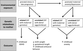
Following children to 2 years of age, higher pregnancy-specific anxiety in the second trimester predicts greater likelihood of increased negative temperament (Blair, Glynn, Sandman, & Davis, Reference Blair, Glynn, Sandman and Davis2011) and fear behavior (Bergman, Sarkar, O'Connor, Modi, & Glover, Reference Bergman, Sarkar, O'Connor, Modi and Glover2007). Prenatal anxiety exposure in the third trimester has been associated with behavioral/emotional problems among 4-year-olds (O'Connor, Heron, Bolding, et al., 2002) and ADHD symptoms among 6-year-olds (O'Connor, Heron, Golding, & Glover, Reference O'Connor, Heron, Golding and Glover2003). For 8- and 9-year-old children, elevated prenatal anxiety in the first and second trimester accounted for 22% and 15% of the variance in teacher- and parent-rated ADHD and externalizing symptoms, respectively (Van den Bergh & Marcoen, Reference Van den Bergh and Marcoen2004). Follow-up studies of this same cohort to 15 years of age showed that prenatal anxiety predicted poorer performance on a continuous performance task for boys (a validated instrument for detection of ADHD symptoms (Van den Bergh et al., Reference Van den Bergh, Mennes, Stevens, Van der Meere, Borger and Stiers2006) and greater impulsivity for both boys and girls (Van den Bergh et al., Reference Van den Bergh, Mennes, Oosterlaan, Stevens, Stiers and Marcoen2005). Finally, depression during pregnancy has been linked to risk for depression in adolescents independent of the impact of the mothers' subsequent depressions throughout the child's life (Pawlby et al., Reference Pawlby, Hay, Sharp, Waters and O'Keane2009). What emerges from these studies is a risk profile associated with prenatal exposure to maternal distress: less adaptive emotion regulation, evidenced in particular by greater reactivity to stimuli, which may be an early marker of stress-linked psychopathology, as well as ADHD symptoms. Animal studies of prenatal stress exposure (which largely control for genetic transmission) produce similar results, in other words, offspring who are more distractible and “anxious” (Clarke & Schneider, Reference Clarke and Schneider1997; Schneider, Reference Schneider1992; Schneider, Roughton, Koehler, & Lubach, Reference Schneider, Roughton, Koehler and Lubach1999; Takahashi, Baker, & Kalin, Reference Takahashi, Baker and Kalin1990; Takahashi, Haglin, & Kalin, Reference Takahashi, Haglin and Kalin1992). In an attempt to tease apart genetic from environmental influence on infant/childhood outcomes in humans, a comparison of children conceived through donor eggs (i.e., having maternal genetic dissimilarity) versus IVF (i.e., having maternal genetic similarity) suggests that the emergence of ADHD was based on shared genetic factors, whereas exposure to prenatal distress could increase the risk of antisocial behavior in children (Rice et al., Reference Rice, Harold, Boivin, Van den Bree, Hay and Thapar2009; see Figure 1).
The prenatal cross-fostering design in the study of the effects of human maternal distress. Although differentiating the impact of genetic and environmental factors in predicting childhood outcomes associated with maternal distress is challenging in humans, studies examining mothers who have used in vitro fertilization (IVF) may provide a useful experimental design. Rice et al. (Reference Rice, Harold, Boivin, Van den Bree, Hay and Thapar2009) examined birth outcomes and childhood psychopathology among infants who were genetically related (IVF using mother's own gametes) or genetically unrelated (IVF with donor eggs/embryo) to their mother. Prenatal stress in late pregnancy was found to predict reduced birth weight and gestational length and a higher incidence of childhood antisocial behavior regardless of genetic relatedness to the mother. Prenatal stress was likewise found to predict increased childhood anxiety regardless of maternal genetic relatedness, although this effect was found to be mediated in part by maternal postnatal anxiety/depression. In contrast, increased risk of attention-deficit/hyperactivity disorder was observed only in response to prenatal stress when there was genetic relatedness between mother and infant, which suggests a significant genetic contribution to this outcome.

It is important to note that, with some exceptions (Bergman et al., Reference Bergman, Sarkar, O'Connor, Modi and Glover2007; Davis et al., Reference Davis, Snidman, Wadhwa, Glynn, Schetter and Sandman2004; Pawlby et al., Reference Pawlby, Hay, Sharp, Waters and O'Keane2009; Van den Bergh & Marcoen, Reference Van den Bergh and Marcoen2004; Van den Bergh et al., Reference Van den Bergh, Mennes, Oosterlaan, Stevens, Stiers and Marcoen2005, 2006; Werner et al., Reference Werner, Myers, Fifer, Cheng, Fang and Allen2007), these studies rely on maternal reports for the predictive variable (prenatal distress) and dependent variable (child behavior). Although some researchers argue that parents are the best source of descriptions of their children (Rettew & McKee, Reference Rettew and McKee2005), others have questioned whether maternal reports of infant behavior are sufficiently unbiased, or reflective of, and, therefore, highly correlated with maternal distress, which often is maintained from the pregnancy to the postnatal period (DiPietro, Hodgson, Costigan, & Johnson, Reference DiPietro, Hodgson, Costigan and Johnson1996a; Pesonen, Raikkonen, Strandberg, & Jarvenpaa, Reference Pesonen, Raikkonen, Strandberg and Jarvenpaa2005; Zeanah, Keener, Stewart, & Anders, Reference Zeanah, Keener, Stewart and Anders1985). Moreover, although these studies control for maternal postnatal distress proximal to the child's birth and still identify prenatal effects, the question of whether the prenatal maternal distress is a marker for compromised maternal care throughout development, which itself is an agent of influence, is only rarely considered (see Bergman, Sarkar, Glover, & O'Connor, Reference Bergman, Sarkar, Glover and O'Connor2010; Grant et al., Reference Grant, McMahon, Austin, Reilly, Leader and Ali2009; Kaplan et al., Reference Kaplan, Evans and Monk2007).
Infant and child biological outcomes
The biological impact of prenatal exposure to maternal distress has also been explored and dysregulation of HPA function has been implicated as a primary outcome. At birth, infants exposed to higher levels of maternal plasma cortisol in the second and third trimesters had a larger cortisol response to a heal-stick procedure (Davis, Glynn, Waffarn, & Sandman, Reference Davis, Glynn, Waffarn and Sandman2011). In a study of 4-month-old infants, exposure to third trimester prenatal depression and anxiety, based on psychiatric interviews, predicted higher cortisol levels upon entering a laboratory setting (Kaplan et al., Reference Kaplan, Evans and Monk2007). Among 7-month-old infants exposed prenatally to maternal anxiety, heightened cortisol activation was observed even after the experimental procedures were completed (Grant et al., Reference Grant, McMahon, Austin, Reilly, Leader and Ali2009). A high-flattened, daytime cortisol profile (Van den Bergh et al., 2008) and higher cortisol levels upon awakening (O'Connor et al., Reference O'Connor, Ben-Shlomo, Heron, Golding, Adams and Glover2005) have been observed in preadolescents and adolescents and are related to prenatal distress exposure. Neurobiological correlates of exposure to prenatal maternal distress have also been identified. In a prospective report, children exposed to maternal prenatal anxiety in the second trimester were found to have reduced gray matter volume in the prefrontal cortex, premotor cortex, the medial temporal lobe, and other areas involved with learning and memory (Buss et al., Reference Buss, Davis, Muftuler, Head and Sandman2010). Although these studies of the biological impact of maternal prenatal stress provide promising insights into the pathophysiology of these early life exposures, the issues of the moderating or mediating effects of postnatal factors are typically not explored within these approaches.
Fetal neurobehavior related to maternal distress
Assessment of fetal neurobehavioral outcomes within the study of prenatal exposures provides an opportunity to examine prenatal effects prior to the potential influence of the postnatal period. During baseline assessments, heightened maternal stress was found to be associated with reduced fetal heart-rate variability (DiPietro, Hodgson, Costigan, & Johnson, Reference DiPietro, Hodgson, Costigan and Johnson1996b) as well as increased fetal movement (DiPietro et al., Reference DiPietro, Bornstein, Costigan, Pressman, Hahn and Painter2002). Maternal prenatal depression was also associated with increased motor activity (Dieter et al., Reference Dieter, Field, Hernandez-Reif, Jones, Lecanuet and Salman2001; Emory & Dieter, Reference Emory and Dieter2006). Higher levels of maternal cortisol have been positively associated with greater amplitude of fetal movement, as well as the amount of time spent moving (DiPietro, Kivighan, Costigan, Laudenslager, Reference DiPietro, Kivlighan, Costigan and Laudenslager2009). In contrast, women who endorsed greater trait anxiety had fetuses that spent more time in quiet sleep and showed less gross body movement in active sleep (Groome, Swiber, Bentz, Holland, & Atterbury, Reference Groome, Swiber, Bentz, Holland and Atterbury1995). Differences in fetal assessment may account for this seemingly conflicting finding (i.e., ultrasound vs. Doppler actigraphy).
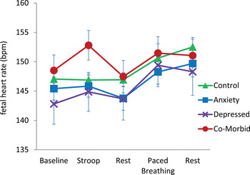
Additional approaches to fetal assessments involve exposing mothers to psychological challenge (e.g., using the Stroop task) or directly stimulating the fetus via a vibroacoustic probe. It has been shown that although fetuses of nonanxious and psychiatrically healthy women do not show a heart-rate change during the Stroop task, fetuses of anxious and depressed women show a significant heart-rate increase (Monk et al., Reference Monk, Fifer, Sloan, Myers, Trien and Hurtado2000, Reference Monk, Myers, Sloan, Werner, Jeon and Tager2004, Reference Monk, Fifer, Myers, Bagiella, Duong and Chen2011; see Figure 2). In the most recent results, maternal cortisol levels were marginally positively associated with fetal heart-rate activity (Monk et al., Reference Monk, Fifer, Myers, Bagiella, Duong and Chen2010). When stimulated directly with a vibroacoustic probe, fetuses of women with elevated levels of corticotropin releasing hormone (CRH; which, during pregnancy, is predominantly of placental origin, and can be increased by maternal cortisol) failed to respond to a novel stimulus in a habituation/dishabituation paradigm. In addition, higher CRH has been related to higher overall fetal heart-rate reactivity to stimulus (Sandman, Wadhwa, Chicz-DeMet, Proto, & Garite, Reference Sandman, Wadhwa, Chicz-DeMet, Porto and Garite1999). Taken together, these findings suggest a similar biobehavioral profile to that of a child exposed to maternal prenatal distress: greater activity and reactivity, and a possible decrement in a rudimentary form of discrimination (e.g., during the habituation studies). Thus, these approaches may provide a valuable new strategy for exploring the early origins of maternal adversity-induced risk of psychopathology.
Fetal heart rate during the laboratory protocol by women's diagnostic status. In a sample of third trimester pregnant women (Monk et al., Reference Monk, Fifer, Myers, Bagiella, Duong and Chen2011), fetuses of women with diagnosed depression and anxiety showed greater heart rate reactivity during Stroop compared to those in the other groups. There was a trend for maternal salivary cortisol to be positively associated with fetal heart rate. [A color version of this figure can be viewed online at http://journals.cambridge.org/dpp]

Effects of Stress, Depression, and Anxiety on Mother–Infant Interactions During the Postpartum Period
Exposure to maternal distress during fetal development is highly correlated with continued exposure during postnatal development, which highlights the importance of considering postnatal factors within the study of prenatal effects. Maternal prenatal depression is strongly associated with risk for postpartum depression (PPD) and one-third of pregnant women with depressive symptomatology have been reported to experience depression during the postpartum period (Beck, Reference Beck1996; Gotlib, Whiffen, Mount, Milnes, Cordy, Reference Gotlib, Whiffen, Mount, Milne and Cordy1989; O'Hara & Swain, Reference O'Hara and Swain1996). Distress during pregnancy is also associated with increased risk for PPD. For example, distress-related risk factors in the prenatal period that predict PPD symptoms include anxiety (Beck, Reference Beck1996; O'Hara & Swain, Reference O'Hara and Swain1996; Sutter-Dallay, Giaconne-Marcesche, Glatigny-Dallay, & Verdoux, Reference Sutter-Dallay, Giaconne-Marcesche, Glatigny-Dallay and Verdoux2004), recent intimate partner violence (Valentine, Rodriguez, Lapeyrouse, & Zhang, Reference Valentine, Rodriguez, Lapeyrouse and Zhang2011), martial adjustment, overall low social support, inadequate support from the baby's father (O'Hara & Swain, Reference O'Hara and Swain1996), and low socioeconomic status (Goyal, Gay, & Lee, Reference Goyal, Gay and Lee2010). When pregnant women were divided into high- and low-risk groups based on depression-related risk factors, 25% of those in the high-risk group (versus 6% in the low-risk group) developed PPD, and depressive symptomatology during pregnancy independently predicted PPD (Verkerk, Pop, Van Son, & Van Heck, Reference Verkerk, Pop, Van Son and Van Heck2003). It seems that when a child experiences an altered in utero environment linked to maternal mood, there likely is continuity to the postpartum environment in the form of maternal PPD or depressive symptoms. This continuity has significant implications for the quality of the postnatal environment, particularly the quality of maternal care.
Healthy mother–infant interactions are critical to the development of healthy children and adults. Specifically, the quality of maternal caregiving behavior (MCB) during infancy has been linked to outcomes such as emotion regulation, social adjustment, stress reactivity, cognitive abilities, and risk for psychopathology. Maternal adversity, in the form of psychosocial distress, negatively affects MCB, and, therefore, results in poor outcomes for children and adolescents. Maternal depression during infancy (or since birth) predicts higher waking cortisol levels during adolescence (Murray, Halligan, Goodyer, & Herbert, Reference Murray, Halligan, Goodyer and Herbert2010), larger amygdala (but not hippocampal) volume and higher cortisol level at 10 years (Lupien et al., Reference Lupien, Parent, Evans, Tremblay, Zelazo and Corbo2011), greater childhood internalizing and total behavior problems (Bagner, Pettit, Lewinsohn, & Seeley, Reference Bagner, Pettit, Lewinsohn and Seeley2010), and greater childhood and adolescent depressive symptoms (Bureau, Easterbrooks, & Lyons-Ruth, Reference Bureau, Easterbrooks and Lyons-Ruth2009). Moreover, adverse outcomes can emerge as early as infancy (Kikkert, Middelburg, & Hadders-Algra, Reference Kikkert, Middelburg and Hadders-Algra2010). Maternal, but not paternal, trait anxiety has been related to less optimal neurological development of the infant as measured by the Towen Infant Neurological Examination at 10 months of age. Even typical variations in MCB predicts differences in infant emotion regulation (Hane & Fox, Reference Hane and Fox2006). Variations in the quality of MCB, assessed in terms of maternal sensitivity and responsiveness during multiple mother–infant interactions, were found to predict infant stress reactivity as indexed by right frontal EEG asymmetry, fear reactivity, and positive sociability with a stranger. Relative to high MCB, low MCB was associated with greater right frontal EEG asymmetry and fear reactivity and less positive sociability, and these findings have been extended to stress reactivity outcomes at 2 and 3 years of age (Hane, Henderson, Reeb-Sutherland, & Fox, Reference Hane, Henderson, Reeb-Sutherland and Fox2010).
Maternal distress and MCB
Assessment of the quality of postnatal mother–infant interactions with a focus on the mother typically measures variables such as maternal sensitivity and responsivity during dyadic interactions such as free play, diaper changing, and feeding. Postnatal maternal depression has been shown to reduce maternal responsivity to infants 6 months of age (Forman et al., Reference Forman, O'Hara, Stuart, Gorman, Larsen and Coy2007) and increase maternal hostility–intrusiveness to infants 18 months of age (Bureau et al., Reference Bureau, Easterbrooks and Lyons-Ruth2009). Mothers with anxiety show hyperarousal during free play with infants 6 months of age (Kaitz, Maytal, Devor, Bergman, & Mankuta, Reference Kaitz, Maytal, Devor, Bergman and Mankuta2010), less sensitivity and reduced emotional tone during free play with infants 10–14 months of age (Nicol-Harper, Harvey, & Stein, Reference Nicol-Harper, Harvey and Stein2007), less maternal sensitivity during free play with infants 9 months of age (Feldman et al., Reference Feldman, Granat, Pariente, Kanety, Kuint and Gilboa-Schechtman2009), and exaggerated behavior during free play and teaching with infants 6 months of age (Kaitz et al., Reference Kaitz, Maytal, Devor, Bergman and Mankuta2010). Finally, maternal stress has been associated with less maternal sensitivity to infant cues in a vocal elicitation paradigm (Crnic, Greenberg, Schettr, Hobel, & Chicz-Demet, 1984).
Maternal distress and infant behavior
Maternal distress can also lead to altered behavioral reactions to mothers. One assessment approach is the use of the Still Face paradigm, during which mothers are asked to become unresponsive to their infants for a brief period of time, and infant reactivity is measured. In a study that compared responses to the Still Face paradigm in 5-month-old infants of depressed versus nondepressed mothers, infants of depressed mothers were found to engage in a self-soothing strategy, whereas infants of nondepressed mothers engaged in an attention strategy (Manian & Bornstein, Reference Manian and Bornstein2009). This finding suggests that infants use varying emotion regulation strategies as a function of maternal depression, and is consistent with earlier work showing that infants of depressed mothers first attempt to use external regulation, and then engage in self-directed regulatory behavior (Gianino & Tronick, Reference Gianino, Tronick and Field1985; Tronick & Gianino, Reference Tronick, Gianino, Tronick and Field1986). Infants have also been observed to display negative affect when interacting with mothers who have depression or with healthy mothers who simulate depression (Gianino & Tronick, Reference Gianino, Tronick and Field1985; Tronick & Gianino, Reference Tronick, Gianino, Tronick and Field1986). Depressed mood in healthy women reduces infant responsivity and infant positive responses (Zekoski, O'Hara, & Wills, Reference Zekoski, O'Hara and Wills1987): infants look less at depressed mothers and appear distressed (Cohn, Matias, Tronick, Connell, & Lyons-Ruth, Reference Cohn, Matias, Tronick, Connell and Lyons-Ruth1986; Tronick, Reference Tronick1989), and infants of depressed mothers show low social engagement (Feldman et al., Reference Feldman, Granat, Pariente, Kanety, Kuint and Gilboa-Schechtman2009). Likewise, maternal anxiety was found to be associated with reduced social engagement among infants (Feldman et al., Reference Feldman, Granat, Pariente, Kanety, Kuint and Gilboa-Schechtman2009), although less negative affect in the Still Face paradigm was observed (Kaitz et al., Reference Kaitz, Maytal, Devor, Bergman and Mankuta2010), and maternal stress was found to be associated with less infant responsivity (Crnic et al., Reference Crnic, Greenberg, Robinson and Ragozin1984). Infant attachment, as measured by the Strange Situation task, also varies significantly in association with maternal distress. Infants of depressed mothers are more likely to show insecure attachment (Martins & Gaffan, Reference Martins and Gaffan2000; McMahon, Barnett, Kowalenko, & Tennant, Reference McMahon, Barnett, Kowalenko and Tennant2006; Teti, Felfand, Messinger, & Isabella, Reference Teti, Felfand, Messinger and Isabella1995), and a disorganized attachment style has been linked to the chronicity of maternal depression (Teti et al., Reference Teti, Felfand, Messinger and Isabella1995). Maternal trauma has likewise been found to predict insecure infant attachment (Lyons-Ruth & Block, Reference Lyons-Ruth and Block1996).
Maternal distress and mother–child interactions
The coordination of behavior between mother and infant may also be sensitive to the effects of maternal distress. For example, dyadic gaze rhythm can be measured to assess the level of predictability and expectancy between a mother and infant when they look at and away from one another's faces. In a study that compared mother–infant dyads with either low- or high-distressed mothers, maternal distress related to less efficient gaze rhythm, suggesting that establishing predictable patterns of eye gaze may be more difficult for dyads of infants and high-distress mothers (Beebe et al., Reference Beebe, Badalamenti, Jaffe, Feldstein, Marquette and Helbraun2008). Predictability of behavioral responses can also be assessed through the observed contingency of behavior between dyads (interactive contingency) or within an individual (self-contingency). Anxious mothers have been found to exhibit heightened (vigilant) interactive and self-contingencies in some modalities and lowered (withdrawn) interactive and self-contingencies in others, whereas infants of anxious mothers exhibited lowered self-contingency (Beebe et al., Reference Beebe, Steele, Jaffe, Buck, Chen and Cohen2011). Anxious mothers were found to engage in heightened visual monitoring, but lowered emotional coordination with their infants. Taken together, these studies suggest that maternal adversity may lead to aberrant forms of social coordination/communication between mothers and infants.
Potential underlying mechanisms
It has been suggested that the primary parenting demands during infancy are (a) to establish a secure attachment with the infant and (b) to foster development of emotion regulation in the infant (Goodman & Gotlib, Reference Goodman and Gotlib1999). In the case of distressed mothers, it appears likely that there is reduced attainment of these goals. Infants of distressed mothers exhibit insecure attachment styles (Martins & Gaffan, Reference Martins and Gaffan2000; McMahon et al., Reference McMahon, Barnett, Kowalenko and Tennant2006; Teti et al., Reference Teti, Felfand, Messinger and Isabella1995) and increased fear reactivity (Feldman et al., Reference Feldman, Granat, Pariente, Kanety, Kuint and Gilboa-Schechtman2009), whereas in healthy participants, the quality of MCB has been linked to reduced fear reactivity (Hane & Fox, Reference Hane and Fox2006). It may also be the case that exposure to maternal depression-associated negative cognition, affect, and behavior affects the infant through social learning or modeling (Goodman & Gotlib, Reference Goodman and Gotlib1999). Infants of depressed mothers tend to show more negative affect, flatter affect, and less positive responsivity (Dawson, Klinger, Panagiotides, Hill, & Spieker, Reference Dawson, Klinger, Panagiotides, Hill and Spieker1992; Gianino & Tronick, Reference Gianino, Tronick and Field1985; Tronick, Reference Tronick1989; Zekoski et al., Reference Zekoski, O'Hara and Wills1987). Infants of depressed mothers may also experience inadequate arousal and stimulation (Field, Reference Field1998). Depressed mothers have been found to be less responsive to verbal utterances (Bettes, Reference Bettes1989) and to provide less tactile stimulation (gentle touching, stroking; Field, Reference Field2002). Tactile experiences during the postnatal period have been demonstrated to influence pain sensitivity, affect, and growth in neonates (Field, Reference Field2010), and the reciprocal tactile stimulation between mother and infant may contribute to increased maternal responsiveness and infant attachment (Bystrova et al., Reference Bystrova, Ivanova, Edhborg, Matthiesen, Ransjo-Arvidson and Mukhamedrakhimov2009; Field, Reference Field2010). Thus, mood-/stress-induced reductions in this form of MCB may serve as a critical mechanism for shifting psychological and physical development in infants.
Epigenetic Perspectives on the Impact of Prenatal Adversity
Studies of the impact of prenatal maternal distress suggest that this form of early life adversity can lead to neurobiological, behavioral, and psychological consequences for infants. Moreover, this distress can also lead to altered mother–infant interactions during the postpartum period, which has been demonstrated to shift developmental trajectories. Thus, it is evident that development is a dynamic process during which shifts in the experiences of the fetus and infant can have profound consequences. In the case of maternal distress, the psychosocial characteristics of the mother can induce these effects, raising the question of how these effects are achieved. An evolving approach that has been applied to address this question involves exploring the biological mechanisms through which environmental exposures shape the activity of genes within the developing organism. This epigenetic perspective has been a significant breakthrough in linking the psychological and physiological experiences of an individual to mechanistic pathways within cells that either enhance or reduce gene expression with consequences for multiple biological and behavioral outcomes.
Epigenetic mechanisms and gene regulation
The term epigenetic is attributed to the developmental biologist Conrad Waddington, who, in the 1940s and 1950s, posited that interactions between genes and their products lead to the diverging characteristics of cells in an organism (Jablonka & Lamb, Reference Jablonka and Lamb2002). This epigenesis of cellular “phenotypes” emphasized the dynamic process of development and the stable maintenance of characteristics once they had emerged. Advances in molecular biology have led to the identification of specific molecular mechanisms through which this process is achieved, and thus the more modern applications of epigenetic refer to those mechanisms. Gene regulation is a complex process and is dependent on a cascade of protein–protein, protein–DNA, and enzymatic interactions that ultimately determine the accessibility of DNA to the process of gene transcription (Turner, Reference Turner2001). DNA methylation and posttranslational histone modifications (Peterson & Laniel, Reference Peterson and Laniel2004; Razin, Reference Razin1998; Umlauf, Goto, & Feil, Reference Umlauf, Goto and Feil2004) are two mechanisms through which this accessibility can be altered (see Figure 3) and have been explored within the context of environmentally induced changes in gene activity that may be relevant to our understanding of the pathways by which prenatal adversity can lead to altered infant and childhood outcomes.
A schematic illustrating epigenetic regulation of gene expression. (a) Gene expression (production of mRNA from DNA) is dependent on the binding of transcription factors to the region upstream of the coding region of the gene. (b) DNA methylation involves the addition of methyl chemical groups (M) to the DNA. This modification reduces accessibility of the DNA to transcription factors, increases the interactions between histone proteins (H) and DNA, and generally leads to decreased gene expression. (c) Modification of histone proteins, such as addition of acetyl chemicals (A), can reduce the interactions between histones and DNA, increase the access of DNA to transcription factors, and lead to increased gene expression. [A color version of this figure can be viewed online at http://journals.cambridge.org/dpp].

DNA methylation and histone modifications
Within DNA, cytosine nucleotides can undergo the process of methylation, which is the addition of a methyl chemical group to the cytosine. This modification is not a mutation and is potentially reversible. When cytosines become methylated, there is generally less accessibility to the DNA (although the location of the methylated cytosine within the gene sequence is an important predictor of this accessibility), so, as a result, DNA methylation is thought to be a process leading to gene silencing (Razin, Reference Razin1998). Methylated cytosines can also attract methyl-binding proteins, which cluster around the DNA and attract enzymes that can shift the state of the histone proteins around which the DNA is wrapped, a process that can further reduce access to the DNA (Fan & Hutnick, Reference Fan and Hutnick2005). The addition of methyl groups to DNA is accomplished via a class of enzymes called DNA methyltransferases (e.g., DNMT1 and DNMT3a/b; Turek-Plewa & Jagodzinski, Reference Turek-Plewa and Jagodzinski2005). Disrupting these enzymes, using targeted gene-deletion techniques, results in genome-wide hypomethylation, impairments in growth, and embryonic or postnatal lethality (Li, Bestor, & Jaenisch, Reference Li, Bestor and Jaenisch1992; Okano, Bell, Haber, & Li, Reference Okano, Bell, Haber and Li1999), which highlights the critical role of DNA methylation in development. In particular, DNA methylation is a process that maintains cellular differentiation. As cells become more specialized, taking on the characteristics of a specific cell type (i.e., muscle cells, neurons, skin cells), DNA methylation patterns must be reliably copied during the process of cell division or the unique characteristics of the cell will be lost when the cell divides. Thus, DNA methylation patterns are heritable during cell division and are also potentially very stable, generating cellular diversity among genetically identical cells (Jones & Taylor, Reference Jones and Taylor1980).
Modification to histone proteins is another critical epigenetic process that can either increase and decrease accessibility to DNA. In order to compact DNA within the cell nucleus, the DNA is wrapped around a core of histone proteins (Turner, Reference Turner2001). The “tails” of these histone proteins can undergo multiple posttranslational modifications that will alter the dynamic interactions between these proteins and the DNA. For example, histone acetylation is a process whereby an acetyl chemical is added to the histone protein tail and is typically associated with increased gene expression due to a looser interaction between the histones and the DNA (Peterson & Laniel, Reference Peterson and Laniel2004; Umlauf et al., Reference Umlauf, Goto and Feil2004). Histone methylation can result in either enhanced or reduced gene expression, depending on the location within the histone tail of the methyl group (Barski et al., Reference Barski, Cuddapah, Cui, Roh, Schones and Wang2007; Koch et al., Reference Koch, Andrews, Flicek, Dillon, Karaoz and Clelland2007). There are many chemicals that can be added or removed from the histone tails, resulting in histone phosphorylation, methylation, and ubiquitination and various other chemical modifications. Taken as a group, these modifications result in a “histone code,” which is a dynamic and complex strategy for reducing or enhancing gene expression (Jenuwein & Allis, Reference Jenuwein and Allis2001). As is the case for DNA methylation, there are specific enzymes that facilitate these epigenetic modifications (Legube & Trouche, Reference Legube and Trouche2003), and disrupting these enzymes can result in significant impairments in development. For example, targeted deletion of a histone acetyltransferase gene can be lethal and induce deficits in neural tube closing (Xu et al., Reference Xu, Edmondson, Evrard, Wakamiya, Behringer and Roth2000; Yao et al., Reference Yao, Oh, Fuchs, Zhou, Ch'ng and Newsome1998); in addition, mutation of the histone methyltransferase euchromatic histone-lysine N-methyltransferase 1 has been found to be associated with severe developmental delay in humans (Kleefstra et al., Reference Kleefstra, Brunner, Amiel, Oudakker, Nillesen and Magee2006).
Epigenetic effects of prenatal adversity
Although epigenetic modifications (particularly DNA methylation) were once thought to be limited in plasticity to early embryonic development, it is becoming evident that both DNA methylation and histone modifications are dynamic and changeable across the life span (Champagne, Reference Champagne2010). Given this plasticity, epigenetic variation may be induced by experiences occurring during prenatal and postnatal development and therefore may be a mechanistic link between maternal adversity and infant outcomes. In humans, the epidemiological studies involving the Dutch Hunger Winter birth cohort have explored whether DNA methylation is altered as a function of this early-life exposure. Analysis of whole blood samples from exposed versus nonexposed sibling pairs has indicated that periconceptual exposure to conditions of famine/stress are associated with differential methylation of several genes among adults (Heijmans et al., Reference Heijmans, Tobi, Stein, Putter, Blauw and Susser2008; Tobi et al., Reference Tobi, Lumey, Talens, Kremer, Putter and Stein2009). In particular, decreased DNA methylation was found within the insulin-like growth factor II (IGF2) gene among exposed compared to nonexposed siblings (Heijmans et al., Reference Heijmans, Tobi, Stein, Putter, Blauw and Susser2008). IGF2 plays a significant role in growth and development (Smith, Garfield, & Ward, Reference Smith, Garfield and Ward2006) and is likely a key factor in regulating many of the outcomes associated with prenatal adversity. Within this cohort, several other genes, including insulin/insulin-like growth factor-1, interleukin-10, leptin, ATP-binding cassette transporter 1, guanine nucleotide binding protein (G protein), alpha stimulating activity polypeptide 1 antisense RNA 1, and maternally expressed 3 were found to have altered DNA methylation levels (Tobi et al., Reference Tobi, Lumey, Talens, Kremer, Putter and Stein2009), suggesting widespread epigenetic effects of this early life exposure. These effects appear to be timing specific, with differential DNA methylation most evident when exposure occurred during the periconceptual period rather than in late gestation.
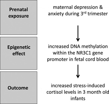
Although sibling studies within the Dutch Hunger Winter cohort did not find significant effects on genes associated primarily with the HPA axis, such as CRH and the glucocorticoid receptor (nuclear receptor subfamily 3, group C, member 1 [NR3C1]), studies of prenatal maternal stress and depression have provided evidence of epigenetic dysregulation within HPA pathways. Analysis of cord blood samples from infants born to mothers with elevated ratings of depression (using the Hamilton Depression Scale) during the third trimester of pregnancy indicated elevated levels of DNA methylation within the NR3C1 gene (Oberlander et al., Reference Oberlander, Weinberg, Papsdorf, Grunau, Misri and Devlin2008). These epigenetic effects were observed in fetal but not maternal blood samples. At 3 months of age, infant HPA reactivity was assessed using a habituation information processing task during which salivary cortisol levels were measured. Levels of NR3C1 DNA methylation in fetal cord blood were found to predict infant cortisol response to stress, suggesting a functional consequence of this epigenetic variation (see Figure 4). The persistence of these prenatal effects on NR3C1 DNA methylation beyond infancy has also been demonstrated. Children and adolescents (age 10–19 years) born to women who had experienced stress in the form of intimate partner violence during pregnancy were found to have elevated NR3C1 DNA methylation levels in whole blood samples (Radtke et al., Reference Radtke, Ruf, Gunter, Dohrmann, Schauer and Elbert2011). As was observed in the case of prenatal maternal depression, these epigenetic effects were present in offspring blood samples, but were not evident in maternal blood samples. Methylation within the gene encoding the serotonin transporter (solute carrier family C6 member 4 [SLC6A4]) gene, has been found to be reduced in cord blood samples in response to increased maternal depressed mood during the second trimester, whereas no effects on brain-derived neurotrophic factor (BDNF) were observed (Devlin, Brain, Austin, & Oberlander, Reference Devlin, Brain, Austin and Oberlander2010).
The impact of prenatal maternal depression/anxiety on DNA methylation and infant stress response. Oberlander et al. (Reference Oberlander, Weinberg, Papsdorf, Grunau, Misri and Devlin2008) found that maternal depression and anxiety assessed during pregnancy (third trimester) was significantly correlated with the degree of DNA methylation within the glucocorticoid receptor gene (nuclear receptor subfamily 3, group C, member 1 [NR3C1]) measured in fetal cord blood. At 3 months of age, this epigenetic effect was found to predict increased infant cortisol response to stress.

Although there has been increasing exploration of epigenetic effects in human cohort studies, the meaning of these epigenetic changes in blood is unclear. However, animal models have demonstrated that, under controlled laboratory conditions, exposure to prenatal stress can induce epigenetic variation within brain regions involved in the behavioral characteristics associated with this form of prenatal adversity. In mice, chronic variable stress exposure during the first gestational week (first trimester) results in increased corticosterone response to stress and increased depressive-like behavior (Mueller & Bale, Reference Mueller and Bale2008). In adult male offspring, prenatal stress is associated with decreased DNA methylation of the CRH gene and increased methylation of the NR3C1 gene in hypothalamic tissue of adult male offspring. Moreover, there is altered expression of these genes (increased CRH expression and decreased glucocorticoid receptor expression) in response to prenatal stress, which probably induces increased HPA reactivity in offspring. Thus, animal studies can be used to provide support for the validity of target genes identified in human studies using peripheral blood samples, which enable us to explore the mechanistic pathways through which prenatal adversity alters the brain and behavior.
Epigenetic Effects During Postnatal Development
The epigenetic impact of postnatal mother–infant interactions has also been explored in both humans and animals and, as has been previously described, may be an important consideration in studies of prenatal adversity. Deprivation of parental care, such as that observed in institutionalized infants, has been found to have broad epigenetic consequences. Among institution reared (since birth) children aged 7–10 years, analysis of blood samples indicates increased DNA methylation throughout the genome when compared to age-matched children reared by their biological parents (Naumova et al., Reference Naumova, Lee, Koposov, Szyf, Dozier and Grigorenko2012). Among the differentially methylated genes are those implicated in brain development, including genes within vasopressinergic, serotonergic, glutamatergic, and GABAergic pathways. The epigenetic effects of childhood abuse have also been observed in human brain tissue (McGowan et al., Reference McGowan, Sasaki, D'Alessio, Dymov, Labonte and Szyf2009). Analysis of gene expression and DNA methylation levels in postmortem hippocampal tissue from adults with or without a documented history of childhood abuse has indicated decreased expression of the NR3C1 gene and increased DNA methylation within the regulatory region of the NR3C1 gene associated with a history of childhood abuse. Decreased levels of glucocorticoid receptors within the hippocampus can lead to a heightened HPA response to stress and, in particular, a decreased capacity to regulate the stress response. As such, the epigenetic effects on NR3C1 induced by postnatal adversity may account for the increased risk of psychopathology and poorer emotional regulation among individuals with a history of abuse.
Variation in maternal care in rodents
Although there is certainly emerging data from human cohorts indicating the impact of postnatal experiences on epigenetic pathways, it is animal studies that have provided a significant basis for this exploration. In particular, studies of laboratory rodents illustrate the neurobiological and behavioral consequences of variations in maternal care within the normal range, and the role of epigenetic mechanisms in the long-term consequences of maternal care on offspring outcomes (Meaney, Reference Meaney2001; Meaney & Szyf, Reference Meaney and Szyf2005). Rodents provide tactile stimulation to offspring through licking/grooming (LG) of pups and this form of maternal care has been found to influence HPA reactivity, cognition, and the reproductive behavior of offspring (Caldji et al., Reference Caldji, Tannenbaum, Sharma, Francis, Plotsky and Meaney1998; Cameron et al., Reference Cameron, Del Corpo, Diorio, McAllister, Sharma and Meaney2008a; Champagne et al., Reference Champagne, Bagot, van Hasselt, Ramakers, Meaney and de Kloet2008; Champagne, Francis, Mar, & Meaney, Reference Champagne, Francis, Mar and Meaney2003; Liu, Diorio, Day, Francis & Meaney, Reference Liu, Diorio, Day, Francis and Meaney2000; Liu et al., Reference Liu, Diorio, Tannenbaum, Caldji, Francis and Freedman1997). Gene expression within the hippocampus and hypothalamus has been found to be altered in adult offspring that experienced low versus high levels of LG during the postnatal period, and may account for the behavioral and physiological effects of LG. Males that have experienced low versus high LG are observed to have reduced levels of hippocampal glucocorticoid receptors within the hippocampus, which probably accounts for the increased HPA reactivity of these offspring (Liu et al., Reference Liu, Diorio, Tannenbaum, Caldji, Francis and Freedman1997). Analysis of DNA methylation within the NR3C1 gene indicates that experiencing low levels of LG is associated with increased DNA methylation at several sites within this gene (Weaver et al., Reference Weaver, Cervoni, Champagne, D'Alessio, Sharma and Seckl2004). These adult patterns of DNA methylation are not observed prenatally or at the time of birth, but instead emerge during the first postnatal week. Histone acetylation (histone H3 lysine 9) is also reduced within the NR3C1 gene in low-LG compared to high-LG offspring. It is worth noting that these early life effects on NR3C1 can be modified in adulthood through pharmacological manipulations that increase histone acetylation or increase the potential for DNA methylation, which suggests a high degree of plasticity within these epigenetic pathways (Weaver et al., Reference Weaver, Cervoni, Champagne, D'Alessio, Sharma and Seckl2004, Reference Weaver, Champagne, Brown, Dymov, Sharma and Meaney2005). Maternal LG also leads to altered levels of DNMT1 in the hippocampus, so that male offspring that receive low levels of LG have elevated levels of DNMT1 mRNA (Zhang et al., Reference Zhang, Hellstrom, Bagot, Wen, Diorio and Meaney2010). In addition to hippocampal glucocorticoid receptors, maternal LG has been found to alter expression of the enzyme glutamic acid decarboxylase gene, and analysis of DNA methylation within the glutamic acid decarboxylase gene indicates that there is heighted DNA methylation and reduced histone acetylation of this gene that can be observed to persist into adulthood among offspring that experience low levels of postnatal LG (Zhang et al., Reference Zhang, Hellstrom, Bagot, Wen, Diorio and Meaney2010). Genome-wide analysis of gene-expression profiles in the hippocampus of male offspring that have received low versus high levels of maternal care indicate that over 900 genes are differentially expressed in the hippocampus as a function of maternal LG, and that, overall, there is increased gene expression among adult offspring of high LG compared to low LG dams (Weaver, Meaney, & Szyf, Reference Weaver, Meaney and Szyf2006). These findings suggest widespread epigenetic consequences of maternal care.
The effects of maternal LG on female offspring have primarily focused on reproductive outcomes such as sexual and maternal behavior (Cameron et al., Reference Cameron, Del Corpo, Diorio, McAllister, Sharma and Meaney2008a; Cameron, Fish, & Meaney, Reference Cameron, Fish and Meaney2008b; Champagne, Francis, et al., 2003), although it should be noted that females display similar stress responsivity profiles to their male siblings as a function of maternal LG (e.g., low LG associated with behavioral inhibition to novelty; Champagne & Meaney, Reference Champagne and Meaney2007). Among female offspring that received low levels of LG during the postnatal period, there is decreased mRNA and protein levels of estrogen receptor alpha (ERα) in the medial preoptic are (MPOA), which may account for the reduced estrogen sensitivity and maternal care evident in these offspring (Champagne, Diorio, Sharma, & Meaney, Reference Champagne, Diorio, Sharma and Meaney2001; Champagne, Weaver, Diorio, Sharma, & Meaney, Reference Champagne, Weaver, Diorio, Sharma and Meaney2003). Analysis of levels of DNA methylation within the ERα gene (ESR1) in MPOA tissue indicates that offspring that experience low LG have elevated ESR1 DNA methylation in adulthood (Champagne et al., Reference Champagne, Weaver, Diorio, Dymov, Szyf and Meaney2006). This persistent epigenetic effect may account for the transmission of maternal LG behavior across generations (Champagne, Reference Champagne2008), and illustrates the profound effect of early rearing experiences on the neurobiological circuits involved in reproductive behavior. Experience-driven epigenetic effects on ESR1 may also lead to sexual dimorphism. Within the MPOA there are sex differences in ESR1 DNA methylation such that males have higher levels of ESR1 DNA methylation in this brain region compared to that of females (Kurian, Olesen, & Auger, Reference Kurian, Olesen and Auger2010). If females are provided with additional tactile stimulation during postnatal days 5–7, however, there are increases in ESR1 DNA methylation, such that females become indistinguishable from males. Previous studies have indicated that male pups receive elevated levels of maternal LG compared to females (Moore, Reference Moore1984; Moore & Morelli, Reference Moore and Morelli1979), so that it may be the case that differential levels of LG can trigger epigenetic pathways that enhance sex differences in the brain. Moreover, the frequency of LG may be an important consideration in studies of prenatal adversity because this form of maternal care has been found to be reduced among females that experienced stress during gestation (Champagne & Meaney, Reference Champagne and Meaney2006).
Epigenetic effects of neglect and abuse in animal models
Maternal separation studies with laboratory primates and rodents have been used as a strategy to model early maternal deprivation/neglect and have demonstrated the impact of this form of postnatal adversity on neurobiological and behavioral outcomes (Lehmann et al., Reference Lehmann, Pryce, Jongen-Relo, Stohr, Pothuizen and Feldon2002; Spinelli et al., Reference Spinelli, Chefer, Suomi, Higley, Barr and Stein2009; Suomi, Eisele, Grady, & Harlow, Reference Suomi, Eisele, Grady and Harlow1975; Suomi, Harlow, & Kimball, Reference Suomi, Harlow and Kimball1971). Rhesus macaques reared under conditions of maternal separation and possess the risk allele of the SLC6A4 gene (the short version which has been associated with increased anxiety-like behavior) were found to have increased DNA methylation of SLC6A4 gene in blood samples (Kinnally et al., Reference Kinnally, Capitanio, Leibel, Deng, LeDuc and Haghighi2010) . These findings highlight the interaction between genetic variation and epigenetic effects in response to early life adversity, which is likely an important consideration in human studies. In mice, maternal separation occurring daily during the postnatal period leads to increased stress sensitivity and an upregulation in the expression of hypothalamic vasopressin (AVP) that can be observed in adulthood (Murgatroyd et al., Reference Murgatroyd, Patchev, Wu, Micale, Bockmuhl and Fischer2009). DNA methylation levels within key regulatory regions of the AVP gene have been found to vary as a function of exposure to postnatal maternal separation, such that maternally separated offspring have decreased hypothalamic AVP DNA methylation compared to normally reared offspring. This separation-induced hypomethylation of AVP was also associated with reduced levels of binding of MeCP2 (a methyl-binding protein) to the AVP gene (Murgatroyd et al., Reference Murgatroyd, Patchev, Wu, Micale, Bockmuhl and Fischer2009).
Maternal abuse of offspring can also be modeled in laboratory rodents and has illustrated the role of epigenetic modifications in maintaining the effects of abuse across the life span. In laboratory rats, aggressive behavior toward pups (such as stepping on pups, aggressive grooming, or transporting of pups by a limb) can be induced by reducing the nesting materials provided to lactating dams during the postnatal period (Roth & Sullivan, Reference Roth and Sullivan2005). This manipulation also reduces the frequency of nurturing behaviors such as maternal LG. When pups are exposed to caregivers (nonbiological mothers) that are provided with limited nesting materials and placed in an unfamiliar environment, there is increased exposure of pups to aggressive encounters (Roth, Lubin, Funk, & Sweatt, Reference Roth, Lubin, Funk and Sweatt2009). Within the prefrontal cortex of abuse-exposed offspring, there is a downregulation of the expression of BDNF. Analysis of DNA methylation within the regulatory regions of the BDNF gene indicates that there are very low levels of DNA methylation among nonabused offspring whereas abused offspring have elevated levels of this repressive epigenetic modification (Roth et al., Reference Roth, Lubin, Funk and Sweatt2009). These epigenetic effects of abuse are also observed in the offspring of abused versus nonabused females, possibly suggesting an epigenetic basis to the multigenerational transmission of abuse and its consequences for neurobiological and behavioral functioning.
Gene Regulation in the Placenta and Fetal Development
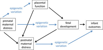
The mechanistic pathways linking prenatal adversity to infant development are likely to be very complex, varying by the timing of exposure, the type of exposure, and underlying genetic and environmental risk factors. However, the functioning of the placenta will probably be of critical importance within these diverse pathways. This temporary endocrine structure regulates the transfer of nutrients to the developing fetus, buffers the fetus from toxins and maternal glucocorticoids, and, by altering maternal hormone levels, can influence maternal mood and the priming of the maternal brain with consequences for the quality of postnatal mother–infant interactions (Cottrell & Seckl, Reference Cottrell and Seckl2009; Desforges & Sibley, Reference Desforges and Sibley2010; Levy, Reference Levy1981; Mann & Bridges, Reference Mann and Bridges2001; see Figure 5). Thus, gene regulation within the placenta can have functional consequences for both fetal and infant development. Gene transcription within the placenta varies during the course of gestation and it is likely that epigenetic mechanisms, such as variation in the levels of placental DNA methyltransferase levels, account for the temporal shifts in placental gene expression profiles (Novakovic et al., Reference Novakovic, Wong, Sibson, Ng, Morley and Manuelpillai2010). Altered gene expression within the placenta has also been associated with adverse birth outcomes, such as intrauterine growth retardation (Angiolini et al., Reference Angiolini, Fowden, Coan, Sandovici, Smith and Dean2006; Lee et al., Reference Lee, Jeon, Lee, Park, Jung and Kim2010; McCarthy et al., Reference McCarthy, Cotter, McElwaine, Twomey, Mooney and Ryan2007; McMinn et al., Reference McMinn, Wei, Schupf, Cusmai, Johnson and Smith2006; Sitras, Paulssen, Leirvik, Vartun, & Acharya, Reference Sitras, Paulssen, Leirvik, Vartun and Acharya2009). Many of the genes identified as differentially expressed in these studies are imprinted genes (McMinn et al., Reference McMinn, Wei, Schupf, Cusmai, Johnson and Smith2006; Tycko, Reference Tycko2006), that is, genes subject to epigenetic silencing mechanisms to achieve parent of origin expression patterns, and there is emerging evidence for altered DNA methylation levels both genome wide and within specific target genes that may account for this altered transcriptional profile (Lambertini et al., Reference Lambertini, Lee, Chan, Lee, Diplas and Wetmur2011; Nelissen, Van Montfort, Dumoulin, & Evers, Reference Nelissen, van Montfoort, Dumoulin and Evers2011; Tabano et al., Reference Tabano, Colapietro, Cetin, Grati, Zanutto and Mando2010).
Pathways linking prenatal maternal distress to infant outcomes. There are both direct and indirect pathways through which epigenetic modifications may shape infant outcomes associated with prenatal maternal distress. Within these pathways, postnatal maternal distress and placental function are critical factors. [A color version of this figure can be viewed online at http://journals.cambridge.org/dpp]

It is interesting that there is significant overlap among genes identified as differentially methylated in the placenta in studies of fetal growth retardation and those genes identified as differentially methylated in blood samples from famine-exposed individuals in the Dutch Famine cohort (e.g., IGF2; guanine nucleotide binding protein, alpha stimulating activity polypeptide 1; leptin; maternally expressed 3; Heijmans et al., Reference Heijmans, Tobi, Stein, Putter, Blauw and Susser2008; McMinn et al., Reference McMinn, Wei, Schupf, Cusmai, Johnson and Smith2006; Tobi et al., Reference Tobi, Lumey, Talens, Kremer, Putter and Stein2009). Thus, it is likely that maternal distress can lead to altered gene expression and epigenetic profiles in placental tissues. In mice, chronic variable stress during the first trimester is associated with altered placental gene expression, including altered mRNA levels of DNMT1 (Mueller & Bale, Reference Mueller and Bale2008). One gene target of particular importance in linking maternal distress to fetal outcomes (particularly the HPA reactivity that is often observed) is the enzyme 11β-hydroxysteroid dehydrogenase 2 gene (11ß-HSD2) encoding the enzyme 11β-hydroxysteroid dehydrogenase 2. 11ß-HSD2 is highly expressed in the placenta and functions to inactivate glucocorticoids (Cottrell & Seckl, Reference Cottrell and Seckl2009). Thus, the degree of expression of 11ß-HSD2 in the placenta is predicted to influence the exposure of the fetus to circulating maternal stress hormones. In humans, heightened maternal anxiety during pregnancy has been found to be negatively correlated with placental 11ß-HSD2 mRNA levels (O'Donnell et al., Reference O'Donnell, Bugge Jensen, Freeman, Khalife, O'Connor and Glover2011), and reduced placental 11ß-HSD2 mRNA levels have also been found to be associated with intrauterine growth retardation and preterm birth (Borzsonyi et al., Reference Borzsonyi, Demendi, Pajor, Rigo, Marosi and Agota2012; Dy, Guan, Sampath-Kumar, & Richardson, & Yang, Reference Dy, Guan, Sampath-Kumar, Richardson and Yang2008). In rats, chronic restraint stress during gestational days 11–20 was found to decrease placental 11ß-HSD2 mRNA levels of this gene (Mairesse et al., Reference Mairesse, Lesage, Breton, Breant, Hahn and Darnaudery2007) and increased DNA methylation within the 11ß-HSD2 gene may account for this downregulation of gene expression. Thus, it is likely that the placenta is a central target of maternal distress effects and a key mechanistic link between maternal distress and infant outcomes.
Future Directions
Although both the acute and long-term consequences of prenatal exposure to maternal distress are evident in humans, the biological mechanisms through which these effects are achieved have yet to be fully elucidated. Exploration of epigenetic factors within the study of maternal distress may provide a promising strategy for identifying the pathways through which development is altered as a function of prenatal adversity, and there is increasing evidence in humans for the epigenetic consequences of the early life environment. From this emerging literature, it is clear that there are critical methodological issues and empirical questions that must be addressed if we are to advance our understanding of the mechanisms of prenatal effects. First, the moderating and mediating influence of postnatal experiences following prenatal adversity must be carefully considered, because prenatal adversity is highly predictive of postnatal adversity and postnatal experiences, particularly those involving variations in mother–infant interactions, which can have significant epigenetic, neurobiological, and behavioral consequences. The investigation of prenatal–postnatal interplay in shaping development may provide insights into the pathways through which fetal exposures lead to childhood and adolescent outcomes, as well as identifying potential interventions by which the long-term consequences of fetal adversity can be attenuated. Second, the validity and interpretation of human peripheral tissue samples in the study of epigenetic effects must be carefully considered. Unlike genetic markers, epigenetic variation is likely to occur within and across tissue types. Although fetal exposures will likely have system-wide consequences for epigenetic modifications and gene expression, the nature of this consequence may differ in brain cells compared to blood cells, thus limiting our capacity to explore the pathophysiology of psychological and behavioral outcomes linked to fetal adversity based exclusively on peripheral tissue samples. To address this issue, animal models in which both brain and blood epigenetic profiles can be compared and responsiveness to environmental exposures assessed, could provide an essential methodological tool. Finally, from both a neuroendocrine and epigenetic perspective, the placenta is a critical interface between maternal adversity and fetal/infant outcomes (see Figure 5). Continued investigation of the impact of prenatal adversity on placental function and gene regulation may contribute significantly to our understanding of the routes through which maternal psychosocial characteristics induce biological effects in the developing fetus. Future studies addressing these issues and incorporating an epigenetic perspective in the study of prenatal maternal distress have the potential to illustrate the complex interplay between genes and environments in shaping infant outcomes and to determine the mechanisms, both genetic and epigenetic, through which the transmission of risk of psychopathology from mothers to infants is achieved.