Introduction
Helminth proteinases in excretory–secretory products (ESP), affect a broad range of biological processes including nutrition, digestion, blood coagulation, host immune evasion, and tissue penetration (Tort et al., Reference Tort, Brindley, Knox, Wolfe and Dalton1999). Most proteinases characterized in parasitic helminths are cysteine proteinases: for example, cathepsin B-like proteinases are expressed in the intestine of Haemonchus contortus and may serve nutrient digestion (Jasmer et al., Reference Jasmer, Roth and Myler2001); cathepsin L- and Z- like proteinase genes are associated with larval moulting in parasitic nematodes, and may have multifunctional roles in worm development (Guiliano et al., Reference Guiliano, Hong, McKerrow, Blaxter, Oksov, Liu, Ghedin and Lustigman2004; Hashmi et al., Reference Hashmi, Zhang, Oksov and Lustigman2004). Serine- and metallo-proteinases may facilitate host tissue penetration and have an anticoagulant function in Strongyloides stercoralis, Toxocara canis and Trichostrongylus vitrinus (McKerrow, Reference McKerrow1989; Robertson et al., Reference Robertson, Bianco, McKerrow and Maizels1989; McKerrow et al., Reference McKerrow, Brindley, Brown, Gam, Staunton and Neva1990; MacLennon et al., Reference MacLennon, Gallagher and Knox1997). Specifically, an astacin-like metallo-proteinase secreted by Ancylostoma caninum larvae, enables tissue migration (Williamson et al., Reference Williamson, Lustigman, Oksov, Deumic, Plieskatt, Mendez, Zhan, Bottazzi, Hotez and Loukas2006). Aspartyl proteinases have been implicated in parasite feeding and/or immune evasion (Geldhof et al., Reference Geldhof, Claerebout, Knox, Agneessens and Vercruysse2000). Secreted nematode immunomodulators such as cysteine protease inhibitors (cystatins) in filarial parasites interfere with different arms of the host immune response (reviewed by Hartmann & Lucius, Reference Hartmann and Lucius2003). As these proteinases serve such significant functions for these parasites, they are appropriate targets for host chemotherapy and anti-parasite vaccines (McKerrow, Reference McKerrow1989, Reference McKerrow1999).
In the free-living nematode Caenorhabditis elegans, cathepsin B-like proteinase genes (CBL) occur as a large multigene family and several CBLs are expressed in the intestine (Ray & McKerrow, Reference Ray and McKerrow1992; Larminie & Johnstone, Reference Larminie and Johnstone1996). A cathepsin-Z like proteinase has a multifunctional role in C. elegans development, and a cathepsin L-like proteinase, essential for early embryogenesis in C. elegans, is functionally conserved in the parasitic nematode H. contortus (Britton & Murray, Reference Britton and Murray2002).
The functional success in parasitic nematodes of their proteinases raises the issue of their origins: are they inherited from free-living ancestral forms, or evolved in whole or in part in response to parasitism? As this issue does not seem to have been addressed experimentally, we investigated the ESP of the genetically-significant and free-living nematode C. elegans, firstly to characterize the C. elegans ESP (CeESP) in terms of subunit composition, pH optima, substrate specificity and inhibitor sensitivity in order to identify the classes and specific proteinases present and secondly to explore the antigenic relatedness of CeESP with that of the well known equine parasite, Strongylus vulgaris (SvESP).
Materials and methods
ESP preparation
Caenorhabditis elegans ESP was collected through the bulk growth method (Epstein & Shakes, Reference Epstein and Shakes1995). Briefly, nematodes were grown in liquid culture (S Medium) containing Escherichia coli strain Na22, for 5 days at 20°C, after which the medium was aspirated, allowed to settle at 4°C, and sterile-filtered through a 0.2 μm Millipore filter. The medium was then concentrated over an YM10 flat ultrafiltration membrane with a 10 kDa cut off in a model 8010-stirred cell. A control incubation was set up with E. coli in S Medium only (C). Protein concentration of the ESP was quantified using a bicinchoninic (BCA) protein assay kit in 96-well microtitre plates with bovine serum albumin as the protein standard.
Synthetic fluorogenic substrates analysis
Hydrolysis of several NMec peptide substrates was determined over a broad pH range (3.0–9.0). 5 μl aliquots (0.74 μg) of C. elegans ESP were assayed for cathepsin-like activity using the fluorogenic substrates, Z-Phe-Arg-NMec and Z-Arg-Arg-NMec, and trypsin- and chymotrypsin-like activity of serine proteinases using the fluorogenic substrates Z-Gly-Gly-Arg-NMec and succinyl-Ala-Ala-ProPhe-NMec, respectively. After preincubating C. elegans ESP for 10 min at 37°C in a designated buffer, the reactions were initiated by introducing the appropriate peptide substrate at a final concentration of 35 μm. Reactions were stopped by the addition of iodoacetic acid (IA) to a final concentration of 10 mm following incubation at 37°C for 12 min in the presence of DTT, a cysteine activator, and for 20 min in its absence. Phenylmethylsulphonyl fluoride (PMSF) (10 mm) was used to stop the reaction with serine substrates. Fluorescence was measured in a final volume of 2 ml (made up with water) using a Perkin Elmer LS-5 luminescence spectrometer (excitation 365 and 455 nm). Proteinase activity was expressed as nmol NMec released min− 1mg− 1 protein, as calculated from a standard curve constructed with free NMec. All activities were corrected for non-enzymatic hydrolysis by subtraction of the appropriate blank. Designated buffers were as follows: 0.1 m sodium citrate (pH 3.0, 4.0, 4.5, 5.0 and 5.5), 0.1 m sodium phosphate (pH 6.0 and 6.5) and 0.1 m Tris-HCl (pH 7.0, 8.0 and 9.0).
Fluorogenic inhibition studies
Inhibitors with specificity for cysteine- (E-64, 30 μm and IA, 1 mm), serine- (PMSF, 2 mm), aspartic- (pepstatin A, 10 μm), and metallo- (EDTA, 10 mm and 1,10-phenanthroline, 1 mm) proteinases were assayed as follows. ESP was preincubated with inhibitors in buffer containing 5 mm DTT for 10 min prior to addition of substrate. Incubation was at 37°C for 12 min (with cysteine substrates Z-Phe-Arg-NMec and Z-Arg-Arg-NMec) or 40 min (with serine substrates Z-Gly-Gly-NMec and N-Suc-Ala-Ala-ProPhe-NMec). Leupeptin, which inhibits serine and cysteine proteinases, was used at a final concentration of 10 μm. Designated buffers were as described above. All substrates and inhibitors were obtained from Sigma-Aldrich Ireland Ltd.
Gelatin-substrate SDS–PAGE and inhibition studies
Electrophoretic separations using 3% stacking gels and 10% resolving gels were performed as described (Laemmli, Reference Laemmli1970) but with the addition of 0.1% bovine skin gelatin (Sigma-Aldrich Ireland Ltd) to the resolving gels. Samples incorporating 10 μl (2.98 μg) ESP and 5 μl sample buffer were applied to the stacking gel without boiling. Mini-gels were run at 22 mA for 2–3 h at 4°C.
After electrophoresis, gels were washed for 30 min at room temperature in buffer containing 2.5% (v/v) Triton X-100 to remove SDS, and incubated for 16 h at 37°C in a similar buffer, but without Triton X-100. The buffers used were: 0.1 m sodium citrate, pH 3.0 and pH 5.5; and 0.1 m sodium phosphate, pH 6.0 and 0.1 m Tris-HCl, pH 8.0. Gels were stained overnight in Coomassie Brilliant Blue stain and destained for approximately 1 h to visualize proteolytic activities, which appear as clear bands on a blue background.
Inhibition of enzyme activity was assayed by preincubating ESP separately with the various inhibitors at room temperature for 30 min at pH 5.5 and 8.0, followed by the addition of sample buffer and electrophoretic analysis. This entailed incubating individual excised gel tracks in buffer containing each inhibitor at the final concentrations detailed above.
SDS–PAGE and immunoblotting
SDS–PAGE was carried out in mini slab gels (Atto Corporation) on C. elegans ESP by: incorporating 10 μl ESP (5.6 μg) with 5 μl sample buffer (incorporating 5 mm DTT) and boiling for 2–3 min before loading onto a 3% stacking gel and resolving through a 12.5% resolving gel (Laemmli, Reference Laemmli1970), for 2–3 h at room temperature. After electrophoresis, gels were stained using Coomassie Brillant Blue stain, and the Biorad® Silver Stain procedure, or electrophoretically transferred to 0.45 μm nitrocellulose membrane (Bio-Rad) in a Bio-Rad Trans-Blot Cell (70 mA at 4°C overnight). Pre-stained molecular weight markers were used to confirm protein transfer.
The membrane was blocked for 1 h in blocking solution (BS, Tris-buffered saline (TBS), pH 7.4, with 5% Marvel, non-fat dried milk). The membrane was then incubated for 1 h at room temperature or overnight at 4°C in BS containing a primary antibody: (i) polyclonal anti-C. elegans Cpr-1 (antiserum to C. elegans cathepsin B, expressed in the gut, and kindly provided by Dr C. Britton, University of Glasgow), diluted 1:500; (ii) mouse polyclonal anti-C. elegans Cpl-1 (antiserum to C. elegans cathepsin L, generated from the mature region of C. elegans cathepsin L gene, and kindly provided by Dr Sarwar Hashmi and Dr J. Zhang of the Laboratory of Molecular Parasitology, Lindsley F. Kimball Research Institute, New York Blood Centre), diluted 1:500; (iii) partially purified HRP-conjugated anti-S. vulgaris (28–30 kDa) diluted 1:100 (see Mobarak & Ryan, Reference Mobarak and Ryan1998); and (iv) monoclonal anti-S. vulgaris 6.5 kDa, both available from this laboratory, courtesy of Dr Wen Chung. The membranes were then washed in TBS-T (Tris-buffered saline containing 0.1% Tween 20, 6 × 5 min) and incubated for 1 h with HRP-conjugated anti-rabbit IgG or HRP-conjugated anti-mouse IgG (Pierce), diluted 1:1500 in BS.
Membranes were then washed, incubated in SuperSignal® West Pico Chemiluminescent substrate (Pierce) and developed using Kodak® BioMax light film.
Results
Synthetic fluorogenic substrates
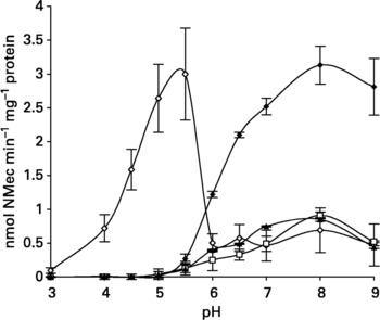
Z-Phe-Arg-NMec preferentially cleaved CeESP over Z-Arg-Arg-NMec, with a maximum of 3 nmol min− 1mg− 1 protein released at the optimal pH of 5.5. This hydrolysis was enhanced up to 12-fold by the addition of DTT at the optimal pH of 5.5, whereas maximum hydrolysis of Z-Arg-Arg-NMec (1.02 nmol min− 1mg− 1 protein released) occurred at pH 9.0, in the absence of DTT (fig. 1).
pH dependence of hydrolysis by Caenorhabditis elegans ESP of the fluorogenic substrates Z-Phe-Arg-NMec (⋄, in the presence of 5 mm DTT), Z-Arg-Arg-NMec (□, in the presence of 5 mm DTT), Z-Gly-Gly-Arg-NMec (▲, in the absence of DTT) and N-Suc-Ala-Ala-ProPhe-NMec (♦, in the absence of DTT). Mean values from three replicates of two independent collections of C. elegans ESP ( ± S.E.M). There was no hydrolysis of fluorogenic substrates by Escherichia coli as control.

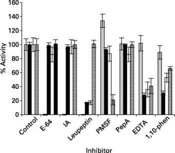
After preincubation at the optimum pH, hydrolysis of Z-Phe-Arg-NMec was abolished by all three cysteine proteinase inhibitors E-64, IA and leupeptin while serine, aspartic and metallo-proteinase inhibitors had little effect ( ≤ 11% inhibition) (fig. 2). Leupeptin, EDTA and 1,10-phenanthroline inhibited hydrolysis of Z-Arg-Arg-NMec by 83, 72 and 69% respectively (fig. 2).
Effects of proteinase inhibitors on hydrolysis of fluorogenic substrates by Caenorhabditis elegans ESP. Hydrolysis of Z-Phe-Arg-NMec () was at pH 5.5, and Z-arg-Arg-NMec (■), Z-Gly-Gly-Arg-NMec (□) and N-Suc-Ala-Ala-NMec () at pH 8.0, the pH optima respectively. Mean values from three replicates of two independent collections of C. elegans ESP ( ± S.E.M). Enzymatic activity is given as a percentage of that in control samples (no inhibitor).

Hydrolysis of Z-Gly-Gly-Arg-NMec, assayed for trypsin-like activity, was optimal at pH 8.0 with 0.85 nmol min− 1mg− 1 protein released. Hydrolysis of N-Suc-Ala-Ala-ProPhe-NMec, assayed for chymotrypsin-like activity, also was optimal at pH 8.0 with 3.13 nmol min− 1mg− 1 protein released; N-Suc-Ala-Ala-ProPhe-NMec was the superior substrate with an almost four-fold higher release rate of NMec than Z-Gly-Gly-Arg-NMec (fig. 1). Cysteine and aspartic proteinase inhibitors had little effect ( ≤ 16% inhibition) on the hydrolysis of both serine fluorogenic substrates. Both EDTA and 1,10-phenanthroline were inhibitory, decreasing hydrolysis of Z-Gly-Gly-Arg-NMec by 45 and 46% respectively, and of N-Suc-Ala-Ala-ProPhe-NMec by 70 and 36% respectively (fig. 2). No fluorogenic hydrolysis occurred with an E. coli incubation, at the same protein concentration as CeESP, as control (C).
SDS–PAGE and gelatin-substrate SDS–PAGE
CeESP resolved into 11 major bands at approximately 159, 100, 82, 71, 55, 40, 25, 22, 19 and 11 kDa, respectively, under reducing conditions. In the absence of 5 mm DTT, eight major bands were resolved within the same range.
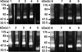
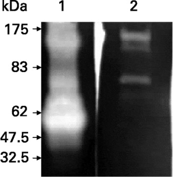
The presence or absence of 5 mm DTT did not affect the gelatinolytic activity of the ESP at any pH value tested. At pH 5.5, 6.0 and 8.0, five bands of proteolytic activity were visible at approximately 115, 74, 66, 51 and 42 kDa, respectively; an additional band at approximately 35 kDa was too weak to reproduce photographically. All bands detected were completely inhibited by the serine proteinase inhibitor, PMSF at pH 5.5. At pH 8.0, there was substantial inhibition of the 74 and 51 kDa bands (gelatinolytic band intensities decreased by 79 and 87% respectively), while the 66 kDa band was completely inhibited (fig. 3). This band was also inhibited by the metallo-proteinase inhibitor EDTA at both pH values, while 1,10-phenanthroline (another metallo-proteinase inhibitor) had no effect. No cysteine proteinase activity was detected by gelatin gel inhibition studies. Gelatinolytic activity by C, occasionally observed at pH 5.5 and increased at pH 8.0, did not correspond to that observed from CeESP and was not considered further (fig. 4).
Gelatin-substrate SDS–PAGE and inhibitor sensitivities of the ESP of Caenorhabditis elegans at pH 5.5 (a, b) and pH 8.0 (c, d). Proteolysis was visualized as clear areas on a Coomassie Brilliant Blue-stained gelatin background after overnight incubation of individual gel slices with appropriate inhibitor in buffer. Lane 1, ESP control; lane 2, ESP +E-64; lane 3, ESP + IA; lane 4, ESP + leupeptin; lane 5, ESP + PMSF; lane 6, ESP control; lane 7, ESP + pepstatinA; lane 8, ESP + EDTA; lane 9, ESP +1,10-phenanthroline. Numbers on the left indicate the position of molecular weight markers (kDa).

Gelatin-substrate SDS–PAGE of the ESP of Caenorhabditis elegans and Escherichia coli as control at pH 8.0. Lane 1, ESP; lane 2, E. coli control.

Immunoblotting
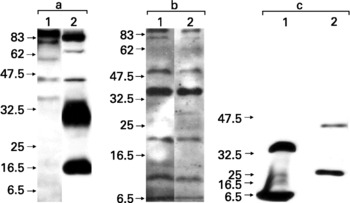
The anti-C. elegans Cpr-l antibody (antiserum to C. elegans cathepsin B) reacted specifically with five subunits (approximately 88, 77, 58, 45 and 35 kDa, respectively) of C. elegans ESP (CeESP) (fig. 5a1). This antibody also recognized subunits in the E. coli control, only one (58 kDa) corresponded to one recognized in the CeESP (fig. 5a2). Anti-C. elegans Cpl-1 (antiserum to C. elegans cathepsin L) reacted strongly with a 6.5 kDa subunit of CeESP and with two subunits of the E. coli control (approximately 40 kDa and 27.5 kDa). At higher CeESP protein concentration (50 ng per 10 μl), a second band at approximately 33.5 kDa was observed (fig. 5c). Preimmune serum or only secondary antibody as controls for both antibodies, did not react with any subunit from CeESP. The HRP-conjugated, partially purified, anti-S. vulgaris (28–30) kDa recognized seven subunits in CeESP with molecular weights within the range 88–6.5 kDa (fig. 5b2), exhibiting strongest recognition of the 35 kDa subunit. The monoclonal anti-6.5 kDa antibody reacted similarly with the subunits of CeESP, in that it recognized the same seven bands (fig. 5b1). Monoclonal antibodies, from five different hybridoma cell lines (LDS 124–128), which had reacted positively to SvESP, all recognized the same bands in CeESP (data not shown) and none reacted with subunits of the E. coli control. Negative controls consisted of secondary antibody only and an irrelevant monoclonal antibody.
Immunoblots of crude Caenorhabditis elegans ESP resolved by 12.5% SDS–PAGE, transferred to 0.45 μm nitrocellulose membrane, and incubated with either polyclonal anti-C. elegans Cpr-1 (antiserum to C. elegans cathepsin B), polyclonal anti-C. elegans Cpl-1 (antiserum to C. elegans cathepsin L), HRP-conjugated anti-(28–30 kDa), or monoclonal antibody anti-6.5 kDa Strongylus vulgaris. a1: CeESP and anti-Cpr-1 C. elegans; a2: Escherichia coli control and anti-Cpr-1 C. elegans; b1: CeESP and monoclonal antibody anti-S. vulgaris 6.5 kDa; b2: CeESP and HRP-conjugated partially purified anti-S. vulgaris (28–30 kDa), c1: CeESP and anti-Cpl-1 C. elegans. c2: E. coli control and anti-Cpl-1. Goat anti-rabbit IgG HRP conjugated antibodies were diluted 1:1000 or 1:1500, anti-mouse IgG HRP conjugated antibodies were diluted 1:1000 or 1:1500 and blots were developed in a chemiluminescent substrate.

Discussion
Thiol proteases have been detected in crude homogenates of C. elegans (Sarkis et al., Reference Sarkis, Kurpiewski, Ashcom, Jen-Jacobson and Jacobson1988) but the present data are the first characterization of the ESP secreted by this species. Essentially, fluorogenic and substrate gel studies establish unequivocally the presence of cysteine and serine proteinases, with rather weaker evidence for the presence of metallo-proteinases, in the ESP from mixed populations of C. elegans. Furthermore, the presence of cysteine proteinases was confirmed by immunoblotting with specific antibodies to C. elegans cathepsin B and cathepsin L proteases. No extract of E. coli, the diet of C. elegans, hydrolysed any fluorogenic substrate tested, indicating that the observed effects may be attributed to CeESP.
Synthetic substrates and inhibitors
Firstly, as Z-Phe-Arg-NMec (widely used to assay for cathepsin B, L and papain) was preferentially cleaved over Z-Arg-Arg-NMec (specific for only cathepsin B), this indicates the presence of cysteine proteinases. Additionally, at the optimal pH (5.5) for hydrolysis of Z-Phe-Arg-NMec, no hydrolysis of Z-Arg-Arg-NMec occurred. Hydrolysis of Z-Arg-Arg-NMec was maximal at pH 9.0 in the absence of DTT, a cysteine activator, and hydrolysis of Z-Phe-Arg-NMec was enhanced up to eight-fold by the addition of DTT. So, C. elegans ESP proteolytic activities are more cathepsin L- or papain-like than cathepsin B-like. Consistent with this is the fact that, following preincubation with class-specific inhibitors, hydrolysis of Z-Phe-Arg-NMec by ESP was abolished by the three cysteine proteinase inhibitors tested (E-64, IA and leupeptin), while serine, aspartic and metallo-proteinase inhibitors had little effect. Most proteinases identified in ESP from adult nematodes are cysteine proteinases, implicated in nutrient digestion (Cox et al., Reference Cox, Pratt, Hageman and Boisvenue1990; Ray & McKerrow, Reference Ray and McKerrow1992; Caffrey & Ryan, Reference Caffrey and Ryan1994; Jasmer et al., Reference Jasmer, Roth and Myler2001). In C. elegans, cathepsin L is also essential for yolk-processing during embryonic development (Britton & Murray, Reference Britton and Murray2004). The presence of cysteine proteinases was confirmed by the enhancing effect of DTT in fluorogenic substrate assays. This was not observed in gelatin substrate SDS–PAGE, which may be attributed to the predominance of serine proteinases.
Although cathepsins are excreted-secreted by C. elegans, the existing nomenclature for proteinases may not always apply to nematode proteinases, as they exhibit a paradoxical substrate specificity (Skuce et al., Reference Skuce, Redmond, Liddell, Stewart, Newlands, Smith and Knox1999). Many nematode cysteine proteinases identified to date are cathepsin B-like when sequenced, despite preferentially cleaving synthetic cathepsin L-like substrates. The substitution of key residues at the active sites could alter substrate specificity from cathepsin B-like to cathepsin L-like (Loukas et al., Reference Loukas, Selzer and Maizels1998).
Secondly, of the two substrates used to assay serine proteinase activity, N-Suc-Ala-Ala-Pro-Phe-NMec (assayed for chymotrypsin-like activity) was superior with an almost four-fold higher release rate of NMec than Z-Gly-Gly-NMec (assayed for trypsin-like activity), at the optimum pH of 8.0. This indicates that the serine proteinase activity present in C. elegans ESP is predominately chymotrypsin-like. Gelatin substrate data indicate that serine proteinases predominate as all bands were completely inhibited by the serine proteinase inhibitor PMSF at pH 5.5 and partial inhibition was observed at pH 8.0, perhaps due to the strength of activity at this pH, and the fact that PMSF has a half-life of 1 h and is inactivated more quickly at pH 8.0 (James, Reference James1978). Alternatively, the bands may represent different forms or complexes of a single major proteinase. Serine activity over a broad pH range (5.0–9.0) has been reported in the parasitic nematodes Toxocara canis (Roberston et al., Reference Robertson, Bianco, McKerrow and Maizels1989), Trichinella spiralis (Todorova et al., Reference Todorova, Knox and Kennedy1995) and Trichostrongylus vitrinus (MacLennon et al., Reference MacLennon, Gallagher and Knox1997). The fact that no cysteine proteinase activity was dectected by gelatin substrate SDS–PAGE could be due to masking or inhibition by the serine proteinases present, as exhibited by metallo-proteinases in the hookworm, Ancylostoma caninum (Dowd et al., Reference Dowd, Dalton, Loukas, Prociv and Brindley1994). Significantly, studies with fluorogenic peptide substrates are more specific than use of the general substrate gelatin (Healer et al., Reference Healer, Ashall and Maizels1991; Geldhof et al., Reference Geldhof, Claerebout, Knox, Agneessens and Vercruysse2000). At both pH 5.5 and 8.0 an additional band at approximately 38 kDa was strongly resolved, only in the presence of PMSF, indicating the presence of a non-serine proteinase. Mass spectrometry might identify the proteinase class to which this band belongs.
Serine proteinases are associated with tissue-invading nematodes especially in their larval stages. Thus, serine proteinases were observed in third stage larvae of Toxocara canis (Robertson et al., Reference Robertson, Bianco, McKerrow and Maizels1989) and Nippostronglyus brasiliensis (Knox & Jones, Reference Knox and Jones1990) and the third/fourth stage larvae of Ascaris suum (Knox & Kennedy, Reference Knox and Kennedy1988). Serine proteinases identified in adult Trichinella spiralis (in which a band of 50 kDa predominated, similar in size to a band in C. elegans ESP) and Trichuris muris ESP might serve digestion (Drake et al., Reference Drake, Bianco, Bundy and Ashall1994; Todorova et al., Reference Todorova, Knox and Kennedy1995).
Thirdly, inhibition of fluorogenesis by the general chelator EDTA and by 1,10-phenanthroline are consistent with the presence of a metallo-proteinase. However, only EDTA inhibited the 66 kDa gelatinolytic band and, as cations activate other types of proteinases such as members of the substilisin family (serine), calpains and clostripain(s) (cysteine), such inhibition by EDTA is not unambiguously indicative of a metallo-proteinase (Barrett, Reference Barrett1994). As PMSF also inhibited this band, it might be a metal-dependent serine proteinase (Distefano et al., Reference Distefano, Palma, Gomez and Del Rio1997) or indeed a moulting residue, as a metallo-protease enables ecdysis in C. elegans (Davis et al., Reference Davis, Birnie, Chan, Page and Jorgensen2004). Overall the inhibition by 1,10-phenanthroline, a specific zinc-chelating metallo-proteinase inhibitor, is a more definitive indicator of metallo-proteinases (Powers & Harper, Reference Powers, Harper, Barrett and Salvesen1986).
Immunoblotting
As polyclonal antibodies raised against C. elegans cathepsin B (anti-Cpr-1) and cathepsin L (anti-Cpl-1) reacted with subunits of CeESP, this verified the presence of cysteine proteinases in C. elegans crude ESP. Anti-Cpr-1 reacted with four subunits of CeESP, although only one band was the expected size of approximately 35 kDa. Cpr-1 is one of six cathepsin B proteins located in the gut of C. elegans (Ray & McKerrow, Reference Ray and McKerrow1992; Britton et al., Reference Britton, McKerrow and Johnstone1998) and an antibody raised against any one of these cpr proteins is likely to cross react with multiple isoforms. Anti-Cpr-1 was raised against recombinant protein expressed in E. coli, and therefore recognized subunits in the E. coli control.
Anti-Cpl-1 reacted strongly with a 6.5 kDa subunit in CeESP and with an additional band of approximately 33.5 kDa at higher protein concentration; the latter is within the expected size range of a cathepsin L protein and the 6.5 kDa band could be due to processing of the protein. Cysteine proteinases are synthesized as zymogens and are converted to mature proteinase forms by proteolytic cleavage (Silva et al., Reference Silva, Batista, Marra, Fragoso, Monteiro, Figueira and Grossi-de-Sa2004).
The partially purified HRP-conjugated anti-S. vulgaris (28–30 kDa) polyclonal antibody recognized seven bands in CeESP. The monoclonal anti-S. vulgaris 6.5 kDa, which recognized two subunits (88 and 48 kDa) in SvESP (Dr Wen Chung, personal communication), and in this study reacted the same as the polyclonal antibody. So, the epitope recognized by the monoclonal antibody is present in both CeESP and SvESP establishing antigenic relatedness of a proteic epitope in the ESP of C. elegans and S. vulgaris.
Although the present data, derived from a mixed population of C. elegans including juveniles, do not allow stage-specific attributions, they are significant in showing that this free-living nematode excretes and secretes cathepsins and other proteinases associated with tissue-penetration and -degradation in parasitic nematodes. Perhaps this is the primordial condition, and evolutionary adaptation to a parasitic life-style provided a new role for such serine and cysteine proteinases, and to a lesser extent metallo-proteinases, without the need to evolve new proteinases.
Acknowledgements
The C. elegans strain used in this work was provided by the Caenorhabditis Genetics Center, University of Minnesota, 1445 Gortner Avenue, St Paul, MN 55 108-1095, USA, which is funded by the NIH National Center for Research Resources (NCRR). The authors also thank Dr Collette Britton, Dr Sarwar Hashmi and Dr Wen Chung for the generous gifts of the antibodies used in this study.