Introduction
Uvulopalatopharyngoplasty (UPPP) is one of the most widely performed surgical procedures for obstructive sleep apnoea (OSA) in the world. Novel techniques have been sought to minimise the potential complications associated with UPPP.Reference Kezirian, Weaver, Yueh, Deyo, Khuri and Daley1,Reference Kezirian, Weaver, Yueh, Khuri, Daley and Henderson2 Barbed pharyngoplasty is an alternative and well accepted technique used in the treatment of OSA, especially in the presence of retropalatal obstruction.Reference Vicini, Hendawy, Campanini, Eesa, Bahgat and AlGhamdi3
In 2015, Vicini et al.Reference Vicini, Hendawy, Campanini, Eesa, Bahgat and AlGhamdi3 proposed the palatal and pharyngeal lifting technique with a barbed suture in OSA, in an attempt to shorten, tense and conglomerate the redundant soft tissues of the soft palate and pharynx, by pulling up the soft palate and lateral pharyngeal structures towards the pterygomandibular raphe and posterior nasal spine. The main objective of this technique is to tighten the redundant soft tissues through the fibromuscular plane of the soft palate with a knotless suture.
Although patients’ outcomes and quality of life are considerably improved, barbed pharyngoplasty may be insufficient in pulling up the uvula and its upper redundant soft tissue, especially in patients with an elongated uvula. In an attempt to overcome this drawback, we proposed a technical update of barbed pharyngoplasty, and compared the outcomes of barbed pharyngoplasty with those of modified barbed pharyngoplasty.
Materials and methods
Prior to initiation of the study, approval from the institutional review board at the Medical University of Yildirim Beyazit was obtained for the study protocol.
This prospective study was performed between November 2015 and May 2016. Thirty-four patients (28 men and 6 women) with OSA confirmed on polysomnography were included. The patients were allocated, in a non-randomised fashion, into two groups based on the surgical procedure performed: barbed pharyngoplasty or modified barbed pharyngoplasty. There were 17 patients in the barbed pharyngoplasty group (mean age of 39.4 years) and 17 patients in the modified barbed pharyngoplasty group (mean age of 40.6 years).
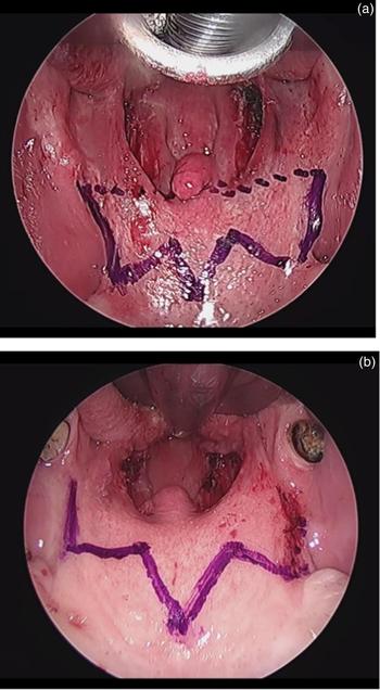
Modified barbed pharyngoplasty is routinely performed in our institution in patients with obstruction at both the lateral pharyngeal wall and retropalatal space. The barbed pharyngoplasty technique is performed in patients with lateral pharyngeal wall obstruction only. The pharyngoplasty technique performed in patients with retropalatal obstruction is shown in Figure 1.
Pre-operative view prior to surgical intervention. (a) Modified barbed pharyngoplasty provides uvula stabilisation via the passing of the suture through the transverse line indicated by the points. (b) In the barbed pharyngoplasty technique, the suture does not pass over the uvula, thus stabilising the soft palate without providing uvula stabilisation.

All patients had retropalatal obstruction, as observed on Müller's manoeuvre under fibre-optic endoscopy and drug-induced sleep endoscopy, and mild to moderate OSA on polysomnography. None of the patients had: nasal obstruction due to a turbinate or septal problem, tonsillar hypertrophy of more than grade 2, morbid obesity (body mass index (BMI) of more than 35 kg/m2), or hypopharyngeal obstruction on Müller's manoeuvre or drug-induced sleep endoscopy.
Patient history focused on snoring and OSA symptoms. All patients underwent overnight polysomnography, and the total and supine apnoea/hypopnea index values were determined. Daytime sleepiness was assessed using the Epworth Sleepiness Scale. Both the patient and partner (if present) were asked to grade snoring severity on a 10-point visual analogue scale (VAS) (0 = no snoring, 10 = extreme snoring). Routine ENT examination was performed, focusing on nasal pathology, tonsil size, uvula and palate, and tongue size. The BMI was calculated.
Post-operatively, ENT examination, snoring VAS and Epworth Sleepiness Scale assessments, and polysomnography were performed, after at least six months following surgery. The mean follow-up period ranged from six to nine months (mean of eight months).
Polysomnography
All patients underwent a full-night comprehensive sleep study using Philips Respironics equipment (Murrysville, Pennsylvania, USA) in the same centre, conducted by experienced technicians. All records were scored by the same physician. Sleep state scoring and the identification of respiratory events were performed according to the American Academy of Sleep Medicine manual for the scoring of sleep and associated events, version 2.2. Apnoeas were defined as the absence of oronasal airflow for at least 10 seconds, with a reduction of more than 90 per cent. Hypopneas were defined as a discernible fall in respiratory effort or flow, associated with a 3 per cent or more oxygen desaturation.
For the objective assessment of the effectiveness of surgical procedures, the pre-operative and six-months’ post-operative polysomnography findings were compared. The primary outcome was surgical response, with ‘success’ defined as a 50 per cent reduction in the baseline apnoea/hypopnea index and a post-operative apnoea/hypopnea index of less than 20 events per hour.
Drug-induced sleep endoscopy
Drug-induced sleep endoscopy was performed in all patients by an ENT specialist within 3–7 days before the surgery. Propofol (1.5 mg/kg, intravenous bolus) was used to induce sleep in the sleep laboratory, under the control of an anaesthesiologist. Sedation level was monitored using electroencephalography, wherein brain waves were examined to assess the effect of anaesthesia on the central nervous system.
Sleep endoscopy was performed towards the end of sleep, when the electroencephalogram showed physiological sleep patterns. The nasal passage, nasopharynx, velum, oropharynx, tongue base, epiglottis and larynx were observed using a flexible endoscope (3.5 mm). Levels of snoring and obstruction were assessed using the velum, oropharynx, tongue base and epiglottis (‘VOTE’) classification.
Surgical technique
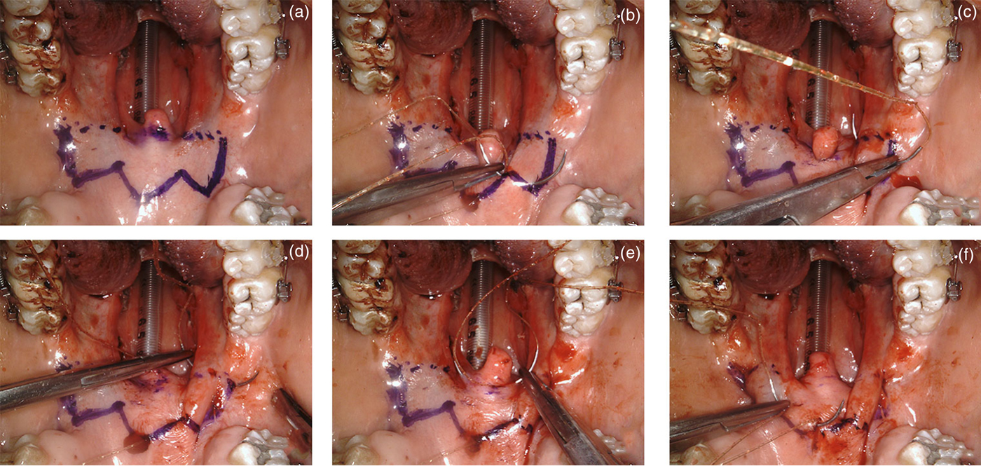
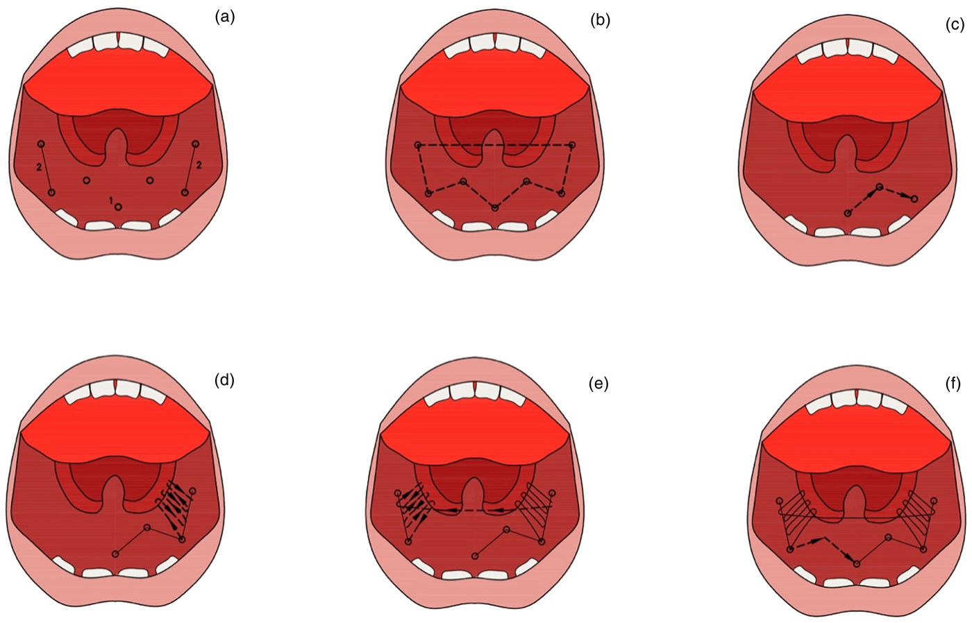
The surgical technique for barbed pharyngoplasty was performed as described by Vicini et al.Reference Vicini, Hendawy, Campanini, Eesa, Bahgat and AlGhamdi3 In the modified barbed pharyngoplasty operation, a bilateral tonsillectomy was performed initially. Then, the inferior portion of palatopharyngeus muscle was partially released using monopolar electrocautery. The suture sites on the soft palate were indicated with a marker pen. A single barbed suture (size 0) was introduced at the centre point (posterior nasal spine), then passed laterally to turn around the pterygomandibular raphe, and then passed back to the tonsillectomy bed to suspend the palatopharyngeus muscle and posterior pillar to the raphe again, at least three times in different directions. When suture was returned to the superior pole of tonsillectomy bed, it was passed horizontally through the base of the uvula to the opposite side of the superior pole of the tonsillectomy bed. The same procedure was then conducted on the opposite side, with the same continuous suture. Finally, the suture was returned to the centre point (posterior nasal spine) and the stitches were locked together (Figures 2 and 3).
(a) Marking the suture lines after bilateral tonsillectomy. (b) The suture is introduced at the posterior nasal spine and passed laterally towards the pterygomandibular raphe. (c) The needle is passed around the pterygomandibular raphe and inferior to the same side of the tonsillectomy bed. (d) The palatopharyngeus muscle and posterior pillar are suspended to the pterygomandibular raphe at least three times. (e) The suture is inserted horizontally through the uvular muscle, to re-emerge at the opposite side of the tonsillectomy bed. (f) The same procedure was conducted on the opposite side, with the same continuous suture, which returns to the centre point for locking.

Schematic illustration of modified barbed pharyngoplasty.

Statistical analysis
All statistical analyses were performed using SPSS version 22.0 software (Mac version; SPSS, Chicago, Illinois, USA). The independent samples t-test and the Mann–Whitney U test were used for the comparisons. The Levine test for equality of variances was used to determine statistically significant variances. Statistical significance was accepted when the p-value was 0.05 or lower.
Results
All patients reported an immediate subjective clinical improvement after the surgical procedures. No major complications were observed. Most of the patients reported mild oropharyngeal pain and swallowing difficulty, which resolved spontaneously within a few days. The most common post-operative complaint for both groups was a foreign body sensation on the soft palate, which resolved spontaneously within one month. Knot extrusion was not seen in any patient. Snoring improvement was confirmed by a reduction in snoring VAS scores, and a reduction in daytime sleepiness as assessed by the Epworth Sleepiness Scale.
Overall, there was no significant differences between the results for the barbed pharyngoplasty and modified barbed pharyngoplasty groups, as determined by the student's t-test (p > 0.05). However, the reductions in snoring VAS and Epworth Sleepiness Scale scores, and in total and supine apnoea/hypopnea index values, were greater in the modified barbed pharyngoplasty group than the barbed pharyngoplasty group (Table 1). The success rates for modified barbed pharyngoplasty and barbed pharyngoplasty were 95 per cent and 82 per cent, respectively.
Patient demographics and surgical outcomes of barbed pharyngoplasty and modified barbed pharyngoplasty

*n = 17; †n = 17. ‡Indicates the group with the better results, for each relevant parameter. SD = standard deviation; BMI = body mass index; ESS = Epworth Sleepiness Scale; pre-op = pre-operative; post-op = post-operative; VAS = visual analogue scale; AHI = apnoea/hypopnea index
Discussion
Refinements in palate surgery are needed to increase the treatment success rates while decreasing morbidities.Reference Kezirian, Weaver, Yueh, Deyo, Khuri and Daley1,Reference Fairbanks4–Reference Friedman, Ibrahim, Vidvasagar and Pomeranz6 One of these refinements is the barbed pharyngoplasty technique, in which the soft palate is advanced, and traction is created in the lateral pharyngeal structures in an attempt to enlarge lateral pharyngeal wall collapse.Reference Vicini, Hendawy, Campanini, Eesa, Bahgat and AlGhamdi3 Similarly, Sorrenti et al.Reference Sorrenti, Pelligra, Albertini, Caccamo and Piccin7 recently proposed a modification of the functional expansion pharyngoplasty technique, which enlarges lateral pharyngeal wall collapse, as in the barbed pharyngoplasty technique. These surgical procedures mainly focus on obstruction at the lateral pharyngeal wall. We propose that our technical update of barbed pharyngoplasty enlarges the retropalatal space in both lateral and anteroposterior directions.
In barbed pharyngoplasty, the soft palate is pulled up in the horizontal plain towards the hard palate using a knotless suture, which is passed from the soft palate to the periosteum of the posterior nasal spine and pterygomandibular raphe, bilaterally. Thus, a resistance occurs in the soft palate by providing stabilisation against negative inspiratory pressure. The oropharyngeal space is enlarged with a transverse cut in the palatopharyngeus muscle and tonsillectomy, and further space is obtained in the lateral retropalatal area by attaching the palatopharyngeus muscle to the pterygomandibular raphe. However, in our experience, adequate stabilisation may not be achieved in the middle area of the soft palate (posterior nasal spine–uvula line). It may also be difficult to facilitate traction in the uvula as well as around the uvula. In an attempt to overcome this problem, we modified the technique.
A few lifting techniques using a suture have been described. In 2008, HurReference Hur8 proposed the use of fibro-osseous structures as anchoring points to increase upper oropharyngeal and retropalatal airway size. In 2012, Mantovani et al.Reference Mantovani, Minetti, Torretta, Pincherle, Tassone and Pignataro9 proposed that anchoring the threads to the strong and deep fibro-osseous structures (posterior nasal spine and pterygoid hamuli) facilitates lifting of the palate and widening of the retropalatal space without surgical resection; they termed this technique the velo-uvulo-pharyngeal lift. In 2015, Vicini et al.Reference Vicini, Hendawy, Campanini, Eesa, Bahgat and AlGhamdi3 reported a new technique, termed barbed pharyngoplasty, inspired by the velo-uvulo-pharyngeal lift and relocation pharyngoplasty, which lifts the posterior pillar and palatopharyngeal muscle. Li et al.Reference Li, Li, Chen and Wang10 reported that the magnitude of forward movement of the soft palate depends on the amplitude of traction of the posterior pillar, which is closer to the root of the uvula as the traction point leads further forward of the soft palate.
In this study, we replicated the barbed pharyngoplasty technique with some modifications. That is, a thread passed horizontally between the superior poles of the tonsillectomy beds through the root of the uvula in order to pull the uvula forward. Thus, the need to resect redundant tissue to enlarge the oropharyngeal inlet was eliminated. Overall, better outcomes were obtained with the modified barbed pharyngoplasty technique than with barbed pharyngoplasty, although the differences were not statistically significant. However, further studies are required using larger series of patients to determine whether statistically significant results can be obtained.
Our technique is essentially a new simple variant of barbed pharyngoplasty. The modified barbed pharyngoplasty technique offers several advantages, as follows. The barbed pharyngoplasty method requires two interventions to the soft palate by using one or two barbed sutures, whereas modified barbed pharyngoplasty requires only a single continuous suture to pull up the uvula and soft palate in both lateral and anteroposterior directions. Modified barbed pharyngoplasty is especially preferable in patients with an elongated uvula; the barbed pharyngoplasty procedure seems less efficient in patients with an elongated uvula.
• This prospective study compared barbed pharyngoplasty with a revised suture technique
• Barbed pharyngoplasty may be inadequate at facilitating traction in the uvula as well as around the uvula
• Modified barbed pharyngoplasty eliminates the need for surgical resection of redundant soft tissues around the uvula while lifting up the uvula base
• The main indication for modified barbed pharyngoplasty is retropalatal obstruction in both lateral and anteroposterior directions
• Barbed pharyngoplasty is appropriate for lateral oropharyngeal narrowing
In barbed pharyngoplasty, it may be difficult to obtain traction on the uvula and the surrounding soft tissues, and a stay suture may be needed in some cases. Two barbed sutures may be required to perform barbed pharyngoplasty, which increases the surgical cost. No resection was required for the modified barbed pharyngoplasty technique. Instead, a suture in the horizontal plain that passes through the uvula facilitates a wider oropharyngeal inlet, with no tissue resection required, and there is no need for full thickness resection in the superolateral triangle of the tonsil bed.
Conclusion
Modified barbed pharyngoplasty is a simple, cost-effective, minimally invasive and well-tolerated technique, with no associated significant morbidity or major complication. However, the major limitation of this technique is that the previous anatomical condition cannot be restored in cases of patient discomfort, because of the knotless feature of the barbed suture. Despite this, none of our patients complained of this discomfort during the six-month follow-up period. Modified barbed pharyngoplasty is a safe and feasible method, especially in cases of retropalatal obstruction with an elongated uvula. Our preliminary findings indicate that the modified barbed pharyngoplasty is a reliable technique.
Competing interests
None declared