Introduction
Intra-cavitary brachytherapy (ICBT) [high-dose rate (HDR) or low-dose rate (LDR)] plays a key role in the management of cervix cancer. For decades, cervix brachytherapy treatment planning was carried out using plain X-ray films on the basis of point-based dose prescriptions and estimates of organ-at-risk (OAR) doses. Point-A, the most frequently referenced point for dose prescription in ICBT treatment planning, is commonly defined as a point 2 cm superior to the cervical os and 2 cm lateral to the os on a line perpendicular to the uterine tandem axis. This reference point is presumed to mark the crossing of ureter and the uterine artery in the para-cervical triangle. The cumulative dose at Point-A (combined external beam + brachytherapy dose) has also guided treatment outcome evaluations in cervix cancer brachytherapy. Eifel et al.Reference Eifel, Thoms and Smith1 correlated pelvic disease recurrence rate and Point-A cumulative dose. The authors reported pelvic disease recurrence rate at 5 years was 33% in patients who received <85 Gy Point-A dose compared with 16% recurrence rate for patients with Point-A dose >85 Gy.
Computed tomography (CT) and magnetic resonance imaging (MRI) compatible intra-cavitary gynaecologic brachytherapy applicators have facilitated three-dimensional (3D) image-based treatment planning, tumour volume-optimised dose prescriptions and dose–volume histogram (DVH)-based evaluation of target and OAR doses. The transition from a two-dimensional (2D) radiograph-based point dose planning to 3D image-based treatment planning has gained momentum in cervix brachytherapy, with a strong drive for MRI-based target volume definition, delineation and treatment planning.Reference Haie-Meder, Potter and van Limbergen2
At the same time as image-guided cervix brachytherapy treatment planning is changing the model for target definition and dose evaluation, the actual clinical practice of dose prescription and OAR dose evaluation itself remains varied as documented in practice patterns.Reference Viswanathan and Erickson3, Reference Pearce, Craighead and Kay4 In the American survey, it is reported that dose to Point-A remains the most frequently used prescription method (76% of the surveyed), and 52% of the surveyed reported using points (ICRU bladder and rectal points) for OAR dose estimation.Reference Viswanathan and Erickson3 Furthermore, the survey also revealed that some practitioners modify treatment plans on the basis of Point-A dose, and majority of these recommended that Point-A reach 100% of the prescription dose.
In the present study, we aimed to investigate whether dose to Point-A necessitates treatment plan modifications in a time of cross-sectional image-based brachytherapy treatment planning and dose evaluation for cervix cancer. This was accomplished by carrying out a comparative assessment of dose distribution for two treatment planning situations. In the first plan, the dose optimisation is based on adjusting individual dwell times to fine tune dose distributions to match implant and tumour geometry while constraining OAR doses to acceptable dose limits without worrying about resulting Point-A dose (PlanTarget). In the second plan, however, the dose is normalised to Point-A (classic pear shape dose distribution) for the same applicator geometry, and therefore the Point-A dose will equal prescription dose (PlanPoint-A). The differences in dose distributions, relative target volume coverage and OAR doses between the two plans were investigated and then compared.
Materials and methods
A total of 125 individual intra-cavitary implants from 25 patients treated in our institution with a combination of external beam radiation therapy (EBRT) and HDR ICBT for cervix carcinoma were randomly selected and retrospectively analysed. Median age was 53 years (range 32–89). The International Federation of Gynaecology and Obstetrics (FIGO) Stage distribution was: IB2 (3), IIA (1), IIB (16), IIIB (4), IVA (1). The EBRT consisted of 45-Gy to the whole pelvis using either 6- or 16-MV energy photon beams with concomitant chemotherapy. In cases with parametrial extension, additional EBRT boost dose of 9–14·4 Gy was given in 1·8-Gy dose fractions.
In the present study, all brachytherapy treatments were accomplished using CT-compatible standard tandem and ovoid (T&O) applicators (Nucletron BV). In our institution, the T&O insertions and HDR brachytherapy treatments are initiated towards the end of the pelvic EBRT treatments and are administered on a weekly basis (until end of EBRT) and twice weekly (upon completion of EBRT). Applicator insertions were performed on an outpatient basis in a CT/simulation room with the patient under conscious sedation. Usually in a separate intra-operative procedure under general anaesthesia, sounding of the uterus, cervical dilation and insertion of CT-compatible cervical sleeve (Smit Sleeve, Nucletron Corporation) were performed. The cervical sleeve was left in place until all brachytherapy applicator insertions and treatments were completed. A Foley balloon catheter filled with 7 cc of diluted radio-opaque contrast was introduced into the bladder for visualisation of the bladder neck. Bladder filling was generally empty unless dictated by the proximity of small bowel to tandem tip in which case the bladder was intentionally filled to push the small bowel away. Rectal separation was achieved by using a rectal retractor. No additional packing was used. Patients underwent CT simulation for brachytherapy treatment planning. A CT scan of pelvis with 2·5-mm slice thickness was performed with the intra-uterine T&Os in place.
In our institution, CT-based brachytherapy treatment planning is carried out for individual insertion. The CT/simulation images were transferred to Plato treatment planning system (Nucletron BV, Elekta AB, Stockholm, Sweden). The clinical target volume (CTV) and OAR structures (bladder and rectum) were contoured on appropriate axial slices. The CTV consisted of high-risk CTV (HR CTV) and included entire uterus, plus any upper/lower vaginal involvement. The CTV was contoured as a single structure. For all cases included this study, target and structure delineation was performed by a single radiation oncologist (S.M.) to remove inter-observer variation.Reference Saarnak, Boersma and van Bunningen5 Following Groupe Europeen de Curietherapie and European Society for Therapeutic Radiology and Oncology (GEC-ESTRO) recommendations, entire organs and not organ walls were contoured for rectum and bladder.Reference Haie-Meder, Potter and van Limbergen2 For bladder, the outer wall was contoured and the rectal contouring was extended superiorly to include sigmoid. Point A (A-Right and A-Left) was identified by measuring 2 cm along the intra-uterine tandem from the cervical os (represented by the Smit Sleeve) and 2 cm laterally in the plane of the intra-cavitary applicator system.
PlanTarget
The treatment planning procedure begins with a customary T&O planning process with the dose normalised and prescribed to applicator points (Points A-Right and A-Left, pear-shaped dose distribution). The resulting isodose distributions are then reviewed on each axial slice by the radiation oncologist, and where necessary the dose distributions are further refined by dynamically changing the individual dwell timings to suit implant geometry, proper target coverage and match patient anatomy while respecting OAR doses. A 6-Gy dose is prescribed to the 100% isodose line, while making every effort to limit when possible combined EBRT + BT bladder and rectal doses to highest irradiated 2-cc volume to receive <80 Gy and 78 Gy [equivalent dose in 2-Gy fractions (EQD2)], respectively. Higher bladder doses are accepted in cases where the tumour has bladder extension. The Points-A doses were recorded but no attempts were made to equate Point-A and prescription doses. This plan (designated as PlanTarget) was used to deliver treatments.
PlanPoint-A
For the study purpose, a repeat hypothetical dosimetry was performed with 100% of the prescription dose prescribed to Point-A. Thus, the average Point-A dose in this case equals the prescription dose of 6 Gy. No additional dose optimisation points were placed along the tandem or ovoid. In essence, this is a renormalisation of PlanTarget to equate prescription and Point-A doses. This prescription and optimisation is similar to the method #3 used in a published planning comparison studyReference Mai, Erickson and Rownd6 and duplicates the planning style of the practitioners who recommended that Point-A reach 100% of the prescription dose in the practice surveys.Reference Viswanathan and Erickson3
Dose calculations
All dose calculations were carried out using a commercial brachytherapy dose planning system (Plato version 14.2.2, Nucletron BV). The treatments were delivered using a micro-selectron HDR brachytherapy treatment unit (version V2, Nucletron BV). Each patient received five separate ICBT insertions in 6-Gy dose fractions. For all 250 treatment plans, DVHs were generated to evaluate target, bladder and rectal doses. From these DVHs, the CTV (%) that is encompassed by the 100% dose (V100), the dose that covered 90% and 100% of the CTV (D90, D100) and minimal doses to highest irradiated 0·1 and 2 cc volumes of rectum and bladder were individually recorded in each case (designated as D0.1 cc and D2 cc, respectively). The cumulative doses to the Point-A, rectum and bladder were calculated by combining contributions from external beam therapy and brachytherapy. In these calculations, the dose contributions to bladder and rectum from parametrial boost with midline shielding were not considered. The total doses were converted to EQD2 using the equation EQD2Total = EQD2External + EQD2Brachy. For this calculation, α/β ratio of 10 for the tumour and 3 for the late effects on the OAR were used. Data were analysed using correlation (Spearman's rank correlation) and two-tailed probability t-tests. All statistical analyses were performed using MedCalc, Version 12 for Microsoft Windows (MedCalc Software, Ostend, Belgium). A p-value of <0·05 was considered statistically significant.
Results
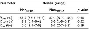
For the study population, the mean CTV was 103·4 (±29·0) cc. Table 1 summarises per fraction differences in dose and volume parameters between PlanTarget and PlanPoint-A. For PlanTarget the median average Point-A dose was 5·4 Gy (range 3·6–7·5), compared with average Point-A dose of 6 Gy for PlanPoint-A. The variation in average Point-A dose for 125 insertions for PlanTarget is shown via a scatter plot in Figure 1. The dotted line in Figure 1 represents the prescription dose of 6 Gy. For PlanTarget, CTV D90 did not correlate with Point-A dose (p = 0·60) (Figure 2). CTV (cc) correlated with Point-A dose (p < 0·0001) (Figure 3); however, for PlanTarget, V100, it did not correlate with Point-A dose (p = 0·50) (Figure 4).
Summary of dose and volume parameters related to CTV for 125 fractions and 25 patients
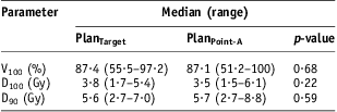
Notes: V100 = per cent volume encompassed by the 100% isodose; D100 and D90 = dose that covered 100% and 90% of the CTV.
Abbreviation: CTV, clinical target volume.
Scatter plot showing variations in average Point-A dose for 125 insertions for PlanTarget.

Correlation of Point-A dose with CTV D90 for PlanTarget.
Correlation of Point-A dose with CTV volume for PlanTarget.
Correlation of Point-A dose with V100 for PlanTarget.
The dose parameters for the highest irradiated 0·1 cc and 2 cc of bladder and rectum were computed separately for each implant, averaged per patient and then for the study population. Bladder and rectal D0.1 cc and D2 cc were higher for PlanPoint-A plans compared with PlanTarget (p < 0·0001). The overall study dose differences for OARs between PlanTarget and PlanPoint-A are summarised in Table 2.
Combined (EBRT + BT) doses to highest irradiated 0·1 cc and 2 cc bladder and rectum for PlanTarget versus PlanPoint-A

Note: Doses are median (range) and values are in EQD2 (Gy).
Abbreviations: EBRT, external beam radiation therapy; BT, brachytherapy; OAR, organ-at-risk.
Discussion
Advances in sectional image-guided cervix brachytherapy have changed the paradigm for target definition and dose evaluation; however, clinical practice of dose prescription and documentation itself remains varied as documented in American and Canadian practice pattern surveys.Reference Viswanathan and Erickson3, Reference Pearce, Craighead and Kay4 Conventional Point-A-based dose prescriptions may lead to inappropriate irradiation of target and normal tissue compared with image-based brachytherapy optimised dose distributions, especially for small tumours.Reference Yoshida, Yamazaki and Takenaka7 In our study, we observed that 0·1 and 2 cc bladder and rectal doses were higher for PlanPoint-A compared with PlanTarget (Table 2). The average OAR sparing factors for bladder and rectum [defined as the dose ratio (PlanTarget/PlanPoint-A) for 0·1 and 2 cc volumes] were in the range 0·82–0·89, respectively. Sparing factor of <1 indicates a reduction in OAR doses. Similar observations can be inferred from the published results of two studies comparing standard versus 3D image-guided dose optimisations.Reference Jurgenliemk-Schulz, Lang and Tanderup8, Reference Tanderup, Nielsen and Nyvang9 In both studies, the OAR doses were reduced for optimised plans as compared with standard plans. On the basis of their data, for limited volume case, the average sparing factors for 2 cc bladder and rectum appear to be in the 0·87–0·96 range.
In our institution, CT-image-based brachytherapy treatment planning was implemented when the traditional simulator was replaced with a CT/simulator. During the initial phases of transition to CT-image-based planning, we continued to rely on our past experience with point dosimetry for ICBT planning and dose evaluation. With accumulation of our own experience and that of published literature, the first step we initiated was in establishing bladder and rectal dose constraints by adapting to volumetric dose evaluations. Similar to reported findings,Reference Barillot, Horiot and Maingon10–Reference Wachter-Gerstner, Wachter and Reinstadler13 our published resultsReference Yaparpalvi, Mutyala and Gorla14 also suggested that the ICRU rectal reference doses correlate significantly with D0.1 cc and D2 cc doses, but bladder ICRU reference doses correlate poorly with D0.1 cc and D2 cc doses. With our current image-based HDR brachytherapy planning and volumetric dose evaluations, we continue to rely on our own past intra-cavitary brachytherapy experience plus literature recommendations and limit wherever possible combined doses to the highest irradiated 2 cc bladder and rectal volumes to below 80 and 78 Gy (EQD2), respectively.Reference Koom, Sohn and Kim15 Roughly, this translates to limiting 2-cc bladder and rectal doses per implant to <75% of the fractional prescription dose. Sigmoid dose (included in rectal contouring in this study) was also restricted to this dose constraint. In calculating cumulative bladder and rectal doses, we have neglected contributions from parametrial boost fields with midline shielding. This is primarily because of lack of treatment planning systems that can radiobiologically integrate EBRT and brachytherapy components. However, it has been shown that external beam parametrial boost with standard midline shielding can add to bladder and rectum cumulative doses, and therefore increases the risk of radiation proctitis in patients with uterine cervix cancer.Reference Huang, Lin and Hsu16, Reference Fenkell, Assenholt and Nielsen17 In addition, in the cumulative bladder and rectal dose calculations, we have assumed that the same volume of OAR is irradiated to the highest dose in each brachytherapy fraction.
A weakness in our study is that the CTV was defined as a single structure on CT-images and were not delineated into separate components gross tumour volume (GTV), HR CTV and intermediate risk clinical target volume (IR CTV) as per GEC ESTRO and other such published recommendations for MRI/CT-based contouring.Reference Haie-Meder, Potter and van Limbergen2, Reference Viswanathan, Dimopoulos and Kirisits18 The main reason for not delineating GTV is primarily because of the inherent difficulty associated with CT-images in accurately distinguishing between tumour and the soft tissue compared with MRI. However, in our CTV contouring, HR CTV and any upper/lower vaginal disease contouring closely matched published CT-image-based contouring guidelineReference Viswanathan, Dimopoulos and Kirisits18 with the exception that contour superiorly was extended to include the entire uterus. In addition, for scheduling logistics and time constraint reasons, we have not implemented MRI-based planning for ICBT in our institution. Recent upgrades to our brachytherapy treatment planning system (Oncentra Brachy, Nucletron BV) offer better image fusion and contouring tools, and we are currently initiating steps to use recommendations for target and OAR definitions, while adapting to CT-based contouring guidelines.Reference Haie-Meder, Potter and van Limbergen2, Reference Viswanathan, Dimopoulos and Kirisits18 In our study, we observed that Point-A dose did not correlate with CTV D90 dose (p = 0·60). As mentioned previously, in our study the CTV was drawn as a single structure and not divided into components. Contouring the entire uterus (rather than just the cervix and tumor extension) has led to much larger target volumes [mean CTV was 103·4 (±29·0) cc] and a wide range of doses for D90 and D100 that are more dependent on the uterine size than on tumour volume. This is probably the reason why we did not see any correlation between Point-A dose and CTV D90 dose. Recent publications have suggested that not dosing the upper part of the intra-uterine tandem does not compromise outcome.Reference Anker, Cachoeira and Boucher19 In cervix brachytherapy literature, the most commonly reported dose–volume parameters for target are the D100, D90 and V100. D90 for HR CTV >87 Gy has been associated with higher 3-year local control rates.Reference Dimopoulos, Lang and Kirisits20, Reference Tanderup, Georg and Potter21 Conceptually, D90 for HR CTV is considered as volumetric–dose equivalent of Point-A dose parameter. In our study context, however, the D100, D90 and V100 parameters for the CTV were quantified for relative target coverage comparison purposes between PlanTarget and PlanPoint-A, and therefore the parameters are not in line with published literature. For PlanTarget, the CTV (cc) correlated with Point-A dose suggesting that as CTV increased laterally, dose delivered at Point-A increased. In our study, the average Point-A dose for PlanTarget was lower compared with prescription dose in 76% of the 125 intra-cavitary insertion plans (Figure 1). Any attempts to increase the Point-A dose in these instances would not have been possible without exceeding bladder and rectal dose constraints or without modifying applicator geometry.
The subject of comparing point and CT/MRI-based cervix treatment plan dose–volume parameters itself is well published in literature (mostly from GEC-ESTRO group experience); however, it is clear from the published surveysReference Viswanathan and Erickson3, Reference Pearce, Craighead and Kay4 that clinical practice does not always reflect the state of the art. Our study aimed to address practitioners who modify their treatment plans on the basis of inadequacy in Point-A dose, and adjust Point-A to reach 100% of the prescription dose. Our data show that depending on applicator geometry, tumour size and patient anatomy, dose to Point-A may vary in magnitude compared with prescription dose. As observed in this study, in cases of inadequate Point-A dose plans, attempts to equate Point-A and prescription doses may result in higher OAR doses and not necessarily improve target coverage (where the increased dose is delivered conundrum). Our limitation of non-standard target volume contouring should not preclude our study message that treatment plan modifications purely based on Point-A dose is unnecessary.