




No CrossRef data available.
Article contents
Ultrastructural studies on the sporogony of Hepatozoon spp. in Culex quinquefasciatus Say, 1823 fed on infected Caiman crocodilus and Boa constrictor from northern Brazil
Published online by Cambridge University Press: 09 October 2003
Abstract
Laboratory reared Culex quinquefasciatus Say, 1823 originating from the vicinity of Belem, in northern Brazil, were allowed to engorge on caimans (Caiman c. crocodilus) infected with Hepatozoon caimani (Carini, 1909) and boas (Boa constrictor) infected with H. cf. terzii (Sambon and Seligmann, 1907) both from Para State. Engorged mosquitoes killed on successive days post-feeding (p.f.) were studied by transmission electron microscopy. Images of oocysts from 13 days p.f., caiman-fed mosquitoes were also examined by scanning electron microscopy. The Hepatozoon spp. from the respective hosts differed in their ability to develop in C. quinquefasciatus. Most female mosquitoes fed on caimans, became fully engorged, and survived beyond 22 days p.f., whereas those engorged on boa became debilitated and did not survive beyond 9 days p.f. In boa-fed mosquitoes oocysts were found on the 6th day p.f. The few mosquitoes surviving to the 9th day p.f. contained either undivided oocysts or those that had already commenced sporogenic division. By 8–10 days p.f. caiman-fed mosquitoes contained uninucleate oocysts. Sporogonic divisions were observed from day 12 p.f. onwards. Although sporogenic development conformed in general with the previously reported accounts, the study allowed a more detailed examination of the plasmalemmal endocytotic system, and the consolidation of the crystalline body in specialized ‘factories’ of cystalline material. Sporozoite differentiation occasionally started on the 18th day p.f., but otherwise was observed only on day 22 p.f.
Keywords
Information
- Type
- Research Article
- Information
- Copyright
- 2003 Cambridge University Press
INTRODUCTION
Caimans (Caiman crocodilus) and the boa (Boa constrictor) in northern Brazil are common hosts of Hepatozoon spp. (Lainson, 1977; Paperna & Lainson, 2003). The species found in the caimans was Hepatozoon caimani (Carini, 1909) Pessoa, de Biasi and de Souza 1972 (Lainson, Paperna & Naiff, 2003). Two species of Hepatozoon, H. terzii (Sambon and Seligman, 1907) and H. juxtanuclearis (Carini, 1947) were described from Brazilian B. constrictor, and a third one, H. fusifex from B. constrictor in Mexico (Ball, Chao & Telford, 1969). Gametocytes of the latter 2 species induce hypertrophy of the infected erythrocytes with conspicuous distortions. Like H. terzii, gametocytes of the presently described species do not distort the host erythrocytes, which might suggest their conspecifity (Paperna & Lainson, 2003). Culicid mosquitoes are natural vectors of reptilian and amphibian Hepatozoon spp. Sporogonic development has been described in experimentally infected laboratory mosquitoes (Culex quinquefasciatus Say, 1823, Mackerras (1962), Culex pipiens, Bashtar, Ghaffar & Mehlhorn (1984), Culex tarsalis, Booden, Chao & Ball (1970), Culex territans, Desser, Hong & Martin (1995); Smith & Desser (1997), Aedes aegyptii, Lowichik et al. (1993), Aedes togoi, Ball et al. (1969) and Anopheles stephensi, Landau et al. (1972)). The fine structure of the gamogonous process and the sporogonic development of several Hepatozoon spp. of reptiles and amphibia has been reported (Vivier, Petitprez & Landau (1972); Bashtar et al. (1984); Lowichik et al. (1993); Desser et al. (1995); Smith & Desser (1997)). Here we describe the fine structure of sporogonic stages developing in mosquitoes, Culex quinquefasciatus which were allowed to engorge on an Hepatozoon-infected caiman, and an abortive development of a Hepatozoon species of the boa in the same species of mosquito.
MATERIALS AND METHODS
Hepatozoon caimani (Carini, 1909)-infected juvenile caiman (Caiman c. crocodilus) were obtained from Barcarena, Para State north Brazil. H. cf. terzii (Sambon and Seligmann, 1907)-infected juvenile boas (Boa constrictor) were obtained from other, unspecified areas of the same region.
Laboratory reared Culex quinquefasciatus originating from the vicinity of Belem, Para were allowed to engorge on the infected caiman and the boa after being deprived of sugar solution for 24 h. Mosquitoes were allowed to feed on caimans twice in 1992 and once again in 1995, and on boas 3 times during 1992 and twice during 1995. Mosquitoes were maintained at an ambient temperature of 24–27 °C.
Engorged mosquitoes were killed with ether fumes at variable days post-feeding (p.f.). For the transmission electron microscopy (TEM) study the gut complex was dissected out from the abdomen, cut into small segments, fixed in 2·5% glutaraldehyde in 0·1 M, pH 7·4 cacodylate buffer for 24 h at 4 °C, then rinsed repeatedly in the same buffer, post-fixed in 1·0% osmium tetroxide in the same buffer for 1 h and, after rinsing in the same buffer, dehydrated in graded alcohols and embedded in Agar 100® resin (Agar Scientific Ltd, Stansted, UK). Thin sections, cut on a Reichert ‘Ultracut’ microtome with a diamond knife, were stained on grids with uranyl acetate and lead citrate and examined in a Jeol 100CX TEM.
For scanning electron microscopy (SEM), an infected gut was fixed in Buffered Neutral Formalin (BNF), dehydrated in an ascending series of alcohols, critical-point-dried with liquid carbon dioxide in a Polaron E-3000 and sputter-coated with gold in a Polaron E-5150. The specimen was examined and photographed using a Jeol JSM 35C SEM.
RESULTS
In all experiments most of the female mosquitoes which fed on caimans became fully engorged and survived until the conclusion of the follow-up, upto 22 days p.f. All engorged mosquitoes dissected by day 8 p.f. and after, were found infected, and none produced eggs. Repeated feeding of C. quinquefasciatus on boa consistently failed to yield a sizeable number of engorged mosquitoes. In most engorged mosquitoes, the blood accumulating in the gut formed a hard clot which distended the intestine and distorted the shape of the insect. Their movements were restricted. When dissected and examined in TEM, their gut and haemolymph were often found to be loaded with bacteria. None of the engorged mosquitoes survived beyond day 9 p.f. The course of sporogenic development in caiman and boa-fed mosquitoes is summarized in Table 1. The blood contents of gut segments taken during the first 24 h compromised their processing, and rendered them inadequate for ultrastructural study; only material taken 6 days p.f., and onwards, was suitable.

Oocysts
In both caiman and boa-fed mosquitoes, the single nucleus of the young oocysts was comprised of a fairly homogenous nucleoplasm, with a large distinct nucleolus of electron-dense fibrillar substance (Fig. 1A–C). The nucleoplasm sometimes contained 1 or 2 chromatoid bodies (Fig. 1A, C). The oocysts were bound by a double membrane, and in some loci the membranes became separated, and the blebs which formed were either empty or filled with medium-density substance (Fig. 1A, F, a processing artifact? – not seen in boa-fed mosquitoes – Fig. 1B, C). The cytoplasm was filled with ribosomes, and traversed by endoplasmic reticulum (ER) and contained a variable number of mitochondria and vacuoles (Fig. 1A, B, D). The cytoplasm of the oocysts found in day 6 p.f. boa-fed mosquitoes was filled with a conspicuous rough ER, and sets of expanded ducts (Fig. 1C) reminiscent of Golgi elements seen in oocysts of other species of Hepatozoon (Vivier et al. (1972); Lowichik et al. (1993)). The cytoplasm of oocysts from caiman-fed mosquitoes, at 8 days p.f., contained electron-dense granules, and tiny vesicles with an electron-dense core which were assembling around one or several clamped ER cisternae (Fig. 1D, E, F). Cytoplasm from oocysts in a 10 day p.f. mosquito contained ER cisternae-like structures, impregnated with electron-dense substance (Fig. 1G).

Fig. 1. (A–F) TEM images of Hepatozoon oocysts on the gut surface of Culex quinqefasciatus. (A) Caiman-fed, 10 days p.f. (arrowhead: chromatoid body). (B) Boa-fed, 6 days p.f. (C) Boa-fed, 6 days p.f., with the cytoplasm filled with dense rough ER (er), arrowhead: chromatoid body. (D–F) Caiman-fed, 8 days p.f.: enlarged views showing electron-dense granules (arrowhead), tiny vesicles with electron-dense core (black-white arrowhead) and their aggregates adjoining one or several cisternae (large arrow). (G) Caiman-fed, 10 days p.f. showing ER cisternae-like structures, filled with electron-dense substance (arrow). e, Enclave (blebs) between wall membranes; G, Golgi elements; M, mitochondria; N, nucleus; n, nucleolus; t, electron-dense ER cisternae-like structures; V, vacuoles.
The cytoplasm of oocysts found in 9 day p.f. boa-fed mosquitoes contained elongate and sometimes branching mitochondria, as well as numerous large vacuoles filled with sparse debris, and small vacuoles with the remains of a lipid-like medium electron-dense substance (Fig. 2A).

Fig. 2. (A–E) TEM images of Hepatozoon oocysts in Culex quinqefasciatus haemocoel. (A) Oocyst from boa-fed, 9 days p.f. showing single nucleus (N), elongate or dichotomous mitochondria (M) and vacuoles filled either with debris (V) or lipid-like substance (L). (B) Divided oocyst from boa-fed mosquitoes, 9 days p.f., showing daughter nuclei (N) and assemblies of crystalline granules (arrow heads). c, Cisternae; G, Golgi elements, and vacuoles filled either with debris (V) or lipid-like substance (L). (C) Enlarged view of (B), showing the assemblies of crystalline granules enclosed in rough ER cisterna (arrowheads), a presumed elongated mitochondrion (arrow), a cisterna (c) and lipid vacuole (L). (D) Caiman-fed mosquitoes, 12 days p.f., dividing oocyst showing daughter nuclei (N), arrays of crystalline substance ‘factories’ (bold arrows) and a sector of the wall forming prolonged plasmalemmal processes (P). (E) Enlarged view of (D), showing the processes (P) arising from gaps (arrow) in an osmiophilic underlining layer (arrowheads). e, Subplasmalemmal vesicles. The cytoplasm contains ‘factories’ of the crystalline material: formations of tubular mitochondria wrapped in rough ER (mr) and aggregates of crystalline granules (f).
Oocyst sporulation
Some oocysts from boa-fed mosquitoes at day 9 p.f. already contained daughter nuclei aligned along their periphery. Their cytoplasm contained electron-dense granules, the precursor substance of the sporozoite's crystalline bodies, assembled inside cisternae of rough ER. Also present were elongate bodies with medium electron-dense granular contents, reminiscent of a mitochondrion (Fig. 2B, C).
In caiman-fed mosquitoes, oocysts with divided nuclei first appeared on day 12 p.f. (Fig. 2D). The wall membrane in some sectors formed numerous round and prolonged processes referred to as ‘blebs’ in previous reports (on sporogony of H. mocassini by Lowichik et al. 1993). These plasmalemmal processes arose from gaps in an osmiophilic underlay. The processes contained flocculent-granular material which was also seen in the vesicles, which assembled beneath the bleb-forming wall zone (Fig. 2D, E).
The cytoplasm contained arrays of crystalline material ‘factory units’, comprised of an elongate tubular mitochondrion with dense granular matrix enclosed by a rough endoplasmic reticulum tubule. Coarse osmiophilic granules accumulated in the cisterna which extended from the ‘factory unit’ (Fig. 2E).
Sporocysts
The SEM image shown in Fig. 3 demonstrates the position of the oocysts on the gut surface of a 13 day p.f. mosquito. A dividing, or already divided sporulating oocyst can be traced beneath a transparent coat (Fig. 3).

Fig. 3. SEM image of the gut of caiman-fed Culex quinqefasciatus, 13 days p.f. carrying 4 Hepatozoon oocysts (arrows).
Viewed in TEM, the cytoplasm of the sporulating oocyst cleaves around the peripherally positioned daughter nuclei; the nucleoplasm contained variable amounts of heterochromatin and a conspicuous nucleolus (Fig. 4A). In mosquitoes at day 15 p.f., oocysts showed separated sporocysts, with 1 or several rounded nuclei (Fig. 4B), and other sporocysts in early stages of sporozoite formation. These possessed a dividing nucleus with an expanded heterochromatous nucleoplasm adjoining rudiments of the emerging sporozoite (Fig. 4D). The aggregates of the electron-dense coarse granules were enclosed inside rough ER (Fig. 4B) and consolidated into crystalline arrays, the precursors of the crystalline bodies (Fig. 4A, C). The rough ER lining was either retained, at least in part, at the periphery of the fully formed crystalline body, or was completely broken off (Fig. 4A, C, D). The consolidation process of the crystalline bodies occurred at a variable pace as some sporulating oocysts in day 12 p.f. mosquitoes already contained bodies with complete crystalline arrays prior to the separation of the sporocyst (Fig. 4A), whereas some sporocysts from days 15 and 18 p.f. mosquitoes still contained non-differentiated granular aggregates. Elongate mitochondria, wrapped in rough ER, remained in variable numbers alongside the granular aggregates, and even where crystalline bodies had become fully formed (Fig. 4A).

Fig. 4. TEM view of Hepatozoon sporulation in caiman-fed Culex quinqefasciatus haemocoel. (A) Day 13 p.f. segmenting oocyst in the process of dividing into sporocysts: cytoplasm contains fully formed crystalline bodies as well as formations of rough ER-wrapped mitochondria (rer). N, Daughter nucleus. (B) Sporocyst 15 days p.f., with daughter nuclei (N), with the crystalline material still not consolidated into arrays (arrow). (C) Sporocyst from same mosquito as in (B), showing fully consolidated crystalline body (arrow). (D) Sporocyst from same mosquito as in (B) showing onset of sporozoite differentiation (S) with early formation of the sporozoite pellicle (p) next to the sporozoite yielding nucleus (N). M, Mitochondrion; R, crystalline body. (E) Sporocyst from a 22 days p.f. mosquito showing premordia of sporozoites (S), pellicle alignments (p) and crystalline bodies (R) in the sporocyst residuum.
TEM-examined oocysts from day 22 p.f. mosquitoes consisted of separated multinucleated sporocysts which already showed budding sporozoites. These buds were bounded by a pellicle and, although some already incorporated a crystalloid body, they lacked rhoptries or micronemes (Fig. 4E).
DISCUSSION
The wide use of laboratory reared mosquitoes (which mostly prefer to feed on mammalian blood) in Hepatozoon life-history studies suggests that mosquito-transmitted species of Hepatozoon are not too fastidious in their compatibility to mosquito hosts. It is noteworthy, however, that some experimental infections fail to reach the final stage of sporozoite formation. Of the 2 haemogregarines found in the monitor lizards studied by Mackerras (1962), Haemogregarina varanicola was much less compatible to Culex quinquefasciatus than Hepatozoon breinli. High mortality occurred, with signs of deformities and bacterial proliferation in the haemocoele also among mosquitoes engorged on blood of monitor lizards heavily infected with H. breinli. Mosquitoes fed on blood with scantier infections by the same species, survived to yield sporozoite stages. In Wozniak & Telford's (1991) experiments with snakes, haemogregarines (unidentified species) from Nerodia fasciata successfully developed in Aedes aegyptii, while those from Coluber constrictor failed to develop in the same mosquito. Incompatibility between the mosquitoes, the boa blood, the parasite, or both, could have been the explanation for the low survival of the boa-engorged mosquitoes in our experiments, in which infected mosquitoes similarly developed deformities and bacterial infection. Some of the boa-fed mosquitoes examined by light microscopy post-feeding (p.f.) were packed with heavily vacuolated oocysts. After subsequent attempts, we did obtain complete development of the boa parasite in C. quinquefasciatus, although only very few mosquitoes survived (Paperna & Lainson, 2003).
Species of Hepatozoon from snake hosts commence oocyst division as early as 6–8 days p.f., form sporocysts at 10 days p.f., and sporozoites at 12–16 days p.f. (Ball et al. 1969; Bashthar et al. 1984, 1991; Lowichik et al. 1993). In H. sipedon, also from snakes development was even slower than in H. caimani from the caiman, reaching sporozoite stages only at day 28 p.f., the ambient temperature being about the same as in the previous studies (~25 °C, Smith & Desser, 1997). In our boa-fed mosquitoes sporogonic division was first detected 9 days p.f., and in caiman-fed mosquitoes only by 12 days p.f. In caiman-fed mosquitoes sporocyst sporulation extended to beyond 22 days p.f., in the last attempted trial (Paperna & Lainson, 2003), the few boa-fed mosquitoes that survived to day 21 p.f. contained sporulated sporocysts.
In addition to interspecific variations in the length of the sporogonous process, we found that the pace of sporogeny of the same Hepatozoon species may vary considerably among mosquitoes fed at the same time and even among the oocysts developing in the same mosquitoes. This is shown in the present study and in our light microscopy studies on the life-history of these Hepatozoon spp.; in a few caiman-fed mosquitoes, some sporocysts had already differentiated into sporozoites by day 18 p.f. (Lainson et al. 2003; Paperna & Lainson, 2003). A similar intraspecific variation has been noted by Mackerras (1962).
The general outline of the fine structural process of the sporogenic developments seems to be repetitive among Hepatozoon spp. from reptile hosts developing in mosquitoes and, in spite of the very different host also among Hepatozoon species developing in ticks (Paperna et al. 2002). In both insect and acrine hosts the sporulation is completed in the haemocoel. Species of haemogregarines of the genus Hemolivia developing in ticks, in their intestinal cells, undergo an entirely different course of development from that of Hepatozoon spp. and exhibit unique ultrastructural features (Smallridge & Paperna 2000 a, b; Boulard et al. 2001).
In the present study the wall membrane of the dividing oocysts was seen to extend numerous elongate processes. Such structures or ‘blebs’ have been mentioned in oocysts of H. mocassini developing in Aedes aegypti (Lowichik et al. 1993) and H. sipedon developing in Culex pipiens (Smith & Desser, 1997). Similar processes are unknown in oocysts of any other haemogregarine or arthropod-transmitted apicomplexan. The plasmalemmal processes, arising from gaps in an osmiophilic underlay (details not visible in the low-magnification images presented by Lowichik et al. 1993), and the vacuoles in the peripheral cytoplasm beneath, have similar contents. This may suggest them to be two components of one system of uptake or excretion.
The thickened ER cisternae seen in the oocysts seem to be the anlagen element of the ER complex which will become involved in the production of the crystalline material (e.g. the future ‘factories’ of the crystalline-body material). They already contained coarse electron-dense granules – the presumed precursor substance for the crystalline material. The small vesicles with the electron-dense core could have been also cross-sections of the thickened ducts. Small aggregates of such granules adjoined the tips of ER cisternae.
The ‘factories’ of crystalline material are comprised from seemingly specialized mitochondria and rough endoplasmic reticulum elements. The crystalline material, initially formed as osmiophilic coarse granules, has been identified as a lipoid-protein (Trefiak & Desser, 1973; Smith & Desser, 1997). There is some variation among the different fine structural reports regarding the relationship between the mitochondria, the ER components and the accumulating crystalline substance (see Vivier et al. 1972; Paperna et al. 2002), in H. catesbianae the deposition of crystalline substance is mediated by the mitochondria alone, without evident participation of ER (Desser et al. 1995). The matrices of granular aggregates prior to their consolidation into crystalloid arrays, are always enclosed in rough ER (Vivier, 1972; Lowichik et al. 1993; Smith & Desser, 1997; Paperna et al. 2002), which either persists (Vivier et al. 1972; Paperna et al. 2002, and present report) or disappears (Smith & Desser, 1997) when the crystalline bodies became fully formed.
References
REFERENCES
BALL, G. H.,
CHAO, J. &
TELFORD, S. Jr
(1969).
Hepatozoon fusifex sp. n., A hemogregarine from Boa constrictor producing marked morphological changes in infected erythrocytes.
Journal of Parasitology 55, 800–813.Google Scholar
BASHTAR, A. R.,
GHAFFAR, F. A. &
MEHLHORN, H.
(1984).
Hepatozoon aegypti nov. sp. 3. Electron microscope studies on the gamogony and sporogony inside the vector Culex pipiens molestus.
Zeitschrift für Parasitenkunde 70, 53–65.Google Scholar
BASHTAR, A. R.,
GHAFFAR, F. A. &
SHAZLY, M. A.
(1991).
Life cycle of Hepatozoon mehlhorni sp. nov. in the viper Echis carinatus and the mosquito Culex pipiens.
Parasitology Research 77, 402–410.Google Scholar
BOODEN, T.,
CHAO, J. &
BALL, G. H.
(1970).
Transfer of Hepatozoon sp. from Boa constrictor to a lizard, Anolis carolinensis, by mosquito vectors.
Journal of Parasitology 56, 832–833.Google Scholar
BOULARD, Y.,
PAPERNA, I.,
PETIT, G. &
LANDAU, I.
(2001).
Ultrastructure of developmental stages of Hemolivia stellata (Apicomplexa: Haemogregarinidae) in the cane toad Bufo marinus and in its vector tick Amblyomma rotondatum.
Parasitology Research 87, 598–604.CrossRefGoogle Scholar
DESSER, S. S.,
HONG, H. &
MARTIN, D. S.
(1995).
The life history, ultrastructure, and experimental transmission of Hepatozoon catesbianae n. comb., an apicomplexan parasite of the bullfrog, Rana catesbeiana and the mosquito Culex territans in Algonquin park, Ontario.
Journal of Parasitology 81, 212–222.Google Scholar
LANDAU, I.,
MICHEL, J. C.,
CHABAUD, A. G. &
BRYGOO, E. R.
(1972).
Cycle biologique d'Hepatozoon domerguei; discussion sur les caracters fondamenteaux d'une cycle de coccidie.
Zeitschrift für Parasitenkunde 38, 250–270.CrossRefGoogle Scholar
LAINSON, R.
(1977).
Trypanosoma cecili n.sp., a parasite of the South American cayman Caiman crocodylus crocodylus (Linnaeus, 1758) (Crocodilia: Alligatoridae). In
Protozoology Vol. III (ed. Canning, E. U.
), pp. 87–93.
Clunbury Cottrell Press,
Berkhampstead, UK.
LAINSON, R.,
PAPERNA, I. &
NAIFF, R. D.
(2003).
Development of an Hepatozoon caimani (Carini, 1909) in the caiman Caiman c. crocodilus, the frog Rana catesbeiana and the mosquito Culex fatigans.
Memorias do Instituto Oswaldo Cruz 98, 103-113.Google Scholar
LOWICHIK, A.,
LANNERS, H. N.,
LOWRIE, JrR. C. &
MEINERS, N. E.
(1993).
Gametogenesis and sporogony of Hepatozoon mocassini (Apicomplexa: Adeleina: Hepatozoidae) in an experimental mosquito host, Aedes aegypti.
Journal of Eukaryotic Microbiology 40, 287–297.CrossRefGoogle Scholar
MACKERRAS, M. J.
(1962).
The life history of a Hepatozoon (Sporozoa: Adeleidea) of varanid lizards in Australia.
Australian Journal of Zoology 10, 35–44.CrossRefGoogle Scholar
PAPERNA, I.,
KREMER-MECABELL, T. &
FINKELMAN, S.
(2002).
Hepatozoon kisrae n. sp. infecting the lizard Agama stellio is transmitted by the tick Hyalomma cf. aegyptium.
Parasite 9, 17–27.Google Scholar
PAPERNA, I. &
LAINSON, R.
(2003).
Hepatozoon cf. terzii (Sambon and Seligmann, 1907) infection in the snake Boa constrictor constrictor from north Brazil: its experimental transmission to the mosquito Culex fatigans and the lizard Tropidurus torquatus.
Memorias do Instituto Oswaldo Cruz. (in the Press).Google Scholar
SMALLRIDGE, C. &
PAPERNA, I.
(2000 a).
Ultrastructure studies on post oocyst development of the lizard hemogregarine Hemolivia mariae in the tick Amblyomma limbatum.
Parasitology Research 86, 467–471.Google Scholar
SMALLRIDGE, C. &
PAPERNA, I.
(2000 b).
Ultrastructure of Hemolivia mariae gamonts in the blood of lizard Tiliqua rugosa and their development to oocyst stages in the tick Amblyomma limbatum.
Parasitology Research 86, 565–585.Google Scholar
SMITH, T. G. &
DESSER, S. S.
(1997).
Ultrastructural features of the gametogonic and sporogonic development of Hepatozoon sipedon (Apicomplexa: Adeleorina). The applicability of ultrastructural data in differeniating among Hepatozoon species.
Parasite 2, 141–151.Google Scholar
TREFIAK, W. D. &
DESSER, S. S.
(1973).
Crystalloid inclusions in species of Leucocytozoon, Parahemoproteus and Plasmodium.
Journal of Parasitology 20, 73–80.Google Scholar
VIVIER, E.,
PETITPREZ, A. &
LANDAU, I.
(1972).
Observasions ultrastructurales sur la sporoblastogenese de l'Hemogregarine Hepatozoon domerguei, Coccidie, Adeleidea.
Protistologica 8, 315–333.Google Scholar
WOZNIAK, E. J. &
TELFORD, S. R. Jr
(1991).
The fate of Hepatozoon species naturally infecting florida black races and watersnakes in potential mosquito and soft tick vectors, and histological evidence of pathogenicity in unnatural host species.
International Journal for Parasitology 21, 511–516.CrossRefGoogle Scholar

Table 1. Course of sporogony of Hepatozoon in Culex quinqefasciatus fed on the caiman C.c. crocodilus, and the boa Boa constrictor

Fig. 1. (A–F) TEM images of Hepatozoon oocysts on the gut surface of Culex quinqefasciatus. (A) Caiman-fed, 10 days p.f. (arrowhead: chromatoid body). (B) Boa-fed, 6 days p.f. (C) Boa-fed, 6 days p.f., with the cytoplasm filled with dense rough ER (er), arrowhead: chromatoid body. (D–F) Caiman-fed, 8 days p.f.: enlarged views showing electron-dense granules (arrowhead), tiny vesicles with electron-dense core (black-white arrowhead) and their aggregates adjoining one or several cisternae (large arrow). (G) Caiman-fed, 10 days p.f. showing ER cisternae-like structures, filled with electron-dense substance (arrow). e, Enclave (blebs) between wall membranes; G, Golgi elements; M, mitochondria; N, nucleus; n, nucleolus; t, electron-dense ER cisternae-like structures; V, vacuoles.
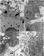
Fig. 2. (A–E) TEM images of Hepatozoon oocysts in Culex quinqefasciatus haemocoel. (A) Oocyst from boa-fed, 9 days p.f. showing single nucleus (N), elongate or dichotomous mitochondria (M) and vacuoles filled either with debris (V) or lipid-like substance (L). (B) Divided oocyst from boa-fed mosquitoes, 9 days p.f., showing daughter nuclei (N) and assemblies of crystalline granules (arrow heads). c, Cisternae; G, Golgi elements, and vacuoles filled either with debris (V) or lipid-like substance (L). (C) Enlarged view of (B), showing the assemblies of crystalline granules enclosed in rough ER cisterna (arrowheads), a presumed elongated mitochondrion (arrow), a cisterna (c) and lipid vacuole (L). (D) Caiman-fed mosquitoes, 12 days p.f., dividing oocyst showing daughter nuclei (N), arrays of crystalline substance ‘factories’ (bold arrows) and a sector of the wall forming prolonged plasmalemmal processes (P). (E) Enlarged view of (D), showing the processes (P) arising from gaps (arrow) in an osmiophilic underlining layer (arrowheads). e, Subplasmalemmal vesicles. The cytoplasm contains ‘factories’ of the crystalline material: formations of tubular mitochondria wrapped in rough ER (mr) and aggregates of crystalline granules (f).

Fig. 3. SEM image of the gut of caiman-fed Culex quinqefasciatus, 13 days p.f. carrying 4 Hepatozoon oocysts (arrows).
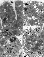
Fig. 4. TEM view of Hepatozoon sporulation in caiman-fed Culex quinqefasciatus haemocoel. (A) Day 13 p.f. segmenting oocyst in the process of dividing into sporocysts: cytoplasm contains fully formed crystalline bodies as well as formations of rough ER-wrapped mitochondria (rer). N, Daughter nucleus. (B) Sporocyst 15 days p.f., with daughter nuclei (N), with the crystalline material still not consolidated into arrays (arrow). (C) Sporocyst from same mosquito as in (B), showing fully consolidated crystalline body (arrow). (D) Sporocyst from same mosquito as in (B) showing onset of sporozoite differentiation (S) with early formation of the sporozoite pellicle (p) next to the sporozoite yielding nucleus (N). M, Mitochondrion; R, crystalline body. (E) Sporocyst from a 22 days p.f. mosquito showing premordia of sporozoites (S), pellicle alignments (p) and crystalline bodies (R) in the sporocyst residuum.
 You have
Access
- 11
- Cited by