Introduction
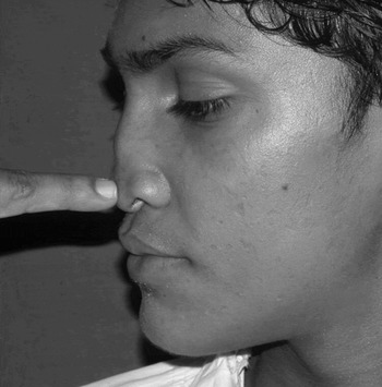
Contour defects of the nose which require dorsal augmentation are seen in patients with septal collapse following submucous resection, post-traumatic defects and congenital deformities. Dorsal augmentation can be performed using alloplastic implants, cartilage grafts or bone grafts. However, the material used for augmentation needs either a structural support or a stable bed to rest upon. For this reason, depressions arising following submucous resection are difficult to correct, as structural support is missing in the lower half of the dorsum. In such cases, the lack of structural support can be easily diagnosed, as the supratip area sinks when pressed by the examining finger (Figure 1). A bone graft fixed at the upper end is a reasonable solution for these deformities.
‘Digital palpation test’ demonstrating lack of structural support for nasal septum.

Graft fixation plays an important role in the final cosmetic outcome. Along with internal shaping, it controls the position of the graft and maintains a good alignment. It has been proven that, in order to decrease the risk of resorption, the fixation must be rigid with good bone-to-bone apposition.Reference Sullivan, Varna and Rozzelle1–Reference Phillips and Rahn7 Furthermore, the recipient site should be immobile and should provide a good blood supply and some functional stress to the graft.Reference Smith and Abramson8
Technique
The technique described below highlights the importance of preparation of the recipient site and graft, and also the importance of graft fixation.
Recipient site preparation
The graft site is prepared as follows. Bilateral alar rim incisions are made, through which the skin and soft tissue envelope of the nose is degloved. Once the inferior edge of the nasal bones is reached, subperiosteal undermining is performed up to and slightly beyond the nasofrontal angle, so that the implant can be easily inserted up to that site and then manipulated in a retrograde fashion towards the nasal tip. The bony dorsum is rasped to remove the remaining periosteum and the superficial layer of bone, thus enabling bone-to-bone contact.
Shaping the graft
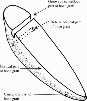
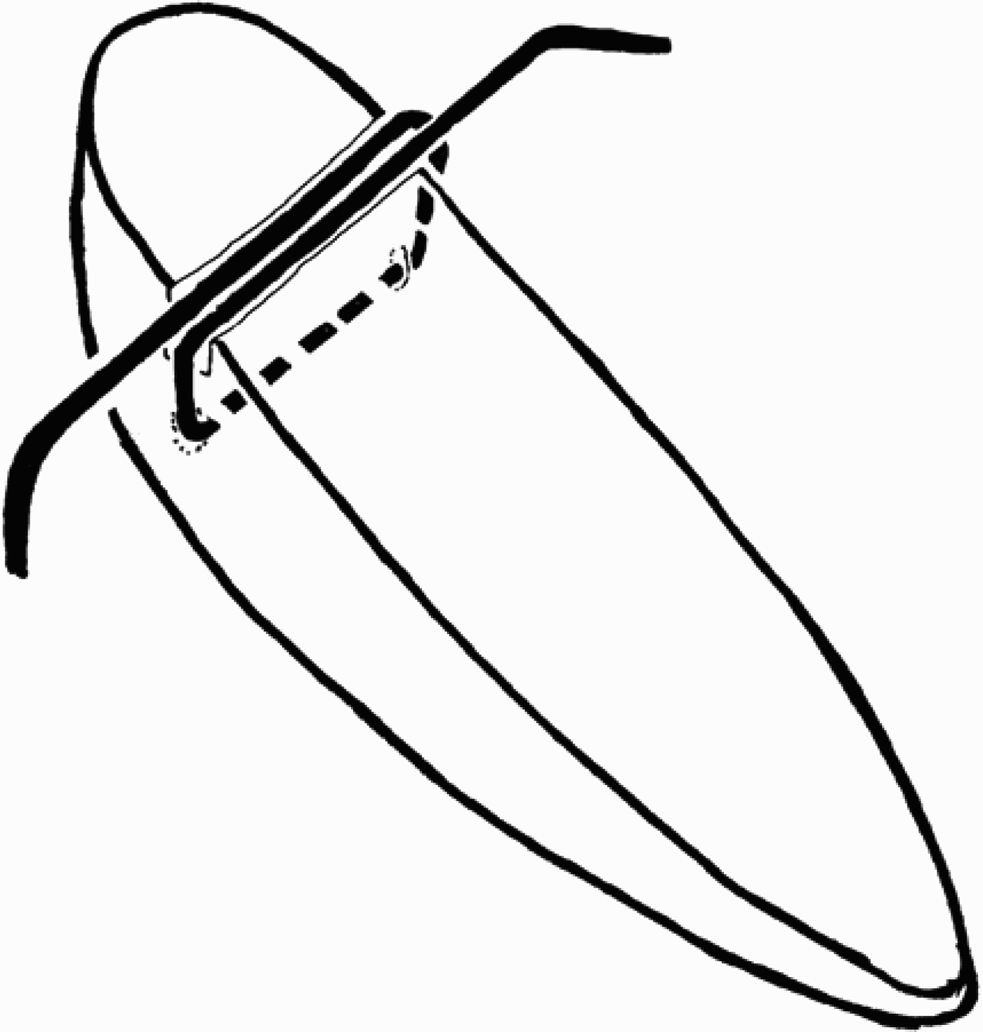
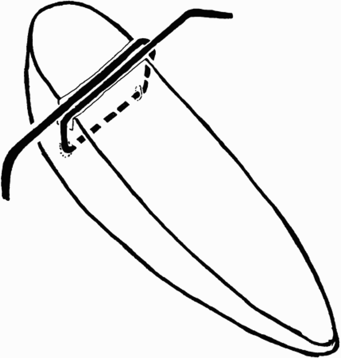
The bone graft is harvested from the iliac crest. This cortico-cancellous iliac crest graft is carved such that the cortex is preserved on one side (the dorsal side) while the cancellous side is shaped to fit the defect, using a number 10 scalpel. The boat-shaped graft should extend up to the tip; depending on the nasal length, it will be approximately 4–4.5 cm in length, 4–6 mm in width and 3–4 mm thick at the site where wire fixation is to be performed (5–10 mm inferior to the nasofrontal angle). At the wire fixation site, a groove is made on the anterior cortical aspect of the graft. Just below this groove, a parallel hole is bored into the cancellous portion using a 20 gauge needle (Figure 2).
Diagram of the bone graft.

The sculpted and prepared graft is inserted into the previously created nasal pocket to test its shape and stability. Cephalad pressure is applied in the radix area to replicate the eventual fixation and to enable assessment of cosmesis. Any additional modifications to the graft can be made at this time.
Graft fixation
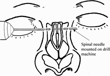
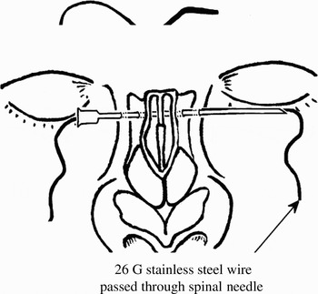
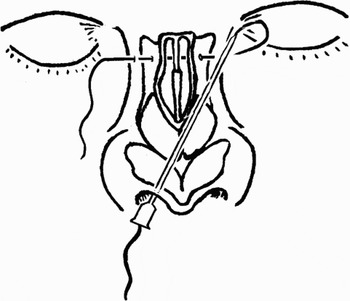
A small stab incision is made in the right medial canthal region and a hole is drilled using a hand drill with a 16 gauge spinal needle attached in lieu of drill bit (Figure 3). The needle passes through the nasal bones and exits the skin at the opposite medial canthus. A 26 gauge, pre-stressed stainless steel wire, approximately 25 cm long, is then threaded through the needle while the needle is withdrawn slowly (Figure 4).
A hole is made with a spinal needle mounted on a hand drill.

A 26 gauge stainless steel wire is passed through the spinal needle.

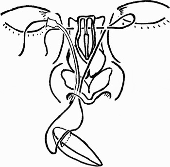
The spinal needle is inserted underneath the skin envelope through the right side of the rim incision to the left medial canthal region. The leading end of the wire is guided through the spinal needle so that it emerges through the rim incision on the right side of the columella (Figure 5). The graft is then mounted onto the wire by passing the wire through the hole in the cancellous bone; the wire is then bent and passed over the groove on the anterior cortical surface (Figure 6). The spinal needle is bent to a minimum curvature and again passed from the stab incision in the right medial canthal region to the right alar rim incision, and the wire guided through it (Figure 7).
The leading edge of the stainless steel wire is brought out through the alar rim incision.

The bone graft is mounted on the stainless steel wire.
The leading edge of the stainless steel wire is brought out through the medial canthal stab incision.

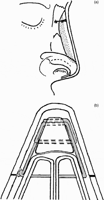
The wire is slowly pulled through the medial canthal stab incision. Both ends of the wire are gradually pulled as the graft is inserted into the pocket and positioned on the nasal bone (Figure 8a). The bevelled graft tip projects beyond the nasal bones, providing tip projection. The wire is then tightened, putting stress on the graft, in order to ensure adequate graft fixation, and the cut ends are buried in the soft tissue in the medial canthal region (Figure 8b).
The bone graft in situ: (a) profile view, (b) transverse section.

Wound closure and splintage
The stab incisions do not require suturing. The rim incision is sutured with 4/0 absorbable suture thread.
The nose is taped and a plaster of Paris splint is placed on the nose for protection and to reduce the risk of haematoma. The splint is removed after three weeks.
Results
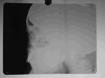
This technique has thus far been used in 16 patients with post-supratip depression after submucous resection. The first 10 patients were followed up for five years. During this time, augmentation was maintained and a straight dorsal line persisted. Routine plain X-rays showed no resorption of bone (Figure 9). The remaining six patients have been followed up for shorter periods (ranging from four months to two years), but no contour changes have been observed thus far.
X-ray showing no bone resorption after three years.

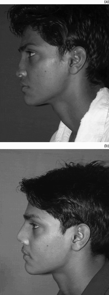
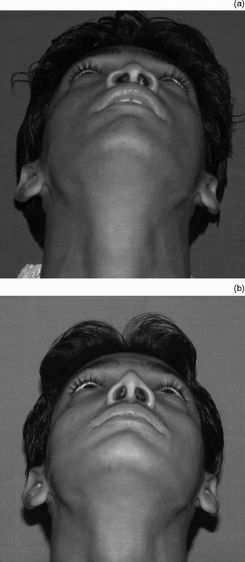
In all these patients, we were able to achieve the desired aesthetic result by correcting the deformity and establishing a pleasant nasal profile (Figure 10). As the graft projects beyond the nasal bones, the resulting cantilever effect raises the nasal tip (Figure 11).
Profile photographs: (a) pre-operative, (b) post-operative.

Basal or ‘worms-eye’ view photographs: (a) pre-operative, (b) post-operative. The cantilever effect of the bone graft raises the nasal tip.

The graft wire was not palpable in any of the cases undertaken, and there has been no apparent fatigue of the wire with time. No post-operative infection was encountered; however, all patients received broad-spectrum antibiotics for two weeks post-operatively.
Discussion
Nasal supratip depression following submucous resection presents a specific challenge, as there is little structural support available for materials used to augment the cartilaginous part of the nose (i.e. the middle and lower third). Because of this, authors have advocated either a bone graft fixed at one end or an L-shaped graft or implant.Reference Sullivan, Varna and Rozzelle1 However, implants are at risk of extrusion, especially in scarred and previously operated-upon noses.Reference Posnick, Seagle and Armstrong2, Reference Wheeler, Kawamoto and Zarem3, Reference Stuzim and Kawamoto9–Reference Jackson, Smith and Mixter12
Various methods have been described for graft fixation, using lag screws, mini-plates and screws, interosseous wiring, and K wires. However, these techniques are complicated and time-consuming, and have the potential for failure due to early malposition, late resorption or poor restoration of normal nasal contour.
Kirschner wires or 30 gauge needles can be passed percutaneously through the bone graft and into the nasal bones, but removal in the early post-operative period leads to malposition and non-union. Thus, K wires are usually used along with another method of fixation.Reference Wheeler, Kawamoto and Zarem3, Reference Neu6, Reference Stuzim and Kawamoto9 Ultra-mini-screws and plates produce rigid fixation but require an incision in the dorsal skin for insertion, and the screws may occasionally be palpable in the frontonasal region.Reference Posnick, Seagle and Armstrong2, Reference Neu6 Lag screws have been used for cantilever grafts; however, a single screw does not provide rigid fixation as rotation around this screw is possible. Bone is also prone to micro-fractures and horizontal cracking as the screw tightens, and if the head is countersunk this weakens the bone. There is also a risk of entry into the nasal cavity.Reference Gruss4
The use of interosseous wire for fixation has been previously described but details of the technique are lacking. Without a dorsal skin incision, it is extremely difficult to guide a wire percutaneously from one canthal region to the other over a bone graft and to make it sit exactly.Reference Stuzim and Kawamoto9 If a dorsal incision is made to expose the nasal bones this facilitates passage of the wire, but it may also result in increased scarring, greater risk of graft exposure and an inferior cosmetic result.
Our method of graft fixation uses interosseous wiring but avoids the need for a dorsal incision by entangling the graft in the wire and by threading the wire through a hollow needle. The graft has a parallel hole beneath a groove, ensuring that the wire sits exactly as intended. As the wire is bent over into the groove, the brunt of compressive forces is borne by the strong cortical bone, thus preventing the wire from cutting through the graft. This rigid fixation of the bone graft allows better ‘take’.
Calvarial bone grafts are superior in terms of lack of resorption but have restricted possibilities as regards graft shaping.Reference Daniel5, Reference Smith and Abramson8, Reference Zins and Whitaker13, Reference Tessier14 The iliac crest provides an abundant source of cortico-cancellous bone graft, and the graft can be surprisingly easily carved with a scalpel and sculpted into the desired shape and contour. It can also be polished with a number 15 blade to make the dorsum smooth. Unfortunately, resorption is unpredictable, leading to loss of shape and volume. However, this can be prevented by rigid fixation and good bone-to-bone apposition. We feel our method offers this, and provides satisfactory results without other complications related to donor site morbidity.
Acknowledgement
We are thankful to our Dean and Director Medical Education (ME) and Municipal Hospitals (MH), Dr Sanjay Oak, for permitting us to publish this paper.