Introduction
Fungal pachymeningitis is an extremely rare opportunistic infection of the central nervous system (CNS). Only two cases have previously been reported. The fungal infection usually spreads to the dura from the ear, nasal cavity, paranasal sinuses or lung. Multiple cranial nerve palsies and headache are the commonest presenting symptoms. Inflammation of the dura may extend beyond the skull case and cause nasopharyngeal swelling. The combination of multiple cranial nerve palsies and nasopharyngeal swelling is also a common feature of nasopharyngeal carcinoma, which must be excluded first in prevalent areas such as Hong Kong and other parts of southern China.
Case report
A 59-year-old Chinese man presented to the otorhinolaryngology and head and neck surgery department of a tertiary referral teaching hospital with a two-month history of hoarseness, diplopia and a left frontal headache. He was a poorly controlled, insulin-dependent diabetic who had impaired renal function and a history of ischaemic heart disease.
On physical examination, the patient was found to have a left abducens nerve palsy, a left hypoglossal nerve palsy and a left vocal fold palsy. He had an automastoid cavity in the right ear and an atelectatic tympanic membrane in the left ear, but no signs of active infection in either ear.
Nasopharyngoscopy showed smooth, bilateral fullness of the nasopharynx, with normal mucosa.
A gadolinium-enhanced magnetic resonance imaging (MRI) scan of the skull base revealed a contrast-enhancing soft tissue lesion which was closely related to the left pharyngeal recess and which encased the left internal carotid artery and extended intracranially (Figure 1).
Axial, T1-weighted, gadolinium-enhanced magnetic resonance image of the nasopharynx, with fat saturation, showing abnormally enhancing soft tissues deep to the left posterolateral nasopharyngeal wall (arrow), with extension into the left parapharyngeal space and carotid space to encase the left internal carotid artery (arrowhead).

The clinical presentation and radiological findings raised suspicion of nasopharyngeal carcinoma (NPC).
Immunofluorescence serological tests for immunoglobulin (Ig) A antibody against Epstein–Barr virus were abnormal: the IgA viral capsid antigen titre was 20 and the early antigen titre was 5.
Multiple deep nasopharyngeal biopsies were taken to exclude NPC; all were negative for malignancy.
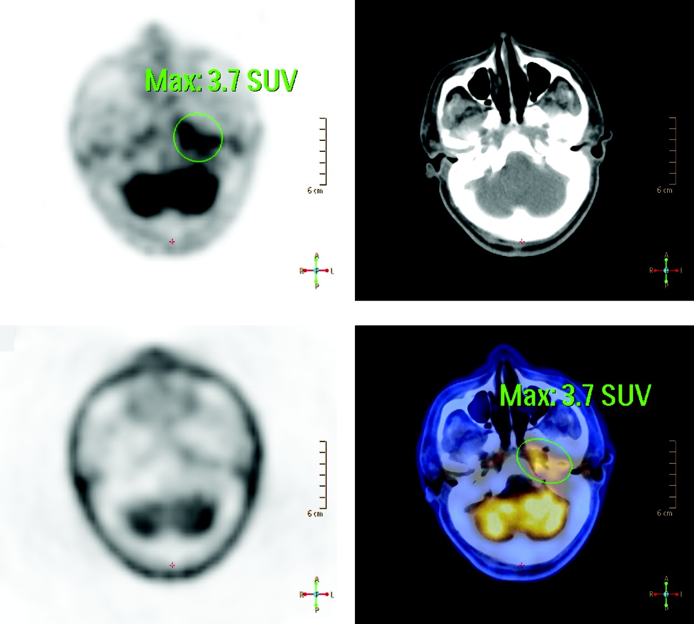
A positron emission tomography (PET) scan of the head and neck showed that increased fluoro-deoxy-glucose (FDG) uptake (maximum standardised uptake value of 4.3) seen in the left nasopharyngeal parapharyngeal space was in continuity with increased uptake in the left posterior fossa dura (Figure 2). Increased FDG uptake in the left mastoid tip (maximum standardised uptake value 4.6) was also seen.
Positron emission tomography scan showing increased fluoro-deoxy-glucose uptake deep to the left posterolateral nasopharyngeal wall (maximum standardised uptake value = 3.7). A = anterior; P = posterior; R = right; L = left

The patient's condition deteriorated, with increasing headache severity and diplopia and decreasing visual acuity bilaterally.
Although he was afebrile, he had a mild leukocytosis of 11.6 × 10⁹/l and an elevated erythrocyte sedimentation rate (ESR) of more than 130 mm/hour. Cerebrospinal fluid (CSF) collected by lumbar puncture was clear, with no organisms seen on microscopy. The CSF glucose level was 5.7 mmol/l and the CSF protein level was elevated, at 1.27 g/l.
The abnormal dural enhancement seen on MRI and PET, together with the elevated inflammatory markers, raised suspicion of pachymeningitis.
This prompted us to undertake an open dural biopsy, taken from the left posterior cranial fossa dura via a retrosigmoid approach. The dura was found to be thickened and to form a plaque-like lesion between the brain and the bone.
Histological examination of the dural biopsy showed fibrous tissue with a dense small lymphocyte infiltrate involving most of its layers. Immunohistochemical studies showed that most of the lymphocytes were T cells. Degenerated fungal filaments with septate branching were also found.
These finding were compatible with a diagnosis of fungal pachymeningitis.
Fungal infection was later confirmed by a finding of elevated serum titres of 1,3-β-d-glucan and galactomannan. The presence of galactomannan suggested an aspergillus infection.
A fungal culture from the dural biopsy grew Aspergillus flavus after two weeks.
The patient's ears were re-examined. An ear swab from deep in the external meatus showed a focus of fungal filaments on microscopy. The infected ear canal was cleaned and miconazole anti-fungal cream instilled. The same ear swab later grew Aspergillus flavus.
Once the diagnosis had been made, systemic antifungal treatment with oral voriconazole 200 mg twice daily was commenced. However, the patient developed a pancytopenic crisis after two weeks of voriconazole therapy, and the antifungal agent was thus changed to intravenous caspofungin 50 mg daily.
After four weeks of antifungal treatment, the patient's ESR had dropped to 35 mm/hour, his headaches had improved and his left hypoglossal nerve function had recovered; however, his vocal fold palsy and ophthalmoplegia persisted.
In addition, during the course of antifungal treatment the patient's visual acuity had deteriorated further bilaterally. A follow-up MRI scan showed possible involvement of both optic nerves by the disease (Figure 3). In view of the continued deterioration of his visual acuity, the patient was rechallenged with voriconazole (after a four-week break from the drug). However, despite this aggressive antifungal treatment, and tight control of blood glucose levels, the patient suffered permanent, bilateral loss of vision.
Coronal, T1-weighted, gadolinium-enhanced magnetic resonance image showing abnormally enhancing dural thickenings over both sphenoid wings, with suspicious compression of the optic nerves at the anterior clinoids especially on the right (arrow).

The patient was subsequently discharged to a convalescent hospital for rehabilitation.
Seven months after initial presentation, a follow-up MRI scan showed persistent dural thickening. As a result, the patient was prescribed lifelong oral voriconazole.
Discussion
Hypertrophic cranial pachymeningitis is a rare clinical condition which is characterised by localised or diffuse thickening of the dura mater.Reference Kupersmith, Martin, Heller, Shah and Mitnick1 Most cases are idiopathic, but rare causes need to be excluded. These include infectious diseases (such as tuberculosis, syphilis, and fungal infections such as aspergillus, candida, cryptococcus and histoplasmosis), inflammatory diseases (including sarcoidosis, rheumatoid arthritis and Wegener's granulomatosis), dural carcinomatosis and intracranial plasmacytosis.Reference Hatano, Behari, Nagatani, Kimura, Ooka and Saito2, Reference Rossi, Giannini, Cerase, Bartalini, Tripodi and Volpi3
Only two previous cases of fungal pachymeningitis have been reported. Murai et al. Reference Murai, Kira, Kobayashi, Goto, Inoue and Hasuo4 reported a possible case of fungal pachymeningitis, which was not confirmed by dural biopsy. Ismail et al. Reference Ismail, Clifford and Meacock5 reported a case in which a positive fungal culture was obtained from the frontal sinus, but they did not state clearly whether or not the fungal infection involved the meninges.
To the best of our knowledge, our patient represents the first reported case of fungal pachymeningitis in which the same fungal species was identified in both an ear culture and a dural culture.
Most CNS fungal infections occur in immunocompromised patients. Aspergillus infections usually spread from the ear, nasal sinuses or lungs to the CNS. The presence of aspergillus in both an ear swab and an ipsilateral dural biopsy would suggest an otogenic source of the CNS infection. Cases of non-fungal pachymeningitis secondary to ear infections are well documented in the literature; most are inflammatory in nature.Reference Iwasaki, Ito and Sugasawa6, Reference Kanzaki, Inoue, Watabe and Ogawa7 Headache is the commonest symptom, and most such headaches are chronic and severe.Reference Kupersmith, Martin, Heller, Shah and Mitnick1, Reference Rossi, Giannini, Cerase, Bartalini, Tripodi and Volpi3 The thickening of the meninges encasing the cranial nerves results in their inflammation and ischaemia, and ultimately in nerve paralysis.Reference Hatano, Behari, Nagatani, Kimura, Ooka and Saito2
Detection of fungal infection in the CNS is difficult. However, immunoassay for fungal cell wall components now provides a sensitive tool for the detection of fungal infections in CSF specimens. Relevant fungal wall components include β-d-glucan, a non-specific polysaccharide present in most fungal cell walls, and galactomannan, which is more specific to aspergillus species.Reference Wasser and Weis8, Reference Bart-Delabesse, Basile, Al Jijakli, Souville, Gay and Philippe9
Gadolinium-enhanced MRI is an indispensable tool which facilitates the diagnosis of pachymenigitis and localises the affected meninges for subsequent dural biopsy.Reference Kupersmith, Martin, Heller, Shah and Mitnick1 More subtle foci of pachymeningitis can be localised using PET, as in our patient. Dural biopsy is the ‘gold standard’ for establishing the diagnosis of fungal pachymeningitis.
The treatment of fungal pachymeningitis is not well established due to the lack of reported cases. The standard steroid regime used for idiopathic cranial pachymeningitis may not be applicable when an infective agent is present. In our patient, antifungal treatment was given empirically to treat fungal meningitis. Surgical decompression of involved cranial nerves has been reported in idiopathic cases, with mixed results.Reference Kupersmith, Martin, Heller, Shah and Mitnick1–Reference Rossi, Giannini, Cerase, Bartalini, Tripodi and Volpi3 The clinical course of fungal pachymeningitis is unpredictable and can be fatal despite antifungal treatment.Reference Murai, Kira, Kobayashi, Goto, Inoue and Hasuo4, Reference Ismail, Clifford and Meacock5
Nasopharyngeal carcinoma (NPC) is prevalent in south-eastern China, and is the commonest head and neck cancer seen in this region.Reference Vlantis, Tsang, Yu, Kam, Tong and Lo10 Most patients with NPC present with the typical symptoms of epistaxis, metastatic cervical lymphadenopathy and otitis media with effusion.Reference Pak, Chan and van Hasselt11 There are however a small group of patients who present with atypical symptoms such as headaches and multiple cranial nerves palsies. As our patient presented with similar symptoms, to a hospital in south-eastern China, NPC had to be excluded before other diagnoses were considered. Although most NPC tumours are obvious on nasopharyngoscopy, we do encounter cases of submucosal NPC in which there is no apparent mucosal abnormality (Figure 4). It is therefore difficult to quickly exclude NPC in this group of atypical patients, unless serology or imaging is grossly abnormal. While PET scanning is a sensitive means of detecting early nasopharyngeal tumours, false positives do occur due to inflammatory conditions, as in our patient. Multiple deep nasopharyngeal biopsies are usually required when a submucosal tumour is suspected. Surgical navigation aids are useful in assisting biopsy in more difficult cases.
Axial, T1-weighted magnetic resonance image of the nasopharynx showing a large submucosal nasopharyngeal carcinoma (arrow) deep to the right fossa of Rosenmüller. The overlying mucosa appeared intact on endoscopy.

• Fungal pachymeningitis should be included in the differential diagnosis of nasopharyngeal carcinoma, especially in immunocompromised patients
• When fungal pachymeningitis is suspected, magnetic resonance imaging is useful in localising the extent of dural involvement and guiding subsequent dural biopsy
• The ear, nasal cavity, paranasal sinuses and chest should be examined to seek a primary source of infection
• In the presented case, treatment of fungal pachymeningitis consisted of eradicating the infective source and commencing lifelong systemic antifungal medication (systemic steroids are indicated in non-infective pachymeningitis but their role in fungal pachymeningitis is yet to be established)
There are similarities between the presenting symptoms and the radiological findings of NPC and pachymeningitis. The clinician should be aware of these similarities when investigating patients with multiple cranial palsies and nasopharyngeal swelling. Fungal pachymeningitis characteristically affects immunocompromised patients, and such patients have more florid CNS symptoms (e.g. headaches). Markedly elevated levels of inflammatory markers and the absence of cervical lymphadenopathy favour a diagnosis of pachymeningitis over NPC, especially in immunocompromised patients. On MRI scans, extensive dural involvement would be more suggestive of fungal pachymeningitis than intracranial NPC.