Takayasu disease is a rare disease mainly affecting women and commonly presenting during the second or third decade. The disease is characterised by chronic inflammatory arteritis affecting preferably the aorta and its main branches. This results in clinical and angiographic signs of arterial stenosis, thrombus formation, and aneurysm formation.Reference Johnston, Lock and Gompels 1 , Reference Gornik and Creager 2
Endovascular angioplasty is often the preferred treatment modality for obstructive long-segment aortitis. Simple balloon angioplasty carries a high risk for dissection and aneurysm formation. Bare or covered stent implantation is often favoured in the presence of such a diseased and fragile aortic wall, to reduce the incidence of those complications.Reference Butera, Manica and Chessa 3
We report a patient with Takayasu arteritis in whom symptomatic obstruction of the descending aorta required, over time, several stent insertions. We encountered, over time, several complications including recoarctation, dissection, and also collapse of a covered stent. This case highlights the challenges of the management of diffuse aortitis.
Case report
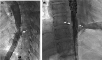
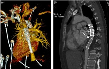
A 31-year-old woman was admitted for acute renal failure. She had been diagnosed with Takayasu arteritis at the age of 14 years. The disease was widespread with brachiocephalic aneurysm, left carotid occlusion, left subclavian artery hypoplasia, and stenosis of the descending aorta, causing severe systemic hypertension. At the age of 15 years, balloon angioplasty of the descending thoracic aorta was performed, followed 6 months later by stent implantation (Palmaz P4014, Johnson & Johnson, United States of America). This procedure allowed appropriate control of the blood pressure. At the age of 29 years, severe arterial hypertension reoccurred and remained resistant to quadri-therapy and immunomodulation. Computed tomography (CT) scan showed recurrent obstruction of the descending aorta. At cardiac catheterisation, intra-stent restenosis was confirmed (peak-to-peak gradient of 70 mmHg). Angiographies (Fig 1a) showed two levels of stenosis: in the middle and immediately below the stent. A mild posterior aneurysm was also observed with diffuse calcifications of the aortic wall. Two covered Cheatham-Platinum stents (CP8Z34TM, each on a 14 mm BIB® balloon, Numed Inc., Hopkinton, New York, United States of America) were inserted, but the procedure was complicated by an extensive dissection of the lower aorta down to the diaphragm (Fig 1b). This was immediately treated by implantation of a 61-mm-long covered AdvantaTM V12 (Atrium, Hudson, New Hampshire, United States of America) stent premounted on a 12 mm balloon. The 12 mm balloon was electively chosen because of the marked narrowing of the abdominal aorta and to avoid overdilation of the dissected segment. No additional ballooning was performed, as the final result was considered satisfactory without residual gradient or dissection seen on the final angiography. A CT-scan performed 24 hours after catheterisation (Fig 2a) confirmed the good result but suggested some discrepancy between the diameter of the aortic segment stented with the AdvantaTM V12 stent and the aorta above and below. Aspirin (160 mg) was added to her usual treatment. During the following months, she remained on triple anti-hypertensive treatment with near-normal arterial blood pressure. After 6 months of stent placement, she acutely presented with pulmonary oedema and renal failure. CT scan showed that the AdvantaTM V12 stent had slided downward and collapsed at its proximal end, obstructing the aortic flow (Fig 2b). There was also some mild recoil seen at the distal end. After stabilisation in the intensive care unit using inotropic support and dialysis, she was brought back to the catheterisation laboratory. The upper infolding was successfully treated by the insertion of a bare Cheatham-PlatinumTM stent mounted on a 14 mm BIB® balloon inside the upper end of the AdvantaTM V12 covered stent. The procedure resulted in marked clinical improvement. She currently remains mildly hypertensive with a clinical upper–lower limb gradient of 20–30 mmHg.
Angiography in the descending aorta. ( a ) Lateral projection. The previously inserted stent is not fully expanded in its middle segment. There is diffuse endothelial proliferation with intra-stent restenosis. This is mostly marked at the mid portion and at the lower part of the stent. There is also a small posterior aneurysm (white arrow). ( b ) Antero-posterior projection. An extensive dissection can be seen below the stents newly inserted.

( a ) Computed tomography (CT) scan of the aorta 24 hours after the stenting of the descending aorta, showing good position of the stents but some diameter discrepancy between the aorta stented with the Advanta stent and the aorta above and below. ( b ) CT scan of the aorta showing infolding of the proximal end of the covered atrium stent, with obstruction of the lumen.

Discussion
Patients with Takayasu aortitis are characterised by extremely abnormal behaviour of the aortic wall. Not only balloon angioplasty but also stent placement may be complicated by extensive dissection and requires the availability of covered stents on the shelf to deal with this potentially lethal complication.
The initial placement of a bare stent was complicated by long-term in-stent stenosis but also aneurysm formation. The insertion of two additional covered stents resulted in acute dissection of the abdominal aorta. The AdvantaTM V12 stent was chosen to deal with this complication. It is an open-cell stent covered on both sides by polytetrafluorothylene. The stent is premounted, has a low profile, and comes in various lengths up to 61 mm, which are strong advantages, especially in emergency cases and extensive wall dissections.Reference Bruckheimer, Birk, Santiago, Dagan, Esteves and Pedra 4 However, very recently, three cases of stent collapse have been described in patients with native coarctation in whom the AdvantaTM V12 stent had been electively inserted.Reference Wan, Lee and Benson 5 , Reference Hayes, Podnar and Qureshi 6 In our patient, the long-covered AdvantaTM stent was definitely helpful for the acute management of this life-threatening complication. Unfortunately, the stent slided slightly downwards during follow-up and the proximal end folded in, causing severe and acute aortic obstruction. The exact mechanism of this severe complication is, as in the previous reports, not entirely clear. It is likely that the downward shift happened early after stent placement, before endothelialisation of the stent. The downward shift may have been favoured by the insufficient overlap between the AdvantaTM V12 and the previously inserted stents, and the diameter discrepancy. The 12 mm stent was chosen because of the smaller diameter of the abdominal aorta, but the downward shift could have been avoided by redilating the overlapping parts with a 14-mm balloon to adjust the sizes and to secure the overlap. The collapse of the proximal end most likely happened later, as clinical signs of aortic obstruction were acute. Several mechanisms may explain this observation but may coexist with: (1) progression of the wall dissection with hematoma pushing the stent inwards; (2) inadequate wall apposition of the stent causing blood to circulate between the wall and the stent, eventually folding the stent inwards; (3) stent fracture resulting in an acute collapse; and (4) insufficient radial force of the stent, unable to resist to wall forces. The Advanta stent is known to have immediate recoil, favoured by the polytetrafluorothylene covering the both sides of the stent.Reference Ohno, Chaturvedi, Lee, Horlick, Osten and Benson 7 However, the contribution of the extremely diseased wall to this complication may not be excluded. This highlights the extreme difficulties that may be encountered when managing those patients.
Acknowledgement
None.
Financial Support
This research received no specific grant from any funding agency, commercial, or not-for-profit sectors.
Conflicts of Interest
None.