Introduction
Pathology of the vestibular and auditory systems can present with debilitating symptoms. Due to anatomical proximity and functional interconnections, auditory and vestibular symptoms can manifest simultaneously, making pathology arising in this region challenging to address.
Vestibular evoked myogenic potentials are biphasic, short latency, inhibitory electrical changes measured at the sternocleidomastoid muscles, which result from sound stimulation of the saccular portion of the vestibular system. Many studies have proven their use in the assessment of saccular (otolithic) function and inferior vestibular nerve involvement in this context.
Relevant physiology
The vestibular apparatus maintains balance and equilibrium, along with the proprioceptive and visual systems. It contributes to balance through the vestibulo-ocular, vestibulo-spinal and vestibulo-collic reflexes.
The vestibulo-ocular reflex maintains a stabilised visual image on the retina during head rotation, by inducing compensatory eye movements.
The vestibulo-spinal reflex stabilises the body in relation to gravity.
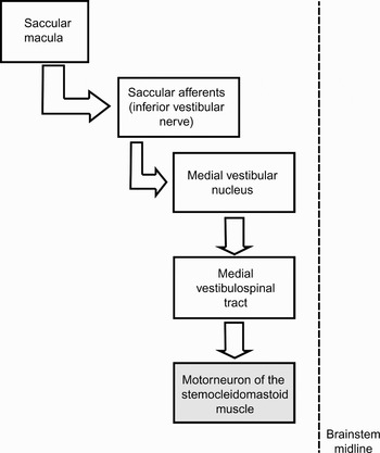
The vestibulo-collic reflex is responsible for the stability of the head in space, and acts specifically on the neck musculature. It also assists the vestibulo-ocular reflex in stabilising visual acuity. This reflex is also indicative of otolith function, specifically that of the saccule, and this is the origin of the synonymous term sacculo-collic reflex. The sacculo-collic reflex forms the basis of the vestibular evoked myogenic potential (Figure 1).
The neural pathway of the sacculo-collic reflex. Adapted with permission.Reference Jacobson, Newman and Kartush1

Vestibular evoked myogenic potential testing
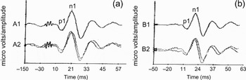
The normal vestibular evoked myogenic potential is biphasic, with a peak and a trough, labelled in terms of their latency and preceded by the lower case letter ‘p’ or ‘n’ for positive and negative amplitudes, respectively (Figure 2).Reference Yoshie and Okudaira2 Wave labelling differentiates them from neural potentials. Colebatch et al. Reference Colebatch, Halmagyi and Skuse3 labelled the normal response as two distinct waves, namely p1n1 (often called p13n23 as it occurs at 13 and 23 ms, respectively) and n34p44. This terminology has been adopted in the majority of later studies. The second wave is inconsistent;Reference Colebatch, Halmagyi and Skuse3–Reference Ferber-Viart, Duclaux, Colleaux and Dubreuil5 it is considered to be of lesser clinical significance and is thought to be non-vestibular in origin. The amplitude of the positive and negative peaks can vary from a few microvolts to several hundred microvolts, and is related to the tension in the muscle.Reference Colebatch, Halmagyi and Skuse3, Reference Robertson and Ireland4, Reference Bickford, Jacobson and Cody6–Reference Ochi, Ohashi and Nishino10 It is therefore necessary to have a relatively constant level of tonicity in the sternocleidomastoid muscle during vestibular evoked myogenic potential testing. Vestibular evoked myogenic potentials are inhibitory to the sternocleidomastoid muscle, and therefore are only detectable when there is electrical activity present within the muscle.
Normal vestibular evoked myogenic potential for (a) left and (b) right sternocleidomastoid muscle. The main potential, p1, is located at about 13 ms. Each side is about 250 µV in amplitude (which is far above the lower limit of normal, approximately 70 µV). (An electrical artefact at 0 ms on the left side can be ignored.) A1, A2, B1, B2 represent stimulation levels in dB nHL

Two methods are generally used for vestibular evoked myogenic potential testing: the head elevation non-rotation method and the head rotation method.
In the first method, the patient lies in a supine position and elevates their head by 30o. This position can result in fatigue, which can influence the contralateral response if each side is recorded separately.Reference Park, Cha, Kim, Kim, Hwang and Hong11
The second method requires the patient to rotate their head towards one shoulder (in either a supine or sitting position). This carries less muscular burden and is better tolerated;Reference Beyea and Zeitouni12 it is also useful in patients with restricted cervical mobility. However, this method is less reliable and produces smaller potentials.Reference Wang and Young13
Attempts have been made to modify the above two methods. A degree of reliability has been demonstrated for the following: instructing patients to press their forehead against a bar; having patients hold a rubber ball between the chin and the manubrium; and monitoring the bulb pressure whilst using a simple blood pressure manometer apparatus and inflatable cuff to keep constant muscle contraction of the sternocleidomastoid muscle.Reference Colebatch, Halmagyi and Skuse3, Reference Robertson and Ireland4, Reference Ferber-Viart, Soulier, Dubreuil and Duclaux14, Reference Vanspauwen, Wuyts and Van de Heyning15 Currently, there is no proven superior method.
Vestibular evoked myogenic potentials arise with a latency of 6 ms, and differ considerably from startle responses and voluntary head movements in response to acoustic stimuli, which have longer latency periods of 50 and 100 ms, respectively.Reference Bickford, Jacobson and Cody6, Reference Landis and Hunt16 Unlike startle responses, vestibular evoked myogenic potentials are unaffected by factors such as habituation, sensitisation and prepulse inhibition, and can be induced repeatedly.
Early studies measured the response at the inion.Reference Bickford, Jacobson and Cody6, Reference Townsend and Cody17–Reference Cody and Bickford19 Some studies have also explored the use of the post-auricular region, trapezius muscle, and the muscles of the arms and legs.Reference Ferber-Viart, Duclaux, Colleaux and Dubreuil5, Reference Bickford, Jacobson and Cody6, Reference Townsend and Cody17, Reference Kiang, Crist, French and Edwards20, Reference Li, Houlden and Tomlinson21 Colebatch and colleagues proposed the use of the sternocleidomastoid muscle, and showed responses in all their subjects.Reference Colebatch, Halmagyi and Skuse3, Reference Colebatch and Halmagyi7 The sternocleidomastoid muscle has since become the standard recording site. Reference electrodes may be placed on the lateral part of the upper sternum, the forehead or the wrists.Reference Li, Houlden and Tomlinson21
There are conflicting findings on laterality. With regards to monaural stimulation, several studies have demonstrated symmetrical responses from both sides;Reference Bickford, Jacobson and Cody6, Reference Townsend and Cody17 however, other authors have reported either a purely ipsilateral response,Reference Li, Houlden and Tomlinson21, Reference Akin and Muranane22 a larger ipsilateral responseReference Colebatch, Halmagyi and Skuse3, Reference Akin, Muranane and Proffitt23, Reference Murofushi, Ochiai, Ozeki and Iwasaki24 or a larger contralateral response.Reference Ferber-Viart, Duclaux, Colleaux and Dubreuil5 Upon comparing monaural with binaural stimulation, Young et al. Reference Young, Huang and Cheng25 did not find any significant difference in measured vestibular evoked myogenic potentials, while Ferber-Viart et al. Reference Ferber-Viart, Duclaux, Colleaux and Dubreuil5 reported potentials of greater amplitude with binaural stimulation. Simultaneous binaural testing has the disadvantage of susceptibility to artefactual electrical activity crossing the midline, due to the close proximity of the sternocleidomastoid muscle and sternum.Reference Li, Houlden and Tomlinson21 The unilateral method of vestibular evoked myogenic potential testing is therefore considered more representative and reliable. However, binaural testing is quicker, and thus more feasible in patients who are unable to endure a longer test.
Several studies have demonstrated the effect of age-related degenerative changes on the vestibular system, and specifically on vestibular evoked myogenic potentials.Reference Welgampola and Colebatch8, Reference Brantberg, Granath and Schart26–Reference Zapala and Brey30 Such latter changes include prolongation of the p13 and n23 latencies, decreased amplitudes, and an increase in the thresholds required to elicit a response. Some authors have demonstrated a sharp decline from the sixth decade,Reference Welgampola and Colebatch8, Reference Su, Huang, Young and Cheng29 while others have found evidence for progressive change throughout the decades,Reference Brantberg, Granath and Schart26–Reference Ochi and Ohashi28, Reference Zapala and Brey30 with a decrease of nearly 0.02 mV per year. Prolongation of n23 latency with age, but not of p13 latency, has also been demonstrated,Reference Welgampola and Colebatch8, Reference Su, Huang, Young and Cheng29 while others have suggested the contrary.Reference Zapala and Brey30 A positive correlation between neck length and waveform latency has recently been reported, with latencies increasing with neck length up to a length of 15.3 cm.Reference Wang, Yeh, Chang and Young31
Vestibular evoked myogenic potentials can be evoked by clicks presented at 90–100 dB normal hearing level (140–145 dB sound pressure level). Several studies have shown similar responses to short tone bursts.Reference Murofushi, Matsuzaki and Wu32–Reference Cheng, Huang and Young34 The presence of an intact middle ear able to conduct sound waves to the saccule is vital when using this method.Reference Bath, Harris and McEwan35, Reference Halmagyi, Yavor and Colebatch36
Bone-conducted tones and skull taps are useful in patients with conductive hearing loss.Reference Halmagyi, Yavor and Colebatch36–Reference Sheykholeslami, Habiby Kermany and Kaga39
The former are delivered using a bone conductor, optimally 3 × 2 cm postero-superior to the external acoustic meatus, using frequencies of 200–250 Hz.Reference Welgampola, Rosengren, Halmagyi and Colebatch38, Reference Sheykholeslami, Habiby Kermany and Kaga40
Tap-evoked vestibular evoked myogenic potentials require either a tendon hammer or an electromechanical ‘skull tapper’ applied either to the forehead or laterally above the ear. The resulting potentials are 1.5 to three times magnified in comparison to those evoked by clicks; this method is therefore useful in subjects with high thresholds.Reference Welgampola and Colebatch8 However, as tap-evoked potentials are operator-dependent, they are difficult to deliver as a calibrated stimulus.Reference Welgampola and Colebatch8
Although the afferent response pathway involved in bone-conducted and tap-evoked vestibular evoked myogenic potentials is not precisely known, involvement of the utricle had been proposed.Reference Welgampola, Rosengren, Halmagyi and Colebatch38, Reference Brantberg and Tribukait41, Reference Brantberg, Tribukait and Fransson42 Brantberg and colleaguesReference Brantberg, Westin, Löfqvist, Verrecchia and Tribukait43, Reference Brantberg, Löfqvist, Westin and Tribukait44 have recently investigated the mechanism for skull tap induced vestibular evoked myogenic potentials. Their findings support a theory of mediation by two different mechanisms. Furthermore, they have stated that skull tap vestibular evoked myogenic potential testing is not equivalent to sound-induced vestibular evoked myogenic potential testing for diagnostic purposes.
Galvanic vestibular evoked myogenic potentials are the result of a short duration (1–2 ms), pulsed current (3–4 mA) delivered to the mastoid, producing a similar response to that evoked by a sound stimulus.Reference Watson and Colebatch45 Such a current would stimulate the most distal part of the vestibular nerve, thus helping to distinguish between labyrinthine and retrolabyrinthine pathology.Reference Watson and Colebatch45, Reference Murofushi, Takegoshi, Ohki and Ozeki46
Clinical applications of vestibular evoked myogenic potential testing
When assessing the vestibular evoked myogenic potential waveform, the three main parameters of interest are the threshold, the amplitude and the latency. Variations in these parameters have been demonstrated in a variety of clinical conditions.
Ménière's disease
The conventional staging of Ménière's disease was proposed by the American Academy of Otolaryngology – Head and Neck Surgery in 1995.47 Patients with Ménière's disease have been demonstrated to have absent vestibular evoked myogenic potentials in between 18 and 54 per cent of cases,Reference Young, Huang and Cheng25, Reference De Waele, Tran Ba Huy, Diard, Fraysse and Vidal48, Reference Murofushi, Shimizu, Takegoshi and Cheng49 with an incidence of abnormal vestibular evoked myogenic potentials of up to 69 per cent, and high rates of asymmetry.Reference Hong, Yeo, Kim and Cha50 This has been proposed to be caused by saccular dilatation with pressure on the footplate leading to increased sensitivity, subsequent atrophy of the sensory epithelium and eventual collapse of the membrane.Reference Young, Huang and Cheng25 In Ménière's disease patients, altered vestibular evoked myogenic potential thresholds have been detected in both the ipsilateral ear and, to a lesser degree, the contralateral ear, supporting the hypothesis that endolymphatic distension has an impact on saccular dynamics.Reference Rauch, Zhou, Kujawa, Guinan and Herrmann51 Although absent or decreased vestibular evoked myogenic potential responses have been demonstrated in Ménière's disease patients, delays in p13n23 complex latencies have rarely been detected.Reference Murofushi, Shimizu, Takegoshi and Cheng49
A relationship has been demonstrated between increasing inter-aural vestibular evoked myogenic potential amplitude difference (i.e. right ear − left ear/right ear + left ear) and Ménière's disease stage progression.Reference Young, Huang and Cheng25, Reference Osei-Lah, Ceranic and Luxon52 Other studies have demonstrated an increase or reappearance of vestibular evoked myogenic potential amplitudes following glycerol loading or intravenous furosemide treatment in some patients with Ménière's disease.Reference Murofushi, Matsuzaki and Takegoshi53, Reference Seo, Node, Yukimasa and Sakagami54
There is a lack of high quality evidence supporting the clinical application of vestibular evoked myogenic potential testing in Ménière's disease patients. Such testing does not presently have a proven role in Ménière's disease diagnosis, but may yet prove useful as a tool for staging and follow up.
Benign paroxysmal positional vertigo
In patients with benign paroxysmal positional vertigo (BPPV), irrespective of the semicircular canal involved, vestibular evoked myogenic potentials have been shown either to have increased latenciesReference Hong, Yeo, Kim and Cha50, Reference Yang, Kim, Lee and Lee55, Reference Hong, Park, Yeo and Cha56 or to be completely absent.Reference Boleas-Aguirre, Sánchez-Ferrándiz, Artieda and Pérez57 Patients with absent vestibular evoked myogenic potentials required a higher number of repeated canalith repositioning manoeuvres, suggesting that vestibular evoked myogenic potential testing could be a useful method of determining the clinical prognosis of patients with BPPV. However, evidence remains minimal, and there are no statistical data available regarding the usefulness of such testing during vestibular rehabilitation therapy.
Superior semicircular canal dehiscence syndrome
Patients with superior semicircular canal dehiscence syndrome present with sound-induced vertigo and nystagmus (Tullio phenomenon).Reference Minor, Solomon, Zinreich and Zee58 Symptoms can also be induced by activities that increase intracranial pressure, by pressure applied to the external auditory canal, and by flying.Reference Banerjee, Whyte and Atlas59 Previously, the mainstays of diagnosis have been demonstration of sound-evoked ocular movements, and visualisation of the dehiscence on high resolution computed tomography (CT); conventional tests of vestibular function have often given normal results in such patients.
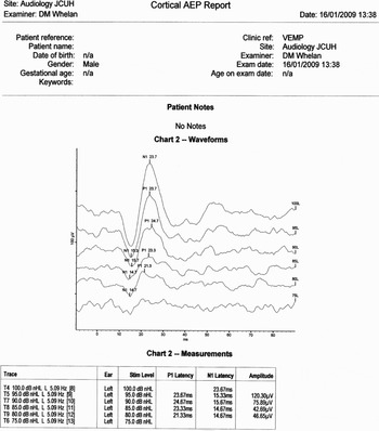
In patients with superior semicircular canal dehiscence, vestibular evoked myogenic potential testing shows decreased thresholds (of 55–70 dB normal hearing level; 100–115 dB sound pressure level) and increased amplitudes (Figure 3).Reference Colebatch, Day, Bronstein, Davies, Gresty and Luxon60–Reference Streubel, Cremer, Carey, Weg and Minor63 Such patients show greatly reduced vestibular evoked myogenic potential amplitudes on skull tap testing, compared with normal subjects; however, on click testing, the vestibular evoked myogenic potential amplitudes of patients and normal subjects are similar.Reference Brantberg, Bergenius and Tribukait64 Galvanic responses remain unchanged.Reference Watson, Halmagyi and Colebatch61 Brantberg et al. Reference Brantberg, Bergenius, Mendel, Witt, Tribukait and Ygge65 and Modugno et al. Reference Modugno, Brandolini, Savastio, Ceroni and Pirodda66 have demonstrated positive CT findings in superior semicircular canal dehiscence syndrome patients with abnormally low vestibular evoked myogenic potential thresholds.
Sample details for vestibular evoked myogenic potential testing in a patient with left-sided superior semicircular canal dehiscence syndrome.

Given the relative ease of vestibular evoked myogenic potential testing, and the high level of reproducibility of results in patients with superior semicircular canal dehiscence syndrome, vestibular evoked myogenic potential testing has now been widely accepted to have a role in the diagnosis of this syndrome.
Acute vestibular neuritis
In patients with vestibular neuritis, clinical examination and caloric tests demonstrate dysfunction of the lateral canal. While either or both of the vestibular nerves can be affected by vestibular neuritis, reports indicate that the superior vestibular nerve is most commonly involved.Reference Halmagyi and Colebatch67–Reference Lesmas Navarro, Pérez Garrigues, Morera Pérez and Piqueras72 Hong et al. Reference Hong, Yeo, Kim and Cha50 found that a third of patients with vestibular neuritis had abnormal vestibular evoked myogenic potentials. Furthermore, Murofushi et al. Reference Murofushi, Halmagyi, Yavor and Colebatch73 noted that the presence or absence of vestibular evoked myogenic potentials (indicating inferior vestibular nerve involvement) in vestibular neuritis patients could predict the risk of subsequent BPPV occurrence.
Therefore, click-evoked vestibular evoked myogenic potential testing may be used in such patients to determine involvement of the inferior vestibular nerve and to assess progress.Reference Ochi, Ohashi and Watanabe68, Reference Chen, Young and Wu70, Reference Murofushi, Halmagyi, Yavor and Colebatch73
Cerebello-pontine angle tumours
Vestibular schwannomas commonly present with unilateral sensorineural hearing loss and tinnitus.Reference Linthicum and Brackmann74 Neurofibromatosis type two is characterised by bilateral vestibular neurofibromas.Reference Rouleau, Wertelecki and Haines75, Reference Martuza and Eldridge76 Of the four nerves passing through the internal auditory canal, all but the inferior vestibular nerve can be assessed by means of auditory brainstem response testing, electroneuronography and caloric testing with videonystagmography.Reference Young, Chiang and Wang77 However, involvement of the inferior vestibular nerve can only ever be determined intra-operatively. Based on the presence of normal vestibular evoked myogenic potentials, Cheng-Ping Wang et al. Reference Wang, Hsu and Young78 have proposed that, in patients with neurofibromatosis type two, the superior vestibular nerve is most commonly involved of the two vestibular nerves. While this finding is similar to the operative findings of Slattery et al.,Reference Slattery, Brackmann and Hitselburger79 a number of studies have shown abnormal amplitudes or complete absence of vestibular evoked myogenic potentials in the majority of patients with vestibular schwannomas.Reference Murofushi, Matsuzaki and Mizuno80–Reference Tsutsumi, Tsunoda, Noguchi and Komatsuzaki85 Patients with large tumours have also been found to have prolonged vestibular evoked myogenic potential latencies.Reference Murofushi, Shimizu, Takegoshi and Cheng49
Vestibular evoked myogenic potential testing alone cannot reliably determine whether such tumours arise from the inferior vestibular nerve or elsewhere.Reference Tsutsumi, Tsunoda, Noguchi and Komatsuzaki85 Although such testing may be useful to detect inferior vestibular nerve involvement and to assess and monitor post-operative residual function, its accuracy and reliability in these respects has yet to be demonstrated.
Acute acoustic trauma and chronic noise-induced hearing loss
The close anatomical proximity of the cochlea and saccule to the stapes footplate exposes them to an increased risk of acoustic trauma.Reference Shupak, Bar-El, Podoshin, Spitzer, Gordon and Ben-David86 It has been shown that industrial workers with noise-induced hearing loss may have sub-clinical balance disorders.Reference Shupak, Bar-El, Podoshin, Spitzer, Gordon and Ben-David86, Reference Osterveld, Polman and Schoonhey87 Long term noise exposure can result in degenerative changes in the auditory sense organs, leading to sensorineural hearing loss.Reference McGill and Schuknecht88 A study involving 20 patients with chronic noise-induced hearing loss found that those with notched thresholds at 4 kHz may exhibit absent or delayed vestibular evoked myogenic potentials.Reference Wang and Young89 Wang et al. Reference Wang, Hsu and Young90 proposed that the absence of vestibular evoked myogenic potentials prior to (dextran) treatment was a predictive factor for treatment response.
There is as yet only limited evidence for the usefulness of vestibular evoked myogenic potential testing in patients with acute or chronic acoustic trauma. However, the link between acoustic insult and vestibular damage is well established, and further studies may find a role for vestibular evoked myogenic potential testing in this clinical context.
Central nervous system disorders
Numerous studies have explored the effect of multiple sclerosis on vestibular evoked myogenic potentials, and have demonstrated decreased amplitudes, prolonged latencies (particularly of p13) and in some cases complete absence.Reference Bickford, Jacobson and Cody6, Reference Shimizu, Murofushi, Sakurai and Halmagyi91–Reference Sartucci and Logi95 It has been proposed that the delay may be due to demyelination of either primary afferent axons at the root entry zone or secondary vestibulo-spinal tract axons, rather than to lesions involving the vestibular nucleus.Reference Shimizu, Murofushi, Sakurai and Halmagyi91 However, when considered alone, the sensitivity of vestibular evoked myogenic potential testing in detecting abnormalities in multiple sclerosis patients appears to vary from 31 to 70 per cent.Reference Versino, Colnaghi, Callieco, Bergamaschi, Romani and Cosi92–Reference Sartucci and Logi95
Vestibular evoked myogenic potentials have also been reported to be altered or absent in patients with other brainstem lesions, such as Wallenberg's syndrome,Reference Itoh, Kim, Yoshioka, Kanaya, Enomoto and Hiraiwa96 Machado–Joseph diseaseReference Takegoshi and Murofushi97 and cerebrovascular accident.Reference Chen and Young98
Therefore, although not diagnostic when used alone, vestibular evoked myogenic potential testing can be a useful test of the integrity of the vestibulo-spinal pathway in patients with suspected multiple sclerosis, and can be considered as a complementary neurophysiological diagnostic tool for patients with other central nervous system lesions.
Gentamicin therapy
Low dose intra-tympanic gentamicin therapy is used to achieve chemical labyrinthectomy, in order to control debilitating vertigo and other peripheral vestibulopathies. In 2002, De Waele et al. Reference De Waele, Meguenni, Freyss, Zamith, Bellalimat and Vidal99 reported that patients with absent vestibular evoked myogenic potentials did not develop recurrent vertigo following such therapy. Further studies are required; however, as vestibular evoked myogenic potentials are reliant on a functioning vestibule, this study demonstrates their potential in monitoring the efficacy of intra-tympanic gentamicin treatment.
Whiplash injury
A recent prospective study by Solarino et al. Reference Solarino, Coppola, Di Vella, Corsalini and Quaranta100 evaluated the role of vestibular evoked myogenic potential testing in the assessment of whiplash injuries, and proposed its use as an important ‘forensic’ diagnostic tool in the assessment of cervical spine injury. On testing subjects with whiplash injury, these authors demonstrated the amplitude of the p1n1 wave to be significantly reduced on day zero but not on day 90, while the vestibular evoked myogenic potential latency was significantly prolonged both on day zero and day 90, on both sides (p < 0.002).
However, this was a small study, and further research is required before vestibular evoked myogenic potential testing can be considered of value in the assessment of whiplash injuries.
Conclusion
Following the initial discovery of vestibular evoked myogenic potentials, it was several years before their possible clinical significance was revisited. Since then, we have developed a much better understanding of the neural pathways involved in this response. Practical aspects of vestibular evoked myogenic potential testing have been refined, and it is now relatively easy and simple to perform. Such testing is very well tolerated by patients, and simple modifications and allowances make it applicable even in the poorly compliant patient.
The current literature lacks consensus regarding the best method of recording vestibular evoked myogenic potentials, and there is a need for more specific research in order to ensure comparability of recordings and to establish a standard for use in clinical practice. The measurable parameters of threshold, amplitude and latency can be influenced by patient factors such as age, neck length and effective activation of the sternocleidomastoid muscle, in addition to any underlying pathology. The majority of research on vestibular evoked myogenic potentials has involved adult subjects. For these reasons, it is important to establish a normative range of data, in order to improve the test's clinical applicability.
A large number of studies have been referenced in this paper, providing an insight into the use of vestibular evoked myogenic potential testing in a variety of vestibular conditions.
There is undisputed consensus regarding the use of vestibular evoked myogenic potential testing in the diagnosis of superior semicircular canal dehiscence syndrome. Abnormally low thresholds and high amplitudes are considered diagnostic, rendering vestibular evoked myogenic potential testing of considerable value in the diagnosis and subsequent monitoring of this condition.
Many other conditions have been found to be associated with vestibular evoked myogenic potentials of prolonged or shortened latency, and often even complete absence. However, current evidence is not sufficiently robust to support the use of vestibular evoked myogenic potential testing to assess the stage or progression of these conditions. The clinical role of vestibular evoked myogenic potential amplitude and threshold levels has also been studied; however, apart from their role in superior semicircular canal dehiscence syndrome, these parameters have not been shown to have a first-line diagnostic use in any other condition. One study demonstrated that the inter-aural amplitude difference may be useful in assessing Ménière's disease stage.
The benefits of vestibular evoked myogenic potential testing have already been established as regards the diagnosis and monitoring of several clinical conditions. Researchers continue to delve deeper into potential new clinical applications, with early results suggesting promising future developments. However, further, high quality evidence is required to confirm the role of this test in routine clinical practice.
Acknowledgement
We thank Professor Timothy C Hain MD, Professor of Neurology, Otolaryngology, and Physical Therapy/Human Movement Science, Northwestern University Medical School, Chicago, Illinois, USA.