Introduction
The vestibular end organs include three semicircular canals, each oriented in a different plane. There are two vertical semicircular planes, the anterior and posterior canals, and one horizontal canal. The vertical canals are oriented roughly at 45° in relation to the sagittal plane, the horizontal canal is tilted upward about 30° from the axial plane, and these canals are orthogonal to each other.Reference Flint and Cummings1 The canals are organised into functional pairs, wherein both members of the pair lie in the same plane. Any rotation in that plane is excitatory to one member of the pair and inhibitory to the other.Reference Wilson and Melvill Jones2 Both horizontal canals form a functional pair, and the anterior canal on one side forms a functional pair with the posterior canal of the contralateral side.Reference Flint and Cummings1, Reference Wilson and Melvill Jones2
In addition to this general knowledge regarding semicircular canals, some reports have provided counter-evidence for the orthogonality of semicircular canals, coplanarity of functional pairs and the well-known angles of semicircular canals to standard planes.Reference Blanks, Curthoys and Markham3, Reference Cox and Jeffery4 Maximal endolymph displacement within a semicircular canal occurs with head rotations in a fixed plane that is close but not necessarily equal to the anatomical semicircular canal plane.Reference Rabbitt5, Reference Curthoys and Oman6 Thus, accurate knowledge of the absolute orientation and position of the semicircular canal is essential for the design of experiments studying the vestibular system and for understanding examination findings during the clinical evaluation of patients with a vestibular disorder.
The present study investigated the angles and orientations of semicircular canals, and the coplanarity of functional canal pairs in magnetic resonance imaging (MRI) scans of normal subjects. The results were applied to the evaluation and treatment of dizzy patients.
Materials and methods
Three-dimensional reconstruction was performed using 20 temporal bone MRI scans from normal subjects. Exclusion criteria were: a history of temporal bone fracture, previous middle-ear or mastoid surgery, pathological findings in the middle ear, congenital anomalies of the ear, and other systemic diseases affecting development of the skull.
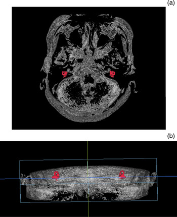
Three-dimensional reconstruction images were obtained using the V-works 4.0 software (Cybermed, Seoul, Korea) and axial scans of T2-weighted constructive interference in the steady state (‘CISS’) images. Enhanced fluid signals in the vestibule and semicircular canals were reconstructed using the surface rendering method (Figure 1). Planes were reconstructed using the three-points method, which was suggested by Aoki et al.Reference Aoki, Takei, Suzuki, Masukawa and Arai7 In this method, the canal plane was determined by three points at the centre of the cross-section of the canal: the ampulla and non-ampulla side ends (bifurcation at the vertical canals) and their approximate midpoint.
Three-dimensional posterior-superior (a) and anterior (b) reconstructions of enhanced fluid signals in the vestibule and semicircular canals (red) using an axial, T2-weighted magnetic resonance image of head structure (white).

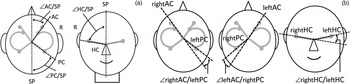
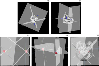
The angles between each semicircular canal plane, the angles between standard planes and canal planes, and the angles between each functional pair of canal planes were measured (Figures 2 and 3).
(a) The angles of the semicircular canal planes to the sagittal plane of the head (right ear).
(‘∠AC/SP’ indicates the anterior open angle of the anterior canal plane to the sagittal plane; ‘∠PC/SP’ reflects the posterior open angle of the posterior canal plane to the sagittal plane; and ‘∠HC/SP’ indicates the superior open angle of the horizontal canal plane to the sagittal plane.) (b) The angular orientation of the contralateral synergistically acting canal pairs. (‘∠rightAC/leftPC’ indicates the angle between the right anterior and left posterior canal planes; ‘∠leftAC/rightPC’ reflects the angle between the left anterior and right posterior canal planes; and ‘∠rightHC/leftHC’ indicates the angle between the right and left horizontal canal planes.) AC = anterior canal; R = right; PC = posterior canal; SP = sagittal plane; HC = horizontal canal. Adapted with permission.7

Three-dimensional planar analysis of enhanced fluid signals of semicircular canals conducted using axial magnetic resonance imaging (MRI), showing a superior axial view (a) and an antero-inferior axial view (b), and synergistic canal planes evaluated by simultaneous reconstruction using MRI, showing a superior view (c), a posterolateral view (d) and a superior sagittal view (e).

Results
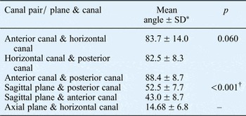
The mean angles between the anterior semicircular canal plane and horizontal semicircular plane, the horizontal semicircular plane and posterior semicircular plane, and the anterior semicircular plane and posterior semicircular plane were 83.7°, 82.5° and 88.4°, respectively; the angles between each canal pair showed no difference (Table I). Measurement of angles between standard planes and canal planes revealed a significantly larger angle between the sagittal plane and posterior canal plane than between the sagittal plane and anterior canal plane (52.5 ± 7.7° vs 43.0 ± 8.7°, respectively). Additionally, the mean angle between the axial plane and horizontal canal plane was measured as 14.68 ± 6.8°.
Mean angles between each semicircular canal pair, and between standard planes and each canal

*n = 20. †p < 0.05 (one-way analysis of variance, Student's t-test). SD = standard deviation
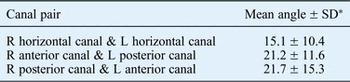
Pairs of contralateral synergistic canal planes formed 15.1° between the right and left horizontal semicircular canal planes, 21.2° between the right anterior semicircular canal and left posterior semicircular canal, and 21.7° between the left anterior semicircular canal and right posterior semicircular canal (Table II).
Mean angles of contralateral synergistic canal pairs

*n = 20. SD = standard deviation; R = right; L = left
Discussion
In 1975, Blanks et al. reported that the angles between human ipsilateral semicircular canals depart significantly from mutual orthogonality, with the angle between the anterior canal and horizontal canal equalling 111.8 ± 7.6° (mean ± standard deviation).Reference Blanks, Curthoys and Markham3 In their study, they used the dissected bony labyrinths of 10 human skulls. These were fixed in a stereotactic frame and the angles of semicircular canals were measured using plain X-ray films. Multiple studies have since been conducted on the inter-semicircular canal angles of human labyrinths using high-resolution histological or radiographic reconstructions. However, subsequent studies have suggested that semicircular canal orientations are much more orthogonal to one another.Reference Aoki, Takei, Suzuki, Masukawa and Arai7–Reference Spoor and Zonneveld12 Our results also showed a much greater degree of semicircular canal orthogonality than those of Blanks et al.,Reference Blanks, Curthoys and Markham3 and the angles between the canals showed no differences.
In analyses of inter-semicircular angles, the curvature of the semicircular canal itself is a major obstacle. One study investigating the torsion of semicircular canal reported a mean torsion range of 8.5° to 16.4°, and the anterior canal showed maximal torsion compared with the other canals.Reference Sato, Sando, Takahashi and Fujita13 A subsequent, well-designed study using reconstructions of high-resolution computed tomography images also reported torsion of semicircular canals (20.9 ± 7.3° to 27.6 ± 5.2°) and maximal non-planarity of the anterior canal.Reference Bradshaw, Curthoys, Todd, Magnussen, Taubman and Aw10 In the present study, we used the three-points method of Aoki et al.Reference Aoki, Takei, Suzuki, Masukawa and Arai7 This method has several advantages: it is easy to perform, takes little time and has high inter-observer reliability. However, it is also associated with some limitations; specifically, it cannot reflect the curvature of the semicircular canal and actual angle of the membranous canal. Because of these limitations, our measurements may not represent precisely the biologically acting canal angles.
However, some reports do not corroborate our reported limitations. First, it has been found that measurements of bony canal angles provide reasonable estimates of the delicate membranous canals – the two differed by only 3.48 ± 1.89°.Reference Ifediba, Rajguru, Hullar and Rabbitt14 Second, some evidence suggests that torsions of the semicircular canals contribute to maintaining the angles between pairs of canals closer to right angles.Reference Sato, Sando, Takahashi and Fujita13 Considering these reports, each semicircular canal seems to make an almost right angle with other canals.
Regarding the analysis of the angles between each semicircular canal and standard plane, our results showed some disagreements with those determined previously. The horizontal canal showed a smaller angle with the axial plane than the known value (14.68 ± 6.8°), and the posterior canal showed a significantly wider mean angle with the sagittal plane than the anterior canal (52.5 ± 7.7° vs 43.0 ± 8.7°, respectively). This wider angle between the posterior canal and sagittal plane marks a departure from coplanarity between the anterior canal of one side and the posterior canal of the other side. Our results indicated an approximately 20° angle difference between the canal planes of the anterior canal of one side and those of the posterior canal of the other side. Several previous studies reported that the range of this angle was 15° to 20°.Reference Blanks, Curthoys and Markham3, Reference Aoki, Takei, Suzuki, Masukawa and Arai7, Reference Della Santina, Potyagaylo, Migliaccio, Minor and Carey9 Additionally, both horizontal canals showed some dis-coplanarity in our study, and the angle between them was 15.1 ± 10.4°.
However, these results would not mean denying the synergistic action of both labyrinths. Della Santina et al.Reference Della Santina, Potyagaylo, Migliaccio, Minor and Carey9 suggested that this departure from coplanarity was negligible because head rotation stimulates canals to a degree dictated by the cosine of the angle between the head rotation axis and the semicircular canal axis. They also stated that rotation of the head about an axis midway between the mean left anterior canal and mean right posterior canal plane axes would simulate rotation about the axis of each semicircular canal to within 0.9 per cent. Otherwise, neural plasticity may overcome the functional consequences of such dis-coplanarity.Reference Bradshaw, Curthoys, Todd, Magnussen, Taubman and Aw10
When we perform the Dix–Hallpike manoeuvre or canalith repositioning manoeuvre, it is noteworthy that the posterior semicircular canal has a wider angle with the sagittal plane than the anterior canal. As previously shown, benign paroxysmal positional vertigo (BPPV) of the posterior canal is the most common type of BPPV.Reference Parnes, Agrawal and Atlas15, Reference Imai, Ito, Takeda, Uno, Matsunaga and Sekine16 During the performance of manoeuvres for the evaluation and treatment of posterior canal BPPV, the exact head rotation to the actual plane of the semicircular canal would be helpful. Our reported angle between the horizontal canal plane and the axial plane, which is smaller than that described previously, is also important for exact stimulation of the horizontal canal when performing the head thrust test or rotation chair test. A previous well-designed study reported results similar to ours, in which the angle between the horizontal canal plane and axial plane was 18.8°.Reference Della Santina, Potyagaylo, Migliaccio, Minor and Carey9 Furthermore, when we encounter patients with intractable BPPV, the variability of the canal plane or curvature of the canal itself could be a cause of unsuccessful positioning. A previous MRI study of patients with intractable BPPV assessed only the shape of the canal, and revealed stenosis and obstruction of the canal as a cause of intractability in some patients; however, the canal plane was not evaluated.Reference Horii, Kitahara, Osaki, Imai, Fukuda and Sakagami17 We believe that analysis of the canal plane may provide additional information concerning unsuccessful treatment, in addition to obstruction or stenosis of the canal.
• Vertical semicircular canals are at 45° in relation to the sagittal plane, and the horizontal semicircular canal is tilted 30° from the axial plane; these canals are orthogonal to each other
• In this study, each of the semicircular canals made an almost right angle with the other two canals
• The synergistically acting functional canal pairs of both ears did not lie in exactly the same plane
• The posterior semicircular canal made a significantly wider angle with the sagittal plane than the anterior canal
• The angle between the horizontal canal plane and axial plane was smaller than reported previously
• Examining individual variability of semicircular canal angles using reconstruction images may be helpful in intractable benign paroxysmal positional vertigo cases
In conclusion, each semicircular canal makes an almost right angle with the other canals, and synergistically acting functional canal pairs of both ears are almost parallel but do not lie in exactly the same plane. The posterior semicircular canal makes a significantly wider angle with the sagittal plane than the anterior canal, and the angle between the horizontal canal plane and the axial plane is smaller than that reported previously. Examining the individual variability of the angle of the semicircular canal using three-dimensional reconstruction images may be helpful in intractable BPPV patients.