Mycobacterium gordonae is ubiquitous in nature and potable water systems and is a cause of endemic contamination of clinical specimens. Nontuberculous mycobacteria proliferate within biofilms in pipes carrying potable water,Reference Falkinham, Norton and LeChevallier 1 , Reference Mullis and Falkinham 2 a phenomenon which is promoted by water stagnancy.Reference Williams, Armbruster and Arduino 3 Previous M. gordonae pseudo-outbreaks have resulted from a variety of contaminated water sources, including drinking water,Reference Arnow, Bakir, Thompson and Bova 4 ice machines,Reference Lalande, Barbut and Varnerot 5 , Reference Panwalker and Fuhse 6 laboratory water,Reference Stine, Harris, Levin, Rivera and Kaplan 7 antimicrobial solutions,Reference Tokars, McNeil and Tablan 8 superoxidized water,Reference Fujita, Nanki, Negayama, Tsutsui, Taminato and Ishida 9 and bronchoscope cleaning fluid.Reference Gubler, Salfinger and von Graevenitz 10 Respiratory specimens contaminated by M. gordonae are more likely to be collected from patients who consumed fluids within 48 hours of sample collection compared with those who had no access to drinking water.Reference Arnow, Bakir, Thompson and Bova 4 Although M. gordonae is a rare cause of disease, the presence of acid-fast bacilli in clinical specimens prior to species identification has potential adverse effects, including prolonged airborne isolation precautions, presumptive anti-mycobacterial therapy, and prolonged hospital stays.
On January 9, 2012, our medical center opened a new 376-bed hospital that houses general medical and surgical units, intensive care units, operating rooms, radiology, and the emergency department. The new hospital is located adjacent to the old hospital but has a separate water distribution system which, as in the old hospital, comes from a Chicago city water supply line. All water pipes in the new hospital were treated with hyperchlorination 6 months prior to opening, and the hot water has been continuously treated with a silver-copper ionization system since opening. A total of 300 patient beds, as well as the central microbiology laboratory, remain in the old hospital. During the pseudo-outbreak, one floor in the new hospital and one floor in the old hospital were unoccupied.
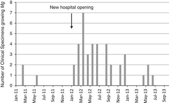
The prevalence of M. gordonae in clinical mycobacterial cultures from inpatients increased from a baseline of 19 of 10,029 samples (0.19%; 95% confidence interval (CI), 0.12–0.3 from 2009 to 2011 and 13 of 771 samples (1.7%; 95% CI, 0.99–2.9) during the first 3 months of 2012 (Figure 1). Infection control staff observed that the most common type of specimen was sputum and that all positive cultures were considered contaminants by treating physicians. The clinical microbiology laboratory did not appear to be the source of the outbreak, as potable water was not used in the processing of mycobacterial specimens and all reagents used were cultured daily as part of the routine laboratory procedure.
Number of clinical specimens growing M. gordonae (Mg) by month, January 2011 to September 2013.

Based on the experience following construction of a neighboring hospital (oral personal communication, Sharon F. Welbel, MD, Robert A. Weinstein, MD, and Kathleen G. Beavis, MD, January 2012), we suspected that the M. gordonae-positive cultures represented endemic contamination of patient specimens from ingestion of water in the new hospital and that water reservoirs in the new water distribution system contained higher concentrations of M. gordonae compared with those of the old hospital. We also hypothesized that (1) water from rooms previously occupied by M. gordonae-positive patients had a higher prevalence and concentration of M. gordonae compared with rooms not previously occupied by these patients, (2) unoccupied floors of the medical center (located in both the new and the old hospitals) had a higher prevalence and concentration of M. gordonae compared with occupied floors, and (3) water from ice and water dispensers had a higher prevalence and concentration of M. gordonae compared with water from other potable water sources.
Methods
Pseudo-outbreak Investigation
We reviewed the results of all mycobacterial cultures from specimens processed at our medical center between January 9, 2012, and December 31, 2012, to identify all hospitalized patients with M. gordonae-positive specimens during the outbreak period. The following data were abstracted from medical records: date of specimen collection, patient location at the time of specimen collection, specimen source, presence of acid fast bacilli by microscopy, patient length of stay, patient age, coexisting chronic lung disease or immunocompromising condition, oral intake within 48 hours of specimen collection, anti-mycobacterial treatment rendered (if any), potential adverse effects of treatment, and institution of airborne isolation precautions. Immunocompromising condition was defined as having human immunodeficiency virus infection, diabetes, active malignancy, or receiving chronic immunosuppressive therapy. Patients with M. gordonae-positive specimens were considered to have mycobacterial contamination, rather than infection, if there was no histopathologic evidence of infection and/or if they did not receive definitive treatment for M. gordonae.
Environmental Cultures and Laboratory Methods
We collected 50 random water samples from sink faucets, shower heads, and drinking fountains in the following 4 medical center areas: unoccupied new hospital, occupied new hospital, unoccupied old hospital, and occupied old hospital. An inventory of all water sources was conducted, and a random number generator was used to select samples. Additionally, we collected water samples from all ice and water dispensers (Follett© Symphony™ ice and water dispenser, Easton, PA) and from all sinks and showers in rooms previously occupied by a subset of patients whose clinical cultures grew M. gordonae between January 9, 2012 and September 30, 2012. A mixture of hot and cold water was collected from shower heads and sink faucets. Cold water was collected from ice and water dispensers. All samples were collected between October 11, 2012, and November 13, 2012.
From each source, 50 mL of water was collected in a sterile 50-mL plastic screw-capped centrifuge tube and transported to the clinical microbiology laboratory, where it was stored at 4°C for up to 24 hours. Each sample was centrifuged, and 10 mL of supernatant was retained for digestion and decontamination with an equal volume of 4% sodium hydroxide, 2.9% sodium citrate, and N-acetyl-L-cysteine solution (NAC-Attack, Thermo Scientific™, Lenexa, KS). Specimens were agitated on a vortex mixer for 30 seconds and allowed to stand for 15 minutes at room temperature. To the tube, 30 mL of M/15 phosphate buffer was then added and centrifuged for 30 minutes. After decanting the supernatant, 1 mL of Bovine Serum Albumin Fraction V, 0.2%, and 0.5 mL of M/15 phosphate buffer were added to the sediment to create a suspension. 0.5 mL of suspension was inoculated to Bactec™ MGIT™ 960 Mycobacterial Detection System (Bactec™, Franklin Lakes, NJ), and 0.25 mL of suspension was inoculated to Middlebrook 7H10 agar plates (Remel™). Cultures were incubated for 8 weeks, after which all Kinyoun stain–positive colonies growing on Middlebrook agar were counted. Broth from MGIT tubes underwent Kinyoun staining at the time of bacterial growth detection. Of each Kinyoun stain-positive specimen, 0.2–0.5 mL was inoculated to Middlebrook 7H10 agar for species identification. Species were presumptively identified by pigment production and morphologic characteristics by two independent laboratory technicians (JC and MLS). DNA probe (Hologic® Gen Probe, San Diego, CA) or DNA sequence analysis were used to identify colonies when lack of agreement between technicians or questionable colony morphology was present. Clinical specimens at our institution are processed in an identical manner to that described for environmental specimens, except that those from normally sterile sites do not undergo decontamination and digestion.
Statistical Analysis
A sample size of 50 water samples per group was chosen to achieve at least 80% power to detect a 25% difference in M. gordonae prevalence between the old hospital and the new hospital, assuming a prevalence of 10% in the old hospital, with an α level of 0.05 in a two-sided Fisher’s exact test for proportions. Comparative analyses were performed using Fisher’s exact test for proportions and Wilcoxon–Mann–Whitney test for continuous variables. P<.05 was considered significant.
Results
Description of Cases
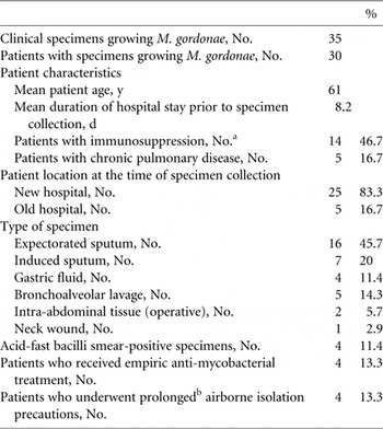
From January 9, 2012, to December 31, 2012, 35 clinical specimens from 30 inpatients grew M. gordonae, with a prevalence of 35 of 3,035 (1.2%, 95% CI [0.83, 1.6]). Between weeks 3 and 6, 74.3% of clinical cultures grew M. gordonae. Most patients (83.3%) were housed in the new hospital at the time of specimen collection (Table 1). All 30 patients had access to potable water at least 48 hours prior to specimen collection. In most cases, specimens were tested to exclude pulmonary infection, particularly Mycobacterium tuberculosis (24 of 30 patients). Four patients were started on empiric treatment for mycobacterial infections (3 for M. tuberculosis and 1 for Mycobacterium avium complex) based on specimens with detectable acid-fast bacilli upon direct microscopy. Duration of treatment ranged from 5 days to 3 months. One patient developed transaminitis and another developed hyperbilirubinemia, both of which were attributed to anti-tuberculous therapy and necessitated early discontinuation. Two other patients continued therapy until their smear-positive specimen either grew M. gordonae or had no growth in culture after 3 months. Airborne isolation precautions for patients suspected of M. tuberculosis infection were continued for 3–7 weeks, until the mycobacterial species was identified as M. gordonae. All cases of M. gordonae were considered specimen contamination, as no patient had histopathologic evidence of infection or received definitive treatment for M. gordonae infection.
Summary of M. gordonae Clinical Cases Between January 9, 2012, and December 31, 2012

a Defined as having human immunodeficiency virus infection, diabetes, active malignancy, or receiving chronic immunosuppressive therapy.
b >7 d.
Environmental Cultures
We sampled potable water from 270 hospital sources: 194 samples from randomly selected sinks, showers, and drinking fountains in patient rooms and common areas of the new and old hospitals, 48 sinks and showers from rooms previously occupied by M. gordonae–positive patients, and 28 ice and water dispensers. Most of the samples from areas previously occupied by M. gordonae–positive patients and from ice and water dispensers were from the new hospital (81.3% and 67.9%, respectively). Of 270 water samples, 194 (71.9%) were from patient rooms (126 sinks and 68 showers) and 76 were from common areas (ice and water dispensers, drinking fountains, eyewash stations, restrooms, scrub sinks, hallway sinks, medication rooms, nourishment rooms, soiled utility rooms, and environmental services areas). There was no clustering of environmental samples around any particular wing of either hospital.
The overall prevalence of mycobacteria was 246 of 270 samples (91.1%). The prevalence of M. gordonae was 238 of 270 samples (88.1%). In the majority of cases (89.1%), liquid media cultures grew M. gordonae between weeks 1 and 4. M. gordonae comprised 75.8% unique mycobacterial isolates (238 of 314 isolates). Other mycobacterial species included M. avium complex (18.2%), Mycobacterium mucogenicum (4.1%), rapidly growing mycobacteria (1.6%), and Mycobacterium xenopi (0.3%). The identities of 34 of 270 mycobacterial isolates (12.6%) were confirmed by DNA probe or DNA sequence analysis.
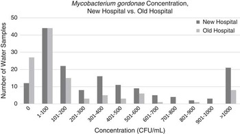
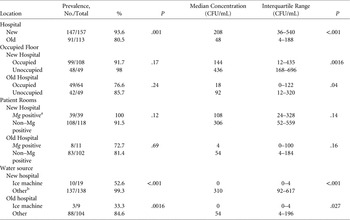
The prevalence and median concentration of M. gordonae were significantly greater in water collected from the new hospital compared with the old hospital (Table 2). The proportion of water samples with >1,000 CFU/mL of M. gordonae was also significantly greater in the new hospital compared with that of the old hospital [21 of 157 samples (13.4%) vs. 8 of 113 samples (7.1%), P=.03]. The distribution of M. gordonae concentration across water sources was skewed toward lower concentrations (0–200 CFU/mL) in both the new and the old hospital (Figure 2). Water from ice and water dispensers had a significantly lower prevalence and concentration of M. gordonae compared with water from sinks, showers, and drinking fountains (Table 2). No significant difference in the prevalence of M. gordonae was noted between occupied versus unoccupied floors or between rooms previously occupied by M. gordonae-positive patients and rooms not previously occupied by those patients. Unoccupied floors, however, had a significantly higher concentration of M. gordonae compared with occupied floors.
Concentration of M. gordonae Among Water Samples in the New Hospital vs. Old Hospital

Prevalence and Median Concentration of M. gordonae (Mg) in Water Samples from Various Locations Within our Medical Center.

a Sinks and showers from rooms previously occupied by patients with Mg-positive cultures between January and September 2012.
b Sink faucets, shower heads, and drinking fountains.
Discussion
We investigated a pseudo-outbreak of M. gordonae among 30 patients at our institution that occurred in association with the opening of a new hospital and found a reservoir of M. gordonae in the water supply, with 88.1% of all water samples tested growing M. gordonae. The highest prevalence of M. gordonae was found in water collected from the new hospital (93.6%), although water from the old hospital had a surprisingly high prevalence of M. gordonae as well (80.5%). The more significant difference was likely the 4-fold greater median concentration of M. gordonae in the water of the new hospital compared with that of the old hospital. Similarly, the median concentration of M. gordonae in water samples from unoccupied floors was 3- to 5-fold greater than those from occupied floors. Rooms previously occupied by M. gordonae–positive patients did not show an increased prevalence or concentration of M. gordonae in potable water samples. Of all hospital water sources included in our study, samples from ice and water dispensers showed the lowest prevalence and concentration of M. gordonae.
These results are consistent with an endemic source of M. gordonae specimen contamination among our 30 case patients. Of the 35 specimens growing M. gordonae, 32 (sputum, BAL, gastric lavage) had potential for contamination via exposure to hospital potable water. A minority of M. gordonae–positive specimens (11.4%) had detectable acid-fast bacilli upon direct microscopy, which is consistent with the low concentration of M. gordonae overall. Although the median concentration of M. gordonae in the new hospital water supply was 208 CFU/mL, 13.4% of water samples contained >1,000 CFU/mL M. gordonae. This is consistent with our findings that 4 of 35 M. gordonae–positive clinical specimens (11.4%) had detectable acid-fast bacilli upon direct microscopy, given that approximately 5,000 organisms are necessary for a positive acid-fast bacilli smear. We believe that this high concentration of M. gordonae in the new hospital water supply was above the threshold required to contaminate the oropharynges or gastrointestinal tracts of case patients and to grow in clinical cultures.
None of our case patients were thought to have M. gordonae infection, and empiric anti-mycobacterial treatment was discontinued after species identification in all cases. Nevertheless, four patients received empiric multi-drug therapy ranging from 5 days to 3 months, and 2 of these patients experienced adverse events (transaminitis and hyperbilirubinemia) from their treatment. The risk of drug-induced liver injury during treatment for active tuberculosis is significant, with a range of 5–33%.Reference Saukkonen, Cohn and Jasmer 11 Of our case patients, 4 were also placed in prolonged airborne isolation for up to 7 weeks as a precaution, which delayed hospital discharge. Although M. gordonae is an infrequent cause of disease,Reference Griffith, Aksamit and Brown-Elliott 12 the presence of acid-fast bacilli in clinical specimens of patients suspected of having mycobacterial disease may lead to unnecessary exposure to hepatotoxic medications and prolonged hospitalization.
Several authors have described outbreaks or pseudo-outbreaks of nontuberculous mycobacteria within hospitals. Most of these, however, have been associated with contamination of specific water sources or apparatuses such as bronchoscope cleaning fluidReference Gubler, Salfinger and von Graevenitz 10 or ice and water dispensers.Reference Lalande, Barbut and Varnerot 5 , Reference Panwalker and Fuhse 6 Like Arnow et al,Reference Arnow, Bakir, Thompson and Bova 4 we found a significant reservoir of M. gordonae within our hospital potable water supply. The prevalence and concentration of M. gordonae at our institution was much higher than that described by Arnow et al, which may have been due to prolonged stasis of water in our hospital water distribution system as well as differences in city water supply and mycobacterial culturing methods.
Interestingly, a similar pseudo-outbreak of M. gordonae occurred in Chicago when the 464-bed county hospital relocated to a new adjacent building in December 2002. Prevalence of M. gordonae in sputum mycobacterial cultures increased from 50 of 26,799 (0.19%) to 397 of 25,536 (1.5%) during the first 12 months after the new hospital opened. Similar to our experience, none of the 292 patient cases had M. gordonae infection. An outbreak investigation performed 3 months after hospital opening revealed that 36 of 85 of hospital potable water sources (42.4%) sampled grew M. gordonae. Rates of M. gordonae remained elevated until April 2005, 28 months after hospital opening, despite hyperchlorination of the hospital water supply (oral personal communication, Sharon F. Welbel, MD, Robert A. Weinstein, MD, and Kathleen G. Beavis, MD, August 2012).
We were unable to find any published reports of outbreaks or pseudo-outbreaks of M. gordonae following new hospital openings, but we suspect that this phenomenon may be underreported. Given that the concentrations of M. gordonae in unoccupied areas of the new and old hospitals were >3 times those in occupied areas in our study, we hypothesize that stagnant water in pipes of new buildings promotes the growth of M. gordonae in biofilm, similar to the promotion of the growth of Legionella species.Reference Doebbeling and Wenzel 13 , Reference Krøjgaard, Krogfelt, Albrechtsen and Uldum 14 In our case, the water distribution system in the new hospital was completed 6 months prior to hospital opening, during which time point-of-use fixtures were turned on biweekly rather than daily.
Our institution’s M. gordonae prevalence returned to the baseline level 1 year after the opening of the new hospital, without any specific intervention other than treatment of the potable hot water with a continuous copper-silver ionization system. With the exception of water from drinking fountains and ice and water dispensers, all samples in our study contained a mixture of hot and cold water. Copper-silver ionization has been shown to control the growth of Legionella and Pseudomonas in biofilm.Reference Shih and Lin 15 We believe that copper-silver ionization may slow the growth of mycobacteria in a similar manner, although we did not specifically assess pipes for biofilm formation. Continuous water flow within pipes also likely contributed to the resolution of the pseudo-outbreak by decreasing the concentration of M. gordonae in biofilm over time.
Another difference between our study and others is that the prevalence and concentration of M. gordonae in ice and water dispensers was significantly lower than in other water sources (i.e., showers and sinks). Several authors have implicated contaminated ice machines as the source of M. gordonae pseudo-outbreaks.Reference Lalande, Barbut and Varnerot 5 , Reference Panwalker and Fuhse 6 One reason for this difference may be that the ice and water dispensers at our medical center contain a silver ion-impregnated 0.5-μm filter system. Similar filters have been used as part of interventions to control mycobacterial outbreaks at other institutions.Reference Gebo, Srinivasan, Perl, Ross, Groth and Merz 16 , Reference Williams, Chen and Keane 17
Several limitations were inherent in our study. First, no environmental samples were collected prior to January 2012, so we could not compare the prevalence or concentration of M. gordonae in our study to a preconstruction period. The inclusion of the “occupied old hospital” group, however, provided a background rate of M. gordonae against which other groups were compared. Additionally, we were unable to differentiate between contamination and colonization of M. gordonae, as we collected only one clinical specimen from most of our case patients.
In conclusion, we identified a reservoir of M. gordonae within the potable water supply at our institution that led to endemic contamination of clinical specimens and a pseudo-outbreak of M. gordonae. Infection control staff should be aware of this phenomenon upon the opening of a new hospital, as this may lower the suspicion for true mycobacterial disease and decrease the number of patients exposed to empiric antimycobacterial treatment and subjected to airborne isolation precautions. Future studies should focus on the potentially protective role of ice and water dispensers, specifically, the silver ion-impregnated 0.5-μm filter system, in mycobacterial contamination of water.
Acknowledgments
We would like to thank Onofre Donceras and Pamela Hagan for their assistance with patient data collection.
Potential conflicts of interest and financial support:
Mary Alice Lavin received financial support from the Association of Practitioners in Infection Control.
All other authors report no relevant conflicts or financial support related to this study.